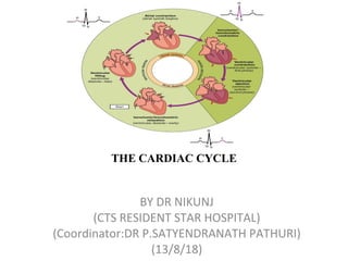
The cardiac cycle consists of mechanical events in the heart that occur in a repeating pattern. It begins with late diastole where the atrioventricular valves open and the heart fills with blood passively. During atrial systole, the atria contract and push a small amount of additional blood into the ventricles. Ventricular systole then begins with isovolumic contraction where the ventricles contract but no blood is ejected due to closed valves. Rapid ejection occurs when the semilunar valves open and blood is forcefully pumped out of the ventricles. The cycle ends with isovolumic relaxation and diastolic filling as the ventricles relax and refill with blood.
Cardiac cycle (The Guyton and Hall physiology)Maryam Fida
Sequence of events from the beginning of one systole to the beginning of next consecutive systole.
One heart beat consists of one systole and one diastole.
Each cardiac cycle is initiated by the cardiac impulse which originates from the SA node.
During each cardiac cycle, certain events occur in the heart and these include pressure changes, volume changes, production of heart sounds, closure and opening of heart valves and electrical changes in the heart.
Useful for medical and biology students who want to study the cardiac cycle in a short time with big benefits !!
CVS physiology - Wigger Diagram - ECG of cardiac cycle - Heart sounds
CARDIAC CYCLE, ECG AND HEART SOUNDS.pptxthiru murugan
CARDIAC CYCLE, ECG AND HEART SOUNDS: BY Wincy Thirumurugan..
“Cardiac cycle refers to the series of events that take place when the heart beats.”
Each cycle is initiated by spontaneous contraction in the SA node and then transmit through the A-V bundle and branches into the ventricles results completion of one cycle.
EVENTS OR PHASES OF CARDIAC CYCLE: Diastolic phase (Diastole) in this phase the heart chamber are in the state of relaxation and fills with blood that receives from the veins [IVC, SVC,PULMONARY VEINS]
Systolic phase (Systole) in this the heart chambers are contracting and pumps the blood towards the periphery via the arteries. [ Pulmonary artery and aorta]
PHASES OF THE CARDIAC CYCLE
The different phases of the cardiac cycle involve:
Atrial diastole - Atrial relaxation
Atrial systole -Atrial contraction
Isovolumic relaxation -ventricular relaxation in the early phase but blood will not move and the Atrio ventricular valves will be closed
Ventricular filling - ventricular relaxation, the Atrio ventricular valves will be open allows filling blood in the ventricles
Isovolumic contraction of ventricle – ventricular systole in the early phase but no movement of the blood. The semilunar valves will be closed.
Ventricular ejection -ventricular contraction and send blood out of the ventricles through opened semilunar valves.
6. Ventricular Filling Stage: second phase. Rapid Filling, Slow Filling & Last Rapid Filling Duration of Cardiac Cycle:
In a normal person, a heartbeat is 72 beats/minute.
An Electrocardiogram (ECG) is a medical test that detects cardiac (heart) abnormalities by measuring the electrical activity generated by the heart as it. The machine that records the patient’s ECG is called an electrocardiograph.
contracts.
PLACEMENT OF ECG LEADS
ECG WAVES:
The P wave is caused by spread of depolarization through the atria, After the onset of the P wave, The QRS waves Occurs as a result of electrical depolarization of the ventricles, the ventricular T wave represents the stage of repolarization of the ventricles, The 'U' wave is a wave comes after the T wave of ventricular repolarization and may not always be observed.
HEART SOUNDS: First Heart Sound (S1)
The first heart sound results from the closing of the mitral and tricuspid valves. Second Heart Sound (S2): The second heart sound is produced by the closure of the aortic and pulmonic valves. Third Heart Sound (S3):
The third heart sound, also known as the “ventricular gallop,” occurs just after S2 when the mitral valve opens, allowing passive filling of the left ventricle. The S3 sound is actually produced by the large amount of blood striking a very compliant LV.
[Compliance heart means how easily the chamber of heart or the lumen of blood vessels expands when it is filling with the blood]
Fourth Heart Sound (S4):
The fourth heart sound, also known as the “atrial gallop,” occurs just before S1 when the atria contract to force blood into the LV.
venous drainage of head and neck and its branches are described in detail along with applied anatomy for better understanding of the anatomy and its application in oral and maxillary surgeries. knowing the anatomy and the course of the veins is crucial and helps in better locating the vein and ligating it to avoid further complications while performing a oral and maxillofacial surgeries such as in trauma fixation, tumor resection and as well as reconstruction of the defect pertaining to the maxillofacial region.
Describe events in cardiac cycle.
Describe atrial, ventricular and aortic pressure changes during cardiac cycle.
Describe the changes in ventricular volume & stroke volume during cardiac cycle.
Relate ECG changes to the phases of cardiac cycle.
Describe the functions of cardiac valves and relate their state to the production of heart sounds during cardiac cycle.
med_students0
Def: The cardiac events that occur from
beginning of one heart beat to the beginning of
the next.
■ first assembled by Lewis in 1920 but first
conceived by Wiggers in 1915 Atria act as PRIMER PUMPS for
ventricles & ventricles provide major
source of power for moving the blood
through the vascular system.
■ Initiated by spontaneous generation of
AP in SA node (located in the superior lateral wall of
the right atrium near the opening of the superior vena cava)
New Directions in Targeted Therapeutic Approaches for Older Adults With Mantl...i3 Health
i3 Health is pleased to make the speaker slides from this activity available for use as a non-accredited self-study or teaching resource.
This slide deck presented by Dr. Kami Maddocks, Professor-Clinical in the Division of Hematology and
Associate Division Director for Ambulatory Operations
The Ohio State University Comprehensive Cancer Center, will provide insight into new directions in targeted therapeutic approaches for older adults with mantle cell lymphoma.
STATEMENT OF NEED
Mantle cell lymphoma (MCL) is a rare, aggressive B-cell non-Hodgkin lymphoma (NHL) accounting for 5% to 7% of all lymphomas. Its prognosis ranges from indolent disease that does not require treatment for years to very aggressive disease, which is associated with poor survival (Silkenstedt et al, 2021). Typically, MCL is diagnosed at advanced stage and in older patients who cannot tolerate intensive therapy (NCCN, 2022). Although recent advances have slightly increased remission rates, recurrence and relapse remain very common, leading to a median overall survival between 3 and 6 years (LLS, 2021). Though there are several effective options, progress is still needed towards establishing an accepted frontline approach for MCL (Castellino et al, 2022). Treatment selection and management of MCL are complicated by the heterogeneity of prognosis, advanced age and comorbidities of patients, and lack of an established standard approach for treatment, making it vital that clinicians be familiar with the latest research and advances in this area. In this activity chaired by Michael Wang, MD, Professor in the Department of Lymphoma & Myeloma at MD Anderson Cancer Center, expert faculty will discuss prognostic factors informing treatment, the promising results of recent trials in new therapeutic approaches, and the implications of treatment resistance in therapeutic selection for MCL.
Target Audience
Hematology/oncology fellows, attending faculty, and other health care professionals involved in the treatment of patients with mantle cell lymphoma (MCL).
Learning Objectives
1.) Identify clinical and biological prognostic factors that can guide treatment decision making for older adults with MCL
2.) Evaluate emerging data on targeted therapeutic approaches for treatment-naive and relapsed/refractory MCL and their applicability to older adults
3.) Assess mechanisms of resistance to targeted therapies for MCL and their implications for treatment selection
Ethanol (CH3CH2OH), or beverage alcohol, is a two-carbon alcohol
that is rapidly distributed in the body and brain. Ethanol alters many
neurochemical systems and has rewarding and addictive properties. It
is the oldest recreational drug and likely contributes to more morbidity,
mortality, and public health costs than all illicit drugs combined. The
5th edition of the Diagnostic and Statistical Manual of Mental Disorders
(DSM-5) integrates alcohol abuse and alcohol dependence into a single
disorder called alcohol use disorder (AUD), with mild, moderate,
and severe subclassifications (American Psychiatric Association, 2013).
In the DSM-5, all types of substance abuse and dependence have been
combined into a single substance use disorder (SUD) on a continuum
from mild to severe. A diagnosis of AUD requires that at least two of
the 11 DSM-5 behaviors be present within a 12-month period (mild
AUD: 2–3 criteria; moderate AUD: 4–5 criteria; severe AUD: 6–11 criteria).
The four main behavioral effects of AUD are impaired control over
drinking, negative social consequences, risky use, and altered physiological
effects (tolerance, withdrawal). This chapter presents an overview
of the prevalence and harmful consequences of AUD in the U.S.,
the systemic nature of the disease, neurocircuitry and stages of AUD,
comorbidities, fetal alcohol spectrum disorders, genetic risk factors, and
pharmacotherapies for AUD.
These simplified slides by Dr. Sidra Arshad present an overview of the non-respiratory functions of the respiratory tract.
Learning objectives:
1. Enlist the non-respiratory functions of the respiratory tract
2. Briefly explain how these functions are carried out
3. Discuss the significance of dead space
4. Differentiate between minute ventilation and alveolar ventilation
5. Describe the cough and sneeze reflexes
Study Resources:
1. Chapter 39, Guyton and Hall Textbook of Medical Physiology, 14th edition
2. Chapter 34, Ganong’s Review of Medical Physiology, 26th edition
3. Chapter 17, Human Physiology by Lauralee Sherwood, 9th edition
4. Non-respiratory functions of the lungs https://academic.oup.com/bjaed/article/13/3/98/278874
Title: Sense of Smell
Presenter: Dr. Faiza, Assistant Professor of Physiology
Qualifications:
MBBS (Best Graduate, AIMC Lahore)
FCPS Physiology
ICMT, CHPE, DHPE (STMU)
MPH (GC University, Faisalabad)
MBA (Virtual University of Pakistan)
Learning Objectives:
Describe the primary categories of smells and the concept of odor blindness.
Explain the structure and location of the olfactory membrane and mucosa, including the types and roles of cells involved in olfaction.
Describe the pathway and mechanisms of olfactory signal transmission from the olfactory receptors to the brain.
Illustrate the biochemical cascade triggered by odorant binding to olfactory receptors, including the role of G-proteins and second messengers in generating an action potential.
Identify different types of olfactory disorders such as anosmia, hyposmia, hyperosmia, and dysosmia, including their potential causes.
Key Topics:
Olfactory Genes:
3% of the human genome accounts for olfactory genes.
400 genes for odorant receptors.
Olfactory Membrane:
Located in the superior part of the nasal cavity.
Medially: Folds downward along the superior septum.
Laterally: Folds over the superior turbinate and upper surface of the middle turbinate.
Total surface area: 5-10 square centimeters.
Olfactory Mucosa:
Olfactory Cells: Bipolar nerve cells derived from the CNS (100 million), with 4-25 olfactory cilia per cell.
Sustentacular Cells: Produce mucus and maintain ionic and molecular environment.
Basal Cells: Replace worn-out olfactory cells with an average lifespan of 1-2 months.
Bowman’s Gland: Secretes mucus.
Stimulation of Olfactory Cells:
Odorant dissolves in mucus and attaches to receptors on olfactory cilia.
Involves a cascade effect through G-proteins and second messengers, leading to depolarization and action potential generation in the olfactory nerve.
Quality of a Good Odorant:
Small (3-20 Carbon atoms), volatile, water-soluble, and lipid-soluble.
Facilitated by odorant-binding proteins in mucus.
Membrane Potential and Action Potential:
Resting membrane potential: -55mV.
Action potential frequency in the olfactory nerve increases with odorant strength.
Adaptation Towards the Sense of Smell:
Rapid adaptation within the first second, with further slow adaptation.
Psychological adaptation greater than receptor adaptation, involving feedback inhibition from the central nervous system.
Primary Sensations of Smell:
Camphoraceous, Musky, Floral, Pepperminty, Ethereal, Pungent, Putrid.
Odor Detection Threshold:
Examples: Hydrogen sulfide (0.0005 ppm), Methyl-mercaptan (0.002 ppm).
Some toxic substances are odorless at lethal concentrations.
Characteristics of Smell:
Odor blindness for single substances due to lack of appropriate receptor protein.
Behavioral and emotional influences of smell.
Transmission of Olfactory Signals:
From olfactory cells to glomeruli in the olfactory bulb, involving lateral inhibition.
Primitive, less old, and new olfactory systems with different path
Tom Selleck Health: A Comprehensive Look at the Iconic Actor’s Wellness Journeygreendigital
Tom Selleck, an enduring figure in Hollywood. has captivated audiences for decades with his rugged charm, iconic moustache. and memorable roles in television and film. From his breakout role as Thomas Magnum in Magnum P.I. to his current portrayal of Frank Reagan in Blue Bloods. Selleck's career has spanned over 50 years. But beyond his professional achievements. fans have often been curious about Tom Selleck Health. especially as he has aged in the public eye.
Follow us on: Pinterest
Introduction
Many have been interested in Tom Selleck health. not only because of his enduring presence on screen but also because of the challenges. and lifestyle choices he has faced and made over the years. This article delves into the various aspects of Tom Selleck health. exploring his fitness regimen, diet, mental health. and the challenges he has encountered as he ages. We'll look at how he maintains his well-being. the health issues he has faced, and his approach to ageing .
Early Life and Career
Childhood and Athletic Beginnings
Tom Selleck was born on January 29, 1945, in Detroit, Michigan, and grew up in Sherman Oaks, California. From an early age, he was involved in sports, particularly basketball. which played a significant role in his physical development. His athletic pursuits continued into college. where he attended the University of Southern California (USC) on a basketball scholarship. This early involvement in sports laid a strong foundation for his physical health and disciplined lifestyle.
Transition to Acting
Selleck's transition from an athlete to an actor came with its physical demands. His first significant role in "Magnum P.I." required him to perform various stunts and maintain a fit appearance. This role, which he played from 1980 to 1988. necessitated a rigorous fitness routine to meet the show's demands. setting the stage for his long-term commitment to health and wellness.
Fitness Regimen
Workout Routine
Tom Selleck health and fitness regimen has evolved. adapting to his changing roles and age. During his "Magnum, P.I." days. Selleck's workouts were intense and focused on building and maintaining muscle mass. His routine included weightlifting, cardiovascular exercises. and specific training for the stunts he performed on the show.
Selleck adjusted his fitness routine as he aged to suit his body's needs. Today, his workouts focus on maintaining flexibility, strength, and cardiovascular health. He incorporates low-impact exercises such as swimming, walking, and light weightlifting. This balanced approach helps him stay fit without putting undue strain on his joints and muscles.
Importance of Flexibility and Mobility
In recent years, Selleck has emphasized the importance of flexibility and mobility in his fitness regimen. Understanding the natural decline in muscle mass and joint flexibility with age. he includes stretching and yoga in his routine. These practices help prevent injuries, improve posture, and maintain mobilit
- Video recording of this lecture in English language: https://youtu.be/lK81BzxMqdo
- Video recording of this lecture in Arabic language: https://youtu.be/Ve4P0COk9OI
- Link to download the book free: https://nephrotube.blogspot.com/p/nephrotube-nephrology-books.html
- Link to NephroTube website: www.NephroTube.com
- Link to NephroTube social media accounts: https://nephrotube.blogspot.com/p/join-nephrotube-on-social-media.html
HOT NEW PRODUCT! BIG SALES FAST SHIPPING NOW FROM CHINA!! EU KU DB BK substit...GL Anaacs
Contact us if you are interested:
Email / Skype : kefaya1771@gmail.com
Threema: PXHY5PDH
New BATCH Ku !!! MUCH IN DEMAND FAST SALE EVERY BATCH HAPPY GOOD EFFECT BIG BATCH !
Contact me on Threema or skype to start big business!!
Hot-sale products:
NEW HOT EUTYLONE WHITE CRYSTAL!!
5cl-adba precursor (semi finished )
5cl-adba raw materials
ADBB precursor (semi finished )
ADBB raw materials
APVP powder
5fadb/4f-adb
Jwh018 / Jwh210
Eutylone crystal
Protonitazene (hydrochloride) CAS: 119276-01-6
Flubrotizolam CAS: 57801-95-3
Metonitazene CAS: 14680-51-4
Payment terms: Western Union,MoneyGram,Bitcoin or USDT.
Deliver Time: Usually 7-15days
Shipping method: FedEx, TNT, DHL,UPS etc.Our deliveries are 100% safe, fast, reliable and discreet.
Samples will be sent for your evaluation!If you are interested in, please contact me, let's talk details.
We specializes in exporting high quality Research chemical, medical intermediate, Pharmaceutical chemicals and so on. Products are exported to USA, Canada, France, Korea, Japan,Russia, Southeast Asia and other countries.
Flu Vaccine Alert in Bangalore Karnatakaaddon Scans
As flu season approaches, health officials in Bangalore, Karnataka, are urging residents to get their flu vaccinations. The seasonal flu, while common, can lead to severe health complications, particularly for vulnerable populations such as young children, the elderly, and those with underlying health conditions.
Dr. Vidisha Kumari, a leading epidemiologist in Bangalore, emphasizes the importance of getting vaccinated. "The flu vaccine is our best defense against the influenza virus. It not only protects individuals but also helps prevent the spread of the virus in our communities," he says.
This year, the flu season is expected to coincide with a potential increase in other respiratory illnesses. The Karnataka Health Department has launched an awareness campaign highlighting the significance of flu vaccinations. They have set up multiple vaccination centers across Bangalore, making it convenient for residents to receive their shots.
To encourage widespread vaccination, the government is also collaborating with local schools, workplaces, and community centers to facilitate vaccination drives. Special attention is being given to ensuring that the vaccine is accessible to all, including marginalized communities who may have limited access to healthcare.
Residents are reminded that the flu vaccine is safe and effective. Common side effects are mild and may include soreness at the injection site, mild fever, or muscle aches. These side effects are generally short-lived and far less severe than the flu itself.
Healthcare providers are also stressing the importance of continuing COVID-19 precautions. Wearing masks, practicing good hand hygiene, and maintaining social distancing are still crucial, especially in crowded places.
Protect yourself and your loved ones by getting vaccinated. Together, we can help keep Bangalore healthy and safe this flu season. For more information on vaccination centers and schedules, residents can visit the Karnataka Health Department’s official website or follow their social media pages.
Stay informed, stay safe, and get your flu shot today!
MANAGEMENT OF ATRIOVENTRICULAR CONDUCTION BLOCK.pdfJim Jacob Roy
Cardiac conduction defects can occur due to various causes.
Atrioventricular conduction blocks ( AV blocks ) are classified into 3 types.
This document describes the acute management of AV block.
Anti ulcer drugs and their Advance pharmacology ||
Anti-ulcer drugs are medications used to prevent and treat ulcers in the stomach and upper part of the small intestine (duodenal ulcers). These ulcers are often caused by an imbalance between stomach acid and the mucosal lining, which protects the stomach lining.
||Scope: Overview of various classes of anti-ulcer drugs, their mechanisms of action, indications, side effects, and clinical considerations.
ARTIFICIAL INTELLIGENCE IN HEALTHCARE.pdfAnujkumaranit
Artificial intelligence (AI) refers to the simulation of human intelligence processes by machines, especially computer systems. It encompasses tasks such as learning, reasoning, problem-solving, perception, and language understanding. AI technologies are revolutionizing various fields, from healthcare to finance, by enabling machines to perform tasks that typically require human intelligence.
3. EVENTS IN LATE DIASTOLE
• A-V valve opens.
• Semilunar valves are closed
• Heart flows in to the diastole,filling the atria and ventricle.
• The rate of filling declines as the ventricles become distended.
4. ATRIAL SYSTOLE (The end of diastole)
• Prior to atrial systole, blood has been
flowing passively from the atrium
into the ventricle through the open
AV valve.
• During atrial systole the atrium
contracts and tops off the volume in
the ventricle with only a small
amount of blood. Atrial contraction
is complete before the ventricle
begins to contract.
5. ATRIAL SYSTOLE
Pressures & Volumes
• The "a" wave occurs when the
atrium contracts, increasing atrial
pressure (yellow).
• Blood arriving at the heart cannot
enter the atrium so it flows back up
the jugular vein, causing the first
discernible wave in the jugular
venous pulse.
• Atrial pressure drops when the atria
stop contracting.
6. ATRIAL SYSTOLE
ECG
• An impulse arising from the SA node results in depolarization and
contraction of the atria (the right atrium contracts slightly before the left
atrium).
• The P wave is due to this atrial depolarization.
• The PR segment is electrically quiet as the depolarization proceeds to the
AV node.
• This brief pause before contraction allows the ventricles to fill completely
with blood.
7. ISOVOLUMETRIC CONTRACTION
(The Beginning of systole)
• The atrioventricular (AV) valves
close at the beginning of this phase.
• Electrically, ventricular systole is
defined as the interval between the
QRS complex and the end of the T
wave (the Q-T interval).
• Mechanically, ventricular systole is
defined as the interval between the
closing of the AV valves and the
opening of the semilunar valves
(aortic and pulmonary valves).
8. ISOVOLUMETRIC CONTRACTION
Pressures & Volumes
• The AV valves close when the
pressure in the ventricles (red)
exceeds the pressure in the atria
(yellow).
• As the ventricles contract
isovolumetrically -- their volume
does not change (white) -- the
pressure inside increases,
approaching the pressure in the aorta
and pulmonary arteries (green).
9. ISOVOLUMETRIC CONTRACTION
ECG
• The electrical impulse propagates from the AV node through the His bundle
and Purkinje system to allow the ventricles to contract from the apex of the
heart towards the base.
• The QRS complex is due to ventricular depolarization, and it marks the
beginning of ventricular systole. It is so large that it masks the underlying
atrial repolarization signal. the ventricles to fill completely with blood.
10. RAPID EJECTION
• The semilunar (aortic and
pulmonary) valves open at the
beginning of this phase.
11. RAPID EJECTION
Pressures & Volumes
• While the ventricles continue
contracting, the pressure in the ventricles
(red) exceeds the pressure in the aorta
and pulmonary arteries (green); the
semilunar valves open, blood exits the
ventricles, and the volume in the
ventricles decreases rapidly (white).
• As more blood enters the arteries,
pressure there builds until the flow of
blood reaches a peak.
• The "c" wave of atrial pressure is not
normally discernible in the jugular
venous pulse. Right ventricular
contraction pushes the tricuspid valve
into the atrium and increases atrial
pressure, creating a small wave into the
jugular vein. It is normally simultaneous
with the carotid pulse.
13. REDUCED EJECTION
(The end of systole)
• At the end of this phase the
semilunar (aortic and pulmonary)
valves close.
14. REDUCED EJECTION
Pressures & Volumes
• After the peak in ventricular and
arterial pressures (red and green),
blood flow out of the ventricles
decreases and ventricular volume
decreases more slowly (white).
• When the pressure in the ventricles
falls below the pressure in the
arteries, blood in the arteries begins
to flow back toward the ventricles
and causes the semilunar valves to
close. This marks the end of
ventricular systole mechanically.
15. REDUCED EJECTION
ECG
• The T wave is due to ventricular repolarization. The end of the T wave
marks the end of ventricular systole electrically.
17. ISOVOLUMETRIC RELAXATION
Pressures & Volumes
• Throughout this and the previous
two phases, the atrium in diastole has
been filling with blood on top of the
closed AV valve, causing atrial
pressure to rise gradually (yellow).
• The "v" wave is due to the back flow
of blood after it hits the closed AV
valve. It is the second discernible
wave of the jugular venous pulse.
• The pressure in the ventricles (red)
continues to drop.
• Ventricular volume (white) is at a
minimum and is ready to be filled
again with blood.
19. RAPID VENTRICULAR FILLING
Heart
• Once the AV valves open, blood that
has accumulated in the atria flows
rapidly into the ventricles.
20. RAPID VENTRICULAR FILLING
Pressures & Volumes
• Ventricular volume (white) increases
rapidly as blood flows from the atria
into the ventricles.
RAPID VENTRICULAR FILLING
ECG
No Deflections
22. REDUCED VENTRICULAR FILLING
Pressures & Volumes
• Ventricular volume (white) increases
more slowly now. The ventricles
continue to fill with blood until they
are nearly full.
REDUCED VENTRICULAR FILLING
ECG
No Deflections