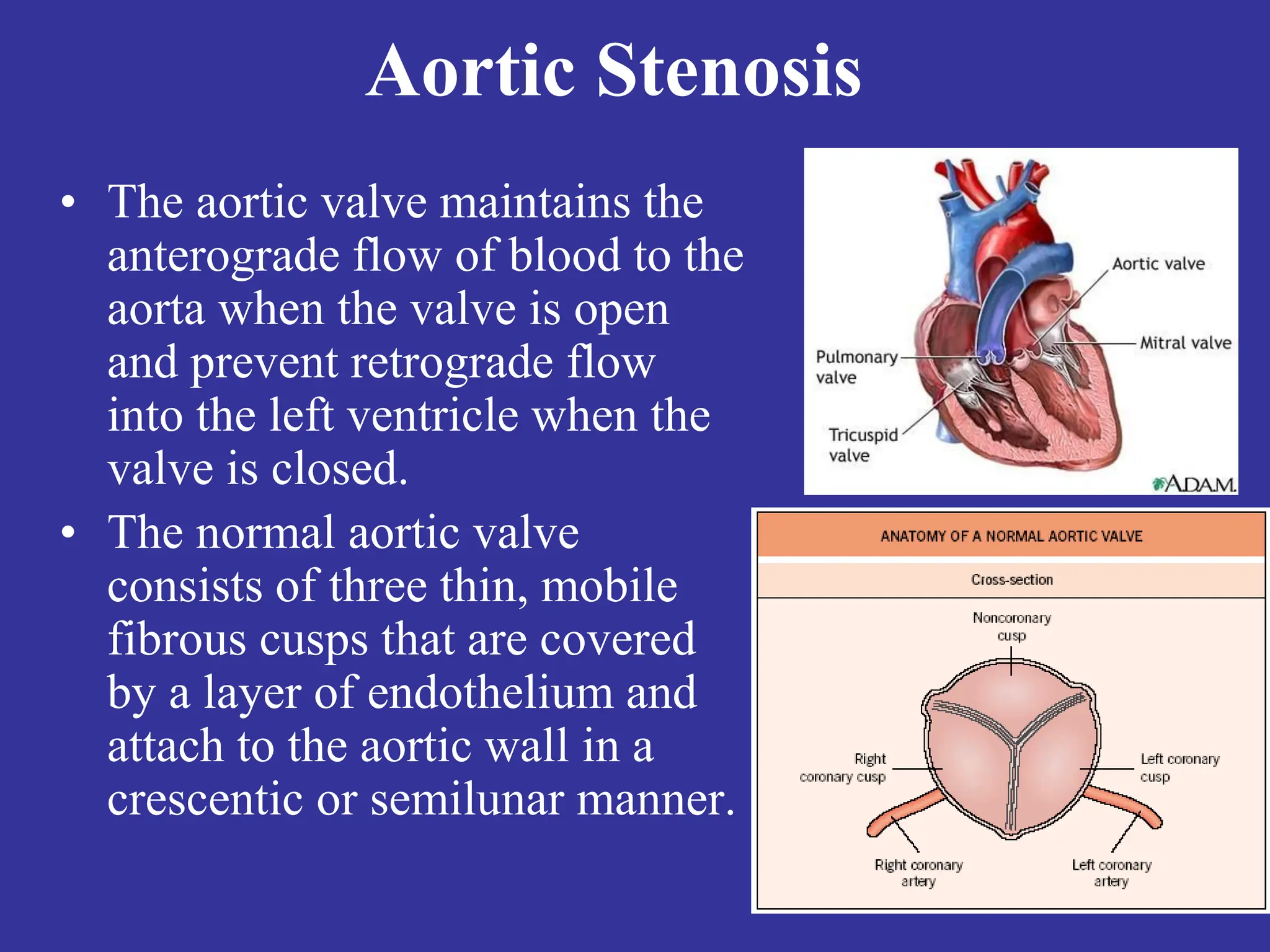
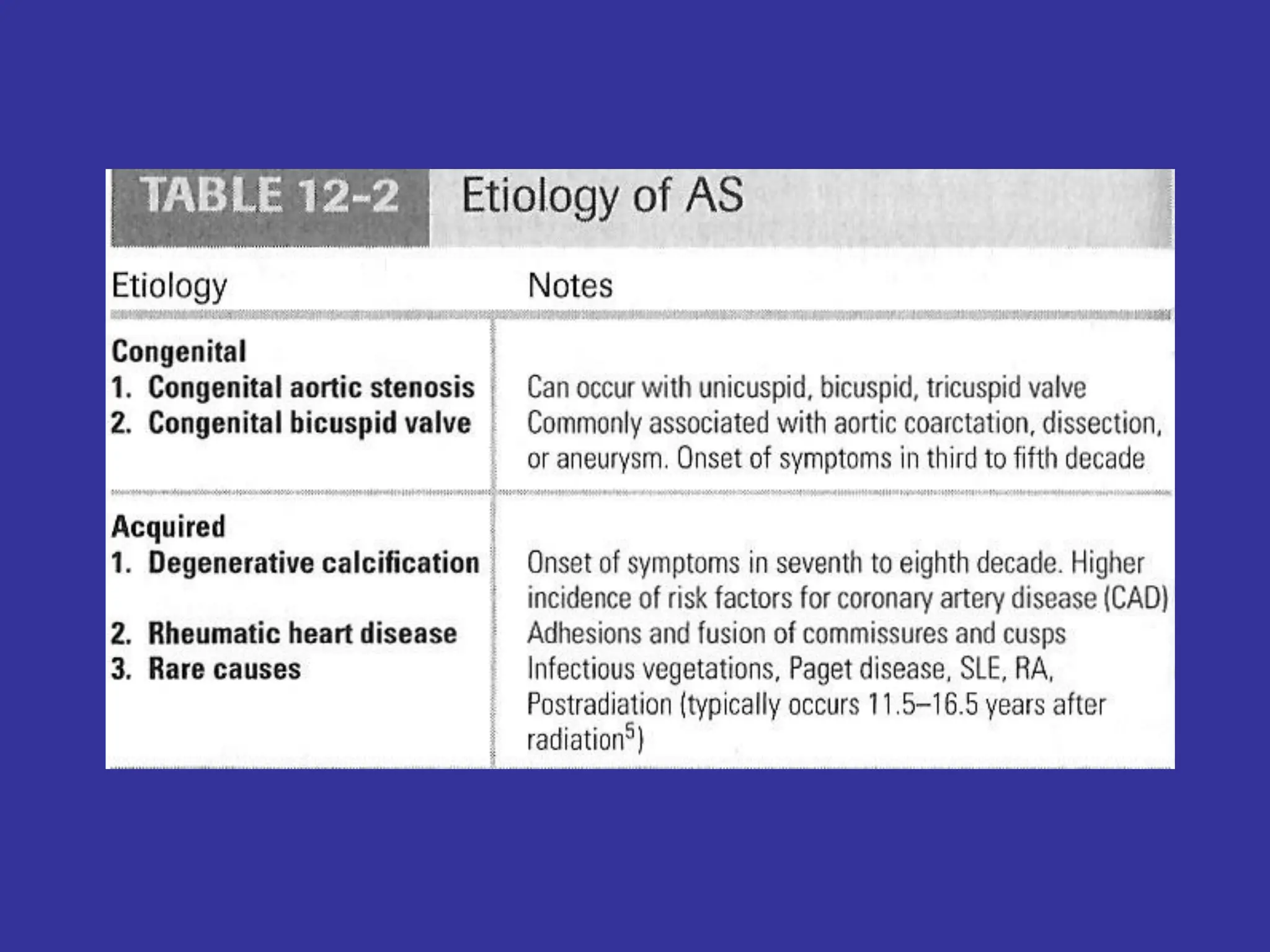
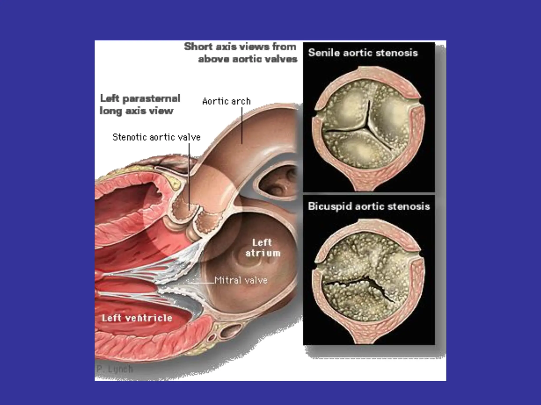
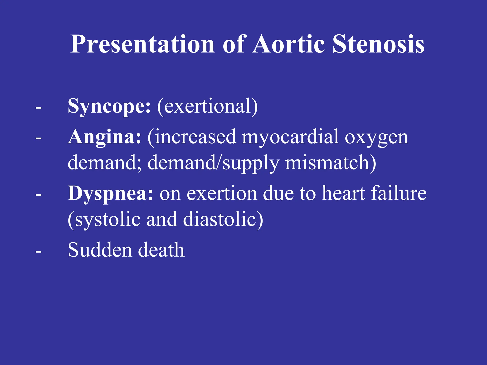
VALVULAR HEART DISEASE
Aortic Valve Diseases
VALVULAR HEART DISEASE
• Etiology
• Pathophysiology
• Physical Exam
• Natural History
• Testing
• Treatment
Aortic Stenosis
• The aortic valve maintains the
anterograde flow of blood to the
aorta when the valve is open
and prevent retrograde flow
into the left ventricle when the
valve is closed.
• The normal aortic valve
consists of three thin, mobile
fibrous cusps that are covered
by a layer of endothelium and
attach to the aortic wall in a
crescentic or semilunar manner.
Aortic Stenosis
• The most frequent valvular heart disease (~25%)
• The most frequent cardiovascular disease after
hypertension and coronary artery disease in Europe
and North America.
• Aortic stenosis is present in 1.3% of people aged
65–74 years and in 4% of people older than 85
years of age
• Aortic sclerosis; A degenerative disease of the
aortic valve most likely represents an early stage of
aortic stenosis. (> 65 years ~ 30%)
Aortic Stenosis
Aortic Stenosis Overview:
• Normal Aortic Valve Area: 3-4 cm2
• Symptoms: Occur when valve area is
1/4th of normal area.
• Types:
– Supravalvular
– Subvalvular
– Valvular
Etiology of Aortic Stenosis
• Congenital
• Rheumatic
• Degenerative/Calcific
Patients under 70: >50% have a congenital
cause
Patients over 70: 50% due to degenerative
Pathophysiology of Aortic Stenosis
• A pressure gradient develops between the
left ventricle and the aorta. (increased
afterload)
• LV function initially maintained by
compensatory pressure hypertrophy
• When compensatory mechanisms exhausted,
LV function declines.
Presentation of Aortic Stenosis
- Syncope: (exertional)
- Angina: (increased myocardial oxygen
demand; demand/supply mismatch)
- Dyspnea: on exertion due to heart failure
(systolic and diastolic)
- Sudden death
Physical Findings in Aortic Stenosis
• Slow rising carotid pulse (pulsus tardus) &
decreased pulse amplitude (pulsus parvus)
• Heart sounds- soft and split second heart
sound, S4 gallop due to LVH.
• Systolic ejection murmur- cresendo-
decrescendo character. This peaks later as
the severity of the stenosis increases.
– Loudness does NOT tell you anything about
severity
Natural History
• Mild AS to Severe AS:
– 8% in 10 years
– 22% in 22 years
– 38% in 25 years
• The onset of symptoms is a poor prognostic
indicator.
Evaluation of AS
• Echocardiography is the most valuable test
for diagnosis, quantification and follow-up
of patients with AS.
• Two measurements obtained are:
a) Left ventricular size and function: LVH,
Dilation, and EF
b) Doppler derived gradient and valve area
(AVA)
Evaluation of AS
Cardiac catheterization: Should only be done for a direct
measurement if symptom severity and echo severity don’t
match OR prior to replacement when replacement is planned.
SYMPTOMATIC AORTIC STENOSIS
• The prognosis of symptomatic patients is extremely poor
without surgical treatment.
• In recent studies, symptomatic patients with aortic
stenosis who have refused surgery have had 5-year
survival rates of only 15–50%.
• Options for valve replacement include the pulmonary
autograft procedure, bioprosthesis and mechanical valves
and recently percutaneous aortic valve replacement.
CoreValve prosthesis
The balloon-expandable prosthesis (Cribier Edwards)
• Stainless steel stent with an attached equine
pericardial trileaflet valve and fabric sealing cuff
(Two sizes: 23- and 26-mm).
Trans-apical Edwards SAPIEN THV
Management of AS
• General- IE prophylaxis in dental procedures
with a prosthetic AV or history of endocarditis.
• Medical - limited role since AS is a mechanical
problem. Vasodilators are relatively
contraindicated in severe AS
• Aortic Balloon Valvotomy- shows little benefit.
• Surgical Replacement/ TAVI: Definitive
treatment
Echo Surveillance
• Mild: Every 5 years
• Moderate: Every 2 years
• Severe: Every 6 months to 1 year
Simplified Indications for Surgery in
Aortic Stenosis
• Any SYMPTOMATIC patient with severe
AS (includes symptoms with exercise)
• Any patient with decreasing EF
• Any patient undergoing CABG with
moderate or severe AS
Summary
• Disease of aging
• Look for the signs on physical exam
• Echocardiogram to assess severity
• Asymptomatic: Medical management and
surveillance
• Symptomatic: AoV replacement (even in
elderly and CHF)
Aortic Regurgitation
Aortic Regurgitation Overview
• Definition: Leakage of blood into LV during
diastole due to ineffective coaptation of the
aortic cusps
Etiology of Acute AR
• Endocarditis
• Aortic Dissection
• Physical Findings:
– Wide pulse pressure
– Diastolic murmur
– Florid pulmonary edema
Treatment of Acute AR
• True Surgical Emergency:
• Positive inotrope: (eg, dopamine,
dobutamine)
• Vasodilators: (eg, nitroprusside)
• Avoid beta-blockers
• Do not even consider a balloon pump
Etiology of Chronic AR
• Bicuspid aortic valve
• Rheumatic
• Infective endocarditis
Pathophysiology of AR
• Combined pressure AND volume overload
• Compensatory Mechanisms: LV dilation,
LVH. Progressive dilation leads to heart
failure
Natural History of AR
• Asymptomatic until 4th or 5th decade
• Rate of Progression: 4-6% per year
• Progressive Symptoms include:
- Dyspnea: exertional, orthopnea, and
paroxsymal nocturnal dyspnea
- Nocturnal angina: due to slowing of heart rate
and reduction of diastolic blood pressure
- Palpitations: due to increased force of
contraction
Physical Exam findings of AR
• Wide pulse pressure: most sensitive
• Hyperdynamic and displaced apical impulse
• Auscultation-
– Diastolic blowing murmur at the left sternal border
– Austin flint murmur (apex): Regurgitant jet
impinges on anterior MVL causing it to vibrate
– Systolic ejection murmur: due to increased flow
across the aortic valve
MRI of the Heart Revealing a Central, High-Velocity Jet Projecting into the Left Ventricular Cavity.
The jet clearly strikes the anterior mitral-valve leaflet, causing distortion and premature closure during
diastole.
The Evaluation of AR
• CXR: enlarged cardiac silhouette and aortic
root enlargement
• ECHO: Evaluation of the AV and aortic root
with measurements of LV dimensions and
function (cornerstone for decision making and
follow up evaluation)
• Aortography: Used to confirm the severity of
disease
Management of AR
• General: IE prophylaxis in dental procedures
with a prosthetic AV or history of endocarditis.
• Medical: Vasodilators (ACEI’s), Nifedipine
improve stroke volume and reduce
regurgitation only if pt symptomatic or HTN.
• Serial Echocardiograms: to monitor
progression.
• Surgical Treatment: Definitive Tx
Simplified Indications for Surgical
Treatment of AR
• ANY Symptoms at rest or exercise
• Asymptomatic treatment if:
– EF drops below 50% or LV becomes
dilated
THANK YOU

1- Aortic Valve Diseases Cardiovascular Module

  • 1.
  • 2.
    VALVULAR HEART DISEASE •Etiology • Pathophysiology • Physical Exam • Natural History • Testing • Treatment
  • 3.
  • 5.
    • The aorticvalve maintains the anterograde flow of blood to the aorta when the valve is open and prevent retrograde flow into the left ventricle when the valve is closed. • The normal aortic valve consists of three thin, mobile fibrous cusps that are covered by a layer of endothelium and attach to the aortic wall in a crescentic or semilunar manner. Aortic Stenosis
  • 6.
    • The mostfrequent valvular heart disease (~25%) • The most frequent cardiovascular disease after hypertension and coronary artery disease in Europe and North America. • Aortic stenosis is present in 1.3% of people aged 65–74 years and in 4% of people older than 85 years of age • Aortic sclerosis; A degenerative disease of the aortic valve most likely represents an early stage of aortic stenosis. (> 65 years ~ 30%) Aortic Stenosis
  • 7.
    Aortic Stenosis Overview: •Normal Aortic Valve Area: 3-4 cm2 • Symptoms: Occur when valve area is 1/4th of normal area. • Types: – Supravalvular – Subvalvular – Valvular
  • 8.
    Etiology of AorticStenosis • Congenital • Rheumatic • Degenerative/Calcific Patients under 70: >50% have a congenital cause Patients over 70: 50% due to degenerative
  • 13.
    Pathophysiology of AorticStenosis • A pressure gradient develops between the left ventricle and the aorta. (increased afterload) • LV function initially maintained by compensatory pressure hypertrophy • When compensatory mechanisms exhausted, LV function declines.
  • 14.
    Presentation of AorticStenosis - Syncope: (exertional) - Angina: (increased myocardial oxygen demand; demand/supply mismatch) - Dyspnea: on exertion due to heart failure (systolic and diastolic) - Sudden death
  • 15.
    Physical Findings inAortic Stenosis • Slow rising carotid pulse (pulsus tardus) & decreased pulse amplitude (pulsus parvus) • Heart sounds- soft and split second heart sound, S4 gallop due to LVH. • Systolic ejection murmur- cresendo- decrescendo character. This peaks later as the severity of the stenosis increases. – Loudness does NOT tell you anything about severity
  • 16.
    Natural History • MildAS to Severe AS: – 8% in 10 years – 22% in 22 years – 38% in 25 years • The onset of symptoms is a poor prognostic indicator.
  • 18.
    Evaluation of AS •Echocardiography is the most valuable test for diagnosis, quantification and follow-up of patients with AS. • Two measurements obtained are: a) Left ventricular size and function: LVH, Dilation, and EF b) Doppler derived gradient and valve area (AVA)
  • 19.
    Evaluation of AS Cardiaccatheterization: Should only be done for a direct measurement if symptom severity and echo severity don’t match OR prior to replacement when replacement is planned.
  • 20.
    SYMPTOMATIC AORTIC STENOSIS •The prognosis of symptomatic patients is extremely poor without surgical treatment. • In recent studies, symptomatic patients with aortic stenosis who have refused surgery have had 5-year survival rates of only 15–50%. • Options for valve replacement include the pulmonary autograft procedure, bioprosthesis and mechanical valves and recently percutaneous aortic valve replacement.
  • 21.
  • 22.
    The balloon-expandable prosthesis(Cribier Edwards) • Stainless steel stent with an attached equine pericardial trileaflet valve and fabric sealing cuff (Two sizes: 23- and 26-mm).
  • 23.
  • 24.
    Management of AS •General- IE prophylaxis in dental procedures with a prosthetic AV or history of endocarditis. • Medical - limited role since AS is a mechanical problem. Vasodilators are relatively contraindicated in severe AS • Aortic Balloon Valvotomy- shows little benefit. • Surgical Replacement/ TAVI: Definitive treatment
  • 25.
    Echo Surveillance • Mild:Every 5 years • Moderate: Every 2 years • Severe: Every 6 months to 1 year
  • 27.
    Simplified Indications forSurgery in Aortic Stenosis • Any SYMPTOMATIC patient with severe AS (includes symptoms with exercise) • Any patient with decreasing EF • Any patient undergoing CABG with moderate or severe AS
  • 28.
    Summary • Disease ofaging • Look for the signs on physical exam • Echocardiogram to assess severity • Asymptomatic: Medical management and surveillance • Symptomatic: AoV replacement (even in elderly and CHF)
  • 29.
  • 31.
    Aortic Regurgitation Overview •Definition: Leakage of blood into LV during diastole due to ineffective coaptation of the aortic cusps
  • 32.
    Etiology of AcuteAR • Endocarditis • Aortic Dissection • Physical Findings: – Wide pulse pressure – Diastolic murmur – Florid pulmonary edema
  • 33.
    Treatment of AcuteAR • True Surgical Emergency: • Positive inotrope: (eg, dopamine, dobutamine) • Vasodilators: (eg, nitroprusside) • Avoid beta-blockers • Do not even consider a balloon pump
  • 35.
    Etiology of ChronicAR • Bicuspid aortic valve • Rheumatic • Infective endocarditis
  • 36.
    Pathophysiology of AR •Combined pressure AND volume overload • Compensatory Mechanisms: LV dilation, LVH. Progressive dilation leads to heart failure
  • 37.
    Natural History ofAR • Asymptomatic until 4th or 5th decade • Rate of Progression: 4-6% per year • Progressive Symptoms include: - Dyspnea: exertional, orthopnea, and paroxsymal nocturnal dyspnea - Nocturnal angina: due to slowing of heart rate and reduction of diastolic blood pressure - Palpitations: due to increased force of contraction
  • 38.
    Physical Exam findingsof AR • Wide pulse pressure: most sensitive • Hyperdynamic and displaced apical impulse • Auscultation- – Diastolic blowing murmur at the left sternal border – Austin flint murmur (apex): Regurgitant jet impinges on anterior MVL causing it to vibrate – Systolic ejection murmur: due to increased flow across the aortic valve
  • 40.
    MRI of theHeart Revealing a Central, High-Velocity Jet Projecting into the Left Ventricular Cavity. The jet clearly strikes the anterior mitral-valve leaflet, causing distortion and premature closure during diastole.
  • 41.
    The Evaluation ofAR • CXR: enlarged cardiac silhouette and aortic root enlargement • ECHO: Evaluation of the AV and aortic root with measurements of LV dimensions and function (cornerstone for decision making and follow up evaluation) • Aortography: Used to confirm the severity of disease
  • 43.
    Management of AR •General: IE prophylaxis in dental procedures with a prosthetic AV or history of endocarditis. • Medical: Vasodilators (ACEI’s), Nifedipine improve stroke volume and reduce regurgitation only if pt symptomatic or HTN. • Serial Echocardiograms: to monitor progression. • Surgical Treatment: Definitive Tx
  • 45.
    Simplified Indications forSurgical Treatment of AR • ANY Symptoms at rest or exercise • Asymptomatic treatment if: – EF drops below 50% or LV becomes dilated
  • 46.