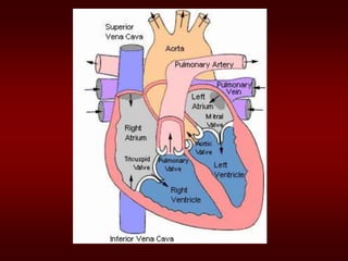
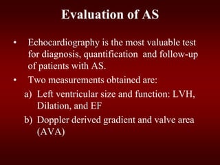
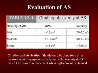
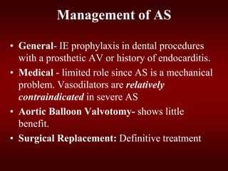
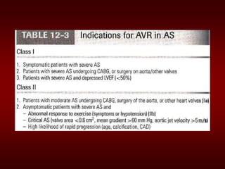
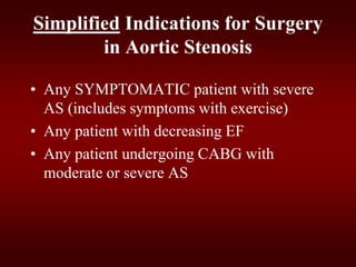
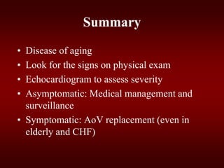
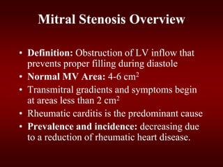
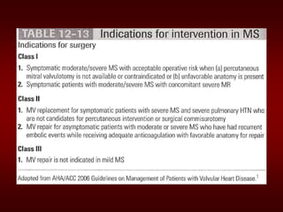
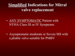
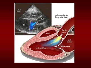
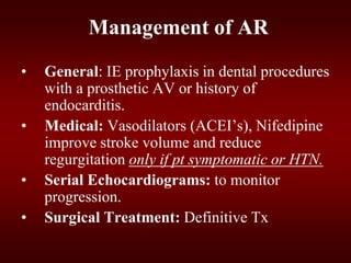
VALVULAR HEART DISEASE
PROF FARHAT BASHIR
UNITED MEDICAL & DENTAL COLLEGE
Goals and Objectives
• Discuss the common etiologies of valvular
stenosis and regurgitation.
• Recognize the signs and symptoms of
severe valvular stenosis and regurgitation
• Be able to quickly identify and treat acute
mitral and aortic regurgitation
• Identify patients who should be referred for
surgical replacement of their valves
Overview
• Aortic Stenosis
• Mitral Stenosis
• Aortic Regurgitation
– Acute and Chronic
• Mitral Regurgitation
– Acute and Chronic
• Etiology
• Pathophysiology
• Physical Exam
• Natural History
• Testing
• Treatment
Aortic Stenosis
Aortic Stenosis Overview:
• Normal Aortic Valve Area: 3-4 cm2
• Symptoms: Occur when valve area is
1/4th of normal area.
• Types:
– Supravalvular
– Subvalvular
– Valvular
Etiology of Aortic Stenosis
• Congenital
• Rheumatic
• Degenerative/Calcific
Patients under 70: >50% have a congenital
cause
Patients over 70: 50% due to degenerative
Pathophysiology of Aortic Stenosis
• A pressure gradient develops between the
left ventricle and the aorta. (increased
afterload)
• LV function initially maintained by
compensatory pressure hypertrophy
• When compensatory mechanisms
exhausted, LV function declines.
Presentation of Aortic Stenosis
- Syncope: (exertional)
- Angina: (increased myocardial oxygen
demand; demand/supply mismatch)
- Dyspnea: on exertion due to heart failure
(systolic and diastolic)
- Sudden death
Physical Findings in Aortic Stenosis
• Slow rising carotid pulse (pulsus tardus) &
decreased pulse amplitude (pulsus parvus)
• Heart sounds- soft and split second heart
sound, S4 gallop due to LVH.
• Systolic ejection murmur- cresendo-
decrescendo character. This peaks later as
the severity of the stenosis increases.
– Loudness does NOT tell you anything about
severity
Natural History
• Mild AS to Severe AS:
– 8% in 10 years
– 22% in 22 years
– 38% in 25 years
• The onset of symptoms is a poor prognostic
indicator.
Evaluation of AS
• Echocardiography is the most valuable test
for diagnosis, quantification and follow-up
of patients with AS.
• Two measurements obtained are:
a) Left ventricular size and function: LVH,
Dilation, and EF
b) Doppler derived gradient and valve area
(AVA)
Evaluation of AS
Cardiac catheterization: Should only be done for a direct
measurement if symptom severity and echo severity don’t
match OR prior to replacement when replacement is planned.
Management of AS
• General- IE prophylaxis in dental procedures
with a prosthetic AV or history of endocarditis.
• Medical - limited role since AS is a mechanical
problem. Vasodilators are relatively
contraindicated in severe AS
• Aortic Balloon Valvotomy- shows little
benefit.
• Surgical Replacement: Definitive treatment
Echo Surveillance
• Mild: Every 5 years
• Moderate: Every 2 years
• Severe: Every 6 months to 1 year
Simplified Indications for Surgery
in Aortic Stenosis
• Any SYMPTOMATIC patient with severe
AS (includes symptoms with exercise)
• Any patient with decreasing EF
• Any patient undergoing CABG with
moderate or severe AS
Summary
• Disease of aging
• Look for the signs on physical exam
• Echocardiogram to assess severity
• Asymptomatic: Medical management and
surveillance
• Symptomatic: AoV replacement (even in
elderly and CHF)
Mitral Stenosis
Mitral Stenosis Overview
• Definition: Obstruction of LV inflow that
prevents proper filling during diastole
• Normal MV Area: 4-6 cm2
• Transmitral gradients and symptoms begin
at areas less than 2 cm2
• Rheumatic carditis is the predominant cause
• Prevalence and incidence: decreasing due
to a reduction of rheumatic heart disease.
Etiology of Mitral Stenosis
• Rheumatic heart disease: 77-99% of all
cases
• Infective endocarditis: 3.3%
• Mitral annular calcification: 2.7%
MS Pathophysiology
• Progressive Dyspnea (70%): LA dilation 
pulmonary congestion (reduced emptying)
– worse with exercise, fever, tachycardia, and pregnancy
• Increased Transmitral Pressures: Leads to left
atrial enlargement and atrial fibrillation.
• Right heart failure symptoms: due to
Pulmonary venous HTN
• Hemoptysis: due to rupture of bronchial vessels
due to elevated pulmonary pressure
Natural History of MS
• Disease of plateaus:
– Mild MS: 10 years after initial RHD insult
– Moderate: 10 years later
– Severe: 10 years later
• Mortality: Due to progressive pulmonary
congestion, infection, and thromboembolism.
Physical Exam Findings of MS
• prominent "a" wave in jugular venous pulsations:
Due to pulmonary hypertension and right ventricular
hypertrophy
• Signs of right-sided heart failure: in advanced
disease
• Mitral facies: When MS is severe and the cardiac
output is diminished, there is vasoconstriction,
resulting in pinkish-purple patches on the cheeks
• Diastolic murmur:
– Low-pitched diastolic rumble most prominent at
the apex.
– Heard best with the patient lying on the left side
in held expiration
– Intensity of the diastolic murmur does not
correlate with the severity of the stenosis
Heart Sounds in MS
• Loud Opening S1 snap: heard at the apex when
leaflets are still mobile
– Due to the abrupt halt in leaflet motion in early
diastole, after rapid initial rapid opening, due to
fusion at the leaflet tips.
– A shorter S2 to opening snap interval indicates
more severe disease.
Heart Sounds in MS
Evaluation of MS
• ECG: may show atrial fibrillation and LA
enlargement
• CXR: LA enlargement and pulmonary
congestion. Occasionally calcified MV
• ECHO: The GOLD STANDARD for
diagnosis. Asses mitral valve mobility,
gradient and mitral valve area
Management of MS
Serial echocardiography:
– Mild: 3-5 years
– Moderate:1-2 years
– Severe: yearly
• Medications: MS like AS is a mechanical
problem and medical therapy does not prevent
progression
– -blockers, CCBs, Digoxin which control heart
rate and hence prolong diastole for improved
diastolic filling
– Duiretics for fluid overload
Management of MS
• Identify patient early who might benefit from
percutaneous mitral balloon valvotomy.
• IE prophylaxis: Patients with prosthetic valves or
a Hx of IE for dental procedures.
Simplified Indications for Mitral
valve replacement
• ANY SYMPTOMATIC Patient with
NYHA Class III or IV Symptoms
• Asymptomatic moderate or Severe MS with
a pliable valve suitable for PMBV
Aortic Regurgitation
Aortic Regurgitation Overview
• Definition: Leakage of blood into LV during
diastole due to ineffective coaptation of the
aortic cusps
Etiology of Acute AR
• Endocarditis
• Aortic Dissection
• Physical Findings:
– Wide pulse pressure
– Diastolic murmur
– Florid pulmonary edema
Treatment of Acute AR
• True Surgical Emergency:
• Positive inotrope: (eg, dopamine,
dobutamine)
• Vasodilators: (eg, nitroprusside)
• Avoid beta-blockers
• Do not even consider a balloon pump
Etiology of Chronic AR
• Bicuspid aortic valve
• Rheumatic
• Infective endocarditis
Pathophysiology of AR
• Combined pressure AND volume overload
• Compensatory Mechanisms: LV dilation,
LVH. Progressive dilation leads to heart
failure
Natural History of AR
• Asymptomatic until 4th or 5th decade
• Rate of Progression: 4-6% per year
• Progressive Symptoms include:
- Dyspnea: exertional, orthopnea, and
paroxsymal nocturnal dyspnea
- Nocturnal angina: due to slowing of heart rate
and reduction of diastolic blood pressure
- Palpitations: due to increased force of
contraction
Physical Exam findings of AR
• Wide pulse pressure: most sensitive
• Hyperdynamic and displaced apical impulse
• Auscultation-
– Diastolic blowing murmur at the left sternal
border
– Austin flint murmur (apex): Regurgitant jet
impinges on anterior MVL causing it to vibrate
– Systolic ejection murmur: due to increased flow
across the aortic valve
MRI of the Heart Revealing a Central, High-Velocity Jet Projecting into the Left Ventricular Cavity.
The jet clearly strikes the anterior mitral-valve leaflet, causing distortion and premature closure
during diastole.
The Evaluation of AR
• CXR: enlarged cardiac silhouette and aortic
root enlargement
• ECHO: Evaluation of the AV and aortic root
with measurements of LV dimensions and
function (cornerstone for decision making and
follow up evaluation)
• Aortography: Used to confirm the severity of
disease
Management of AR
• General: IE prophylaxis in dental procedures
with a prosthetic AV or history of
endocarditis.
• Medical: Vasodilators (ACEI’s), Nifedipine
improve stroke volume and reduce
regurgitation only if pt symptomatic or HTN.
• Serial Echocardiograms: to monitor
progression.
• Surgical Treatment: Definitive Tx
Simplified Indications for Surgical
Treatment of AR
• ANY Symptoms at rest or exercise
• Asymptomatic treatment if:
– EF drops below 50% or LV becomes
dilated
Mitral Regurgitation
• Definition: Backflow of blood from the LV to
the LA during systole
• Mild (physiological) MR is seen in 80% of
normal individuals.
Chronic Mitral Regurgitation
Overview
Acute MR
• Endocarditis
• Acute MI
• Malfunction or disruption of prosthetic
valve
Management of Acute MR
• Myocardial infarction: Cardiac cath or
thrombolytics
• Most other cases of mitral regurgitation
is afterload reduction:
– Diuretics and nitrates
– nitroprusside, even in the setting of a
normal blood pressure.
Management of Acute MR
• Do not attempt to alleviate tachycardia
with beta-blockers. Mild-to-moderate
tachycardia is beneficial in these patients
because it allows less time for the heart to
have backfill, which lowers regurgitant
volume.
Treatment of Acute MR
• Balloon Pump
• Nitroprusside even if hypotensive
• Emergent Surgery
• Myxomatous degeneration (MVP)
• Ischemic MR
• Rheumatic heart disease
• Infective Endocarditis
Etiologies of Chronic Mitral
Regurgitation
Pathophysiology of MR
• Pure Volume Overload
• Compensatory Mechanisms: Left atrial
enlargement, LVH and increased
contractility
– Progressive left atrial dilation and right
ventricular dysfunction due to pulmonary
hypertension.
– Progressive left ventricular volume overload
leads to dilation and progressive heart failure.
Physical Exam findings in MR
• Auscultation: soft S1 and a holosystolic
murmur at the apex radiating to the axilla
– S3 (CHF/LA overload)
– In chronic MR, the intensity of the murmur
does correlate with the severity.
• Exertion Dyspnea: ( exercise intolerance)
• Heart Failure: May coincide with
increased hemodynamic burden e.g.,
pregnancy, infection or atrial fibrillation
The Natural History of MR
• Compensatory phase: 10-15 years
• Patients with asymptomatic severe MR have
a 5%/year mortality rate
• Once the patient’s EF becomes <60%
and/or becomes symptomatic, mortality
rises sharply
• Mortality: From progressive dyspnea and
heart failure
Imaging studies in MR
• ECG: May show, LA enlargement, atrial
fibrillation and LV hypertrophy with severe MR
• CXR: LA enlargement, central pulmonary
artery enlargement.
• ECHO: Estimation of LA, LV size and
function. Valve structure assessment
– TEE if transthoracic echo is inconclusive
Management of MR
• Medications
a) Vasodilator such as hydralazine
b) Rate control for atrial fibrillation with -
blockers, CCB, digoxin
c) Anticoagulation in atrial fibrillation and flutter
d) Diuretics for fluid overload
Management of MR
• Serial Echocardiography:
– Mild: 2-3 years
– Moderate: 1-2 years
– Severe: 6-12 months
• IE prophylaxis: Patients with prosthetic
valves or a Hx of IE for dental procedures.
Simplified Indications for MV
Replacement in Severe MR
• ANY Symptoms at rest or exercise with
(repair if feasible)
• Asymptomatic:
– If EF <60%
– If new onset atrial fibrillation
THANK YOU

Valvular diseases

  • 1.
    VALVULAR HEART DISEASE PROFFARHAT BASHIR UNITED MEDICAL & DENTAL COLLEGE
  • 2.
    Goals and Objectives •Discuss the common etiologies of valvular stenosis and regurgitation. • Recognize the signs and symptoms of severe valvular stenosis and regurgitation • Be able to quickly identify and treat acute mitral and aortic regurgitation • Identify patients who should be referred for surgical replacement of their valves
  • 3.
    Overview • Aortic Stenosis •Mitral Stenosis • Aortic Regurgitation – Acute and Chronic • Mitral Regurgitation – Acute and Chronic
  • 4.
    • Etiology • Pathophysiology •Physical Exam • Natural History • Testing • Treatment
  • 5.
  • 7.
    Aortic Stenosis Overview: •Normal Aortic Valve Area: 3-4 cm2 • Symptoms: Occur when valve area is 1/4th of normal area. • Types: – Supravalvular – Subvalvular – Valvular
  • 8.
    Etiology of AorticStenosis • Congenital • Rheumatic • Degenerative/Calcific Patients under 70: >50% have a congenital cause Patients over 70: 50% due to degenerative
  • 13.
    Pathophysiology of AorticStenosis • A pressure gradient develops between the left ventricle and the aorta. (increased afterload) • LV function initially maintained by compensatory pressure hypertrophy • When compensatory mechanisms exhausted, LV function declines.
  • 14.
    Presentation of AorticStenosis - Syncope: (exertional) - Angina: (increased myocardial oxygen demand; demand/supply mismatch) - Dyspnea: on exertion due to heart failure (systolic and diastolic) - Sudden death
  • 15.
    Physical Findings inAortic Stenosis • Slow rising carotid pulse (pulsus tardus) & decreased pulse amplitude (pulsus parvus) • Heart sounds- soft and split second heart sound, S4 gallop due to LVH. • Systolic ejection murmur- cresendo- decrescendo character. This peaks later as the severity of the stenosis increases. – Loudness does NOT tell you anything about severity
  • 16.
    Natural History • MildAS to Severe AS: – 8% in 10 years – 22% in 22 years – 38% in 25 years • The onset of symptoms is a poor prognostic indicator.
  • 18.
    Evaluation of AS •Echocardiography is the most valuable test for diagnosis, quantification and follow-up of patients with AS. • Two measurements obtained are: a) Left ventricular size and function: LVH, Dilation, and EF b) Doppler derived gradient and valve area (AVA)
  • 19.
    Evaluation of AS Cardiaccatheterization: Should only be done for a direct measurement if symptom severity and echo severity don’t match OR prior to replacement when replacement is planned.
  • 20.
    Management of AS •General- IE prophylaxis in dental procedures with a prosthetic AV or history of endocarditis. • Medical - limited role since AS is a mechanical problem. Vasodilators are relatively contraindicated in severe AS • Aortic Balloon Valvotomy- shows little benefit. • Surgical Replacement: Definitive treatment
  • 21.
    Echo Surveillance • Mild:Every 5 years • Moderate: Every 2 years • Severe: Every 6 months to 1 year
  • 23.
    Simplified Indications forSurgery in Aortic Stenosis • Any SYMPTOMATIC patient with severe AS (includes symptoms with exercise) • Any patient with decreasing EF • Any patient undergoing CABG with moderate or severe AS
  • 24.
    Summary • Disease ofaging • Look for the signs on physical exam • Echocardiogram to assess severity • Asymptomatic: Medical management and surveillance • Symptomatic: AoV replacement (even in elderly and CHF)
  • 25.
  • 26.
    Mitral Stenosis Overview •Definition: Obstruction of LV inflow that prevents proper filling during diastole • Normal MV Area: 4-6 cm2 • Transmitral gradients and symptoms begin at areas less than 2 cm2 • Rheumatic carditis is the predominant cause • Prevalence and incidence: decreasing due to a reduction of rheumatic heart disease.
  • 27.
    Etiology of MitralStenosis • Rheumatic heart disease: 77-99% of all cases • Infective endocarditis: 3.3% • Mitral annular calcification: 2.7%
  • 32.
    MS Pathophysiology • ProgressiveDyspnea (70%): LA dilation  pulmonary congestion (reduced emptying) – worse with exercise, fever, tachycardia, and pregnancy • Increased Transmitral Pressures: Leads to left atrial enlargement and atrial fibrillation. • Right heart failure symptoms: due to Pulmonary venous HTN • Hemoptysis: due to rupture of bronchial vessels due to elevated pulmonary pressure
  • 33.
    Natural History ofMS • Disease of plateaus: – Mild MS: 10 years after initial RHD insult – Moderate: 10 years later – Severe: 10 years later • Mortality: Due to progressive pulmonary congestion, infection, and thromboembolism.
  • 34.
    Physical Exam Findingsof MS • prominent "a" wave in jugular venous pulsations: Due to pulmonary hypertension and right ventricular hypertrophy • Signs of right-sided heart failure: in advanced disease • Mitral facies: When MS is severe and the cardiac output is diminished, there is vasoconstriction, resulting in pinkish-purple patches on the cheeks
  • 35.
    • Diastolic murmur: –Low-pitched diastolic rumble most prominent at the apex. – Heard best with the patient lying on the left side in held expiration – Intensity of the diastolic murmur does not correlate with the severity of the stenosis Heart Sounds in MS
  • 36.
    • Loud OpeningS1 snap: heard at the apex when leaflets are still mobile – Due to the abrupt halt in leaflet motion in early diastole, after rapid initial rapid opening, due to fusion at the leaflet tips. – A shorter S2 to opening snap interval indicates more severe disease. Heart Sounds in MS
  • 37.
    Evaluation of MS •ECG: may show atrial fibrillation and LA enlargement • CXR: LA enlargement and pulmonary congestion. Occasionally calcified MV • ECHO: The GOLD STANDARD for diagnosis. Asses mitral valve mobility, gradient and mitral valve area
  • 39.
    Management of MS Serialechocardiography: – Mild: 3-5 years – Moderate:1-2 years – Severe: yearly • Medications: MS like AS is a mechanical problem and medical therapy does not prevent progression – -blockers, CCBs, Digoxin which control heart rate and hence prolong diastole for improved diastolic filling – Duiretics for fluid overload
  • 40.
    Management of MS •Identify patient early who might benefit from percutaneous mitral balloon valvotomy. • IE prophylaxis: Patients with prosthetic valves or a Hx of IE for dental procedures.
  • 45.
    Simplified Indications forMitral valve replacement • ANY SYMPTOMATIC Patient with NYHA Class III or IV Symptoms • Asymptomatic moderate or Severe MS with a pliable valve suitable for PMBV
  • 46.
  • 48.
    Aortic Regurgitation Overview •Definition: Leakage of blood into LV during diastole due to ineffective coaptation of the aortic cusps
  • 49.
    Etiology of AcuteAR • Endocarditis • Aortic Dissection • Physical Findings: – Wide pulse pressure – Diastolic murmur – Florid pulmonary edema
  • 50.
    Treatment of AcuteAR • True Surgical Emergency: • Positive inotrope: (eg, dopamine, dobutamine) • Vasodilators: (eg, nitroprusside) • Avoid beta-blockers • Do not even consider a balloon pump
  • 52.
    Etiology of ChronicAR • Bicuspid aortic valve • Rheumatic • Infective endocarditis
  • 53.
    Pathophysiology of AR •Combined pressure AND volume overload • Compensatory Mechanisms: LV dilation, LVH. Progressive dilation leads to heart failure
  • 54.
    Natural History ofAR • Asymptomatic until 4th or 5th decade • Rate of Progression: 4-6% per year • Progressive Symptoms include: - Dyspnea: exertional, orthopnea, and paroxsymal nocturnal dyspnea - Nocturnal angina: due to slowing of heart rate and reduction of diastolic blood pressure - Palpitations: due to increased force of contraction
  • 55.
    Physical Exam findingsof AR • Wide pulse pressure: most sensitive • Hyperdynamic and displaced apical impulse • Auscultation- – Diastolic blowing murmur at the left sternal border – Austin flint murmur (apex): Regurgitant jet impinges on anterior MVL causing it to vibrate – Systolic ejection murmur: due to increased flow across the aortic valve
  • 57.
    MRI of theHeart Revealing a Central, High-Velocity Jet Projecting into the Left Ventricular Cavity. The jet clearly strikes the anterior mitral-valve leaflet, causing distortion and premature closure during diastole.
  • 58.
    The Evaluation ofAR • CXR: enlarged cardiac silhouette and aortic root enlargement • ECHO: Evaluation of the AV and aortic root with measurements of LV dimensions and function (cornerstone for decision making and follow up evaluation) • Aortography: Used to confirm the severity of disease
  • 60.
    Management of AR •General: IE prophylaxis in dental procedures with a prosthetic AV or history of endocarditis. • Medical: Vasodilators (ACEI’s), Nifedipine improve stroke volume and reduce regurgitation only if pt symptomatic or HTN. • Serial Echocardiograms: to monitor progression. • Surgical Treatment: Definitive Tx
  • 63.
    Simplified Indications forSurgical Treatment of AR • ANY Symptoms at rest or exercise • Asymptomatic treatment if: – EF drops below 50% or LV becomes dilated
  • 64.
  • 65.
    • Definition: Backflowof blood from the LV to the LA during systole • Mild (physiological) MR is seen in 80% of normal individuals. Chronic Mitral Regurgitation Overview
  • 66.
    Acute MR • Endocarditis •Acute MI • Malfunction or disruption of prosthetic valve
  • 67.
    Management of AcuteMR • Myocardial infarction: Cardiac cath or thrombolytics • Most other cases of mitral regurgitation is afterload reduction: – Diuretics and nitrates – nitroprusside, even in the setting of a normal blood pressure.
  • 68.
    Management of AcuteMR • Do not attempt to alleviate tachycardia with beta-blockers. Mild-to-moderate tachycardia is beneficial in these patients because it allows less time for the heart to have backfill, which lowers regurgitant volume.
  • 69.
    Treatment of AcuteMR • Balloon Pump • Nitroprusside even if hypotensive • Emergent Surgery
  • 70.
    • Myxomatous degeneration(MVP) • Ischemic MR • Rheumatic heart disease • Infective Endocarditis Etiologies of Chronic Mitral Regurgitation
  • 71.
    Pathophysiology of MR •Pure Volume Overload • Compensatory Mechanisms: Left atrial enlargement, LVH and increased contractility – Progressive left atrial dilation and right ventricular dysfunction due to pulmonary hypertension. – Progressive left ventricular volume overload leads to dilation and progressive heart failure.
  • 72.
    Physical Exam findingsin MR • Auscultation: soft S1 and a holosystolic murmur at the apex radiating to the axilla – S3 (CHF/LA overload) – In chronic MR, the intensity of the murmur does correlate with the severity. • Exertion Dyspnea: ( exercise intolerance) • Heart Failure: May coincide with increased hemodynamic burden e.g., pregnancy, infection or atrial fibrillation
  • 73.
    The Natural Historyof MR • Compensatory phase: 10-15 years • Patients with asymptomatic severe MR have a 5%/year mortality rate • Once the patient’s EF becomes <60% and/or becomes symptomatic, mortality rises sharply • Mortality: From progressive dyspnea and heart failure
  • 74.
    Imaging studies inMR • ECG: May show, LA enlargement, atrial fibrillation and LV hypertrophy with severe MR • CXR: LA enlargement, central pulmonary artery enlargement. • ECHO: Estimation of LA, LV size and function. Valve structure assessment – TEE if transthoracic echo is inconclusive
  • 76.
    Management of MR •Medications a) Vasodilator such as hydralazine b) Rate control for atrial fibrillation with - blockers, CCB, digoxin c) Anticoagulation in atrial fibrillation and flutter d) Diuretics for fluid overload
  • 77.
    Management of MR •Serial Echocardiography: – Mild: 2-3 years – Moderate: 1-2 years – Severe: 6-12 months • IE prophylaxis: Patients with prosthetic valves or a Hx of IE for dental procedures.
  • 80.
    Simplified Indications forMV Replacement in Severe MR • ANY Symptoms at rest or exercise with (repair if feasible) • Asymptomatic: – If EF <60% – If new onset atrial fibrillation
  • 81.