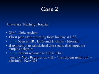
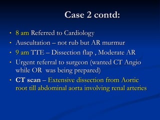
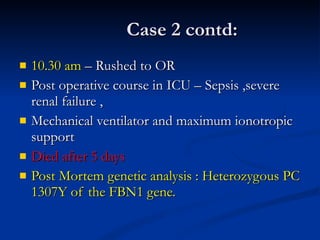
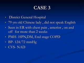
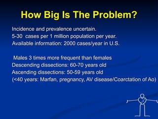
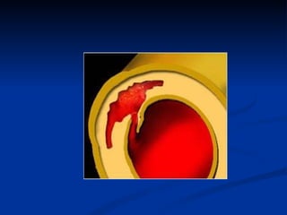
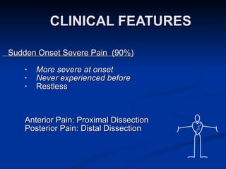
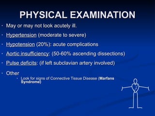
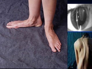
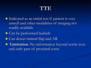
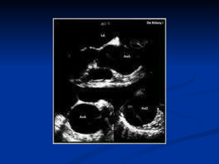
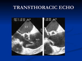
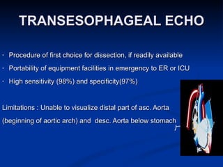
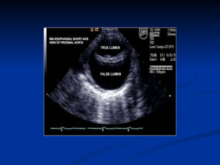
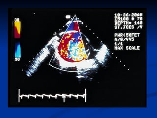
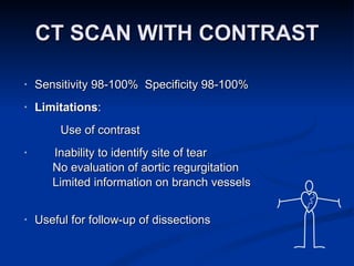
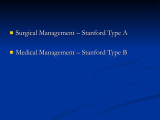
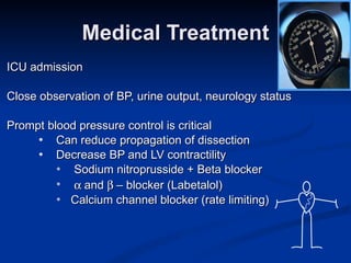
AORTIC DISSECTION SYED  RAZA
CASE 1 University Teaching Hospital 53/f  admitted to Heart Emergency Centre Chest pain/discomfort – 1 hour right leg numbness PMH- Hypertension – not on medication Smoker – 5 cigs/day BP 170/80 mmHg Power RLL 4/5 , Grade 2 AR murmur .
Case 1 contd: ECG- Ischemic changes Inferior leads. CXR – Normal Troponin – Negative Routine blood tests - normal
Case 1 contd: MRA  –  Aortic aneurysm (6.5 cms).Aortic Dissection from aortic root, extending to ascending Aorta and arch involving the left common carotid artery. Small area of infarct Left MCA territory . Urgent Surgery –  Patient did not survive
Case 2 University Teaching Hospital 26/f , Univ. student Chest pain after returning from holiday in USA 2 pm  Seen in ER , ECG and D-dimer - Normal diagnosed- musculoskeletal chest pain, discharged on simple analgesics 8 pm  Patient returned to ER in 6 hrs  Seen by Med. Registrar on call – ‘ heard pericardial rub’ – admitted , NSAIDS
Case 2 contd: 8 am  Referred to Cardiology Auscultation – not rub but AR murmur 9 am  TTE – Dissection flap , Moderate AR Urgent referral to surgeon (wanted CT Angio while OR  was being prepared) CT scan  –  Extensive dissection from Aortic root till abdominal aorta involving renal arteries
Case 2 contd: 10.30 am  – Rushed to OR Post operative course in ICU – Sepsis ,severe renal failure ,  Mechanical ventilator and maximum ionotropic support Died after 5 days Post Mortem genetic analysis : Heterozygous PC 1307Y of the FBN1 gene.
CASE 3 District General Hospital 79 yrs old Chinese lady , did not speak English Seen in ER with chest pain , anterior , on and off  for more than 2 weeks PMH- HPN,DM, End stage COPD  BP- 124/72 mmHg CVS- NAD
ECG-non specific T wave changes. Trop I – normal  Hb 10.1 Discharged as Musculo skeletal chest pain. Anaemia for Inv.
CASE 3 contd: Returned to ER following day – continuing chest pain. Admitted as Atypical chest pain. CXR-unfolded aorta Repeat Trop I-normal , D-dimer –elevated(>500 ng/dl) CTPA – Requested . Anticoagulant commenced. ECG - <1 mm ST elevation in Inferior leads. Referred to Cardiology
Case 3 contd: History from grand son Admitted in hospital in China for 1 day just before coming to the UK. EX- BP right arm 170/96  left arm 122/ 70 CXR- Widened mediastinum
CASE 3 contd: CTPA  – Dissection Asc. Aorta involving RCA , Arch of aorta involving left sub clavian artery extending just beyond the diaphragm. Small to moderate pericardial effusion. Discussed with surgeon- High risk for surgery. Patient and family not keen for intervention. Medical management – aggressive BP control Anticoagulant  stopped
OBJECTIVES  1.Recognition of Aortic Dissection 2.Prevalance 3.Clinical features 4.Investigation and Management
Acute Aortic Dissection is a medical emergency High mortality rate Mortality rate rises at 1% per hour if left untreated. Atypical presentations are very uncommon Painless AAD have been reported
Most important factor leading to a correct diagnosis is a high clinical suspicion!
How Big Is The Problem? Incidence and prevalence uncertain. 5-30  cases per 1 million population per year. Available information: 2000 cases/year in U.S. Males 3 times more frequent than females Descending dissections: 60-70 years old Ascending dissections: 50-59 years old (<40 years: Marfan, pregnancy, AV disease/Coarctation of Ao)
CLASSIFICATION D E BAKEY Type I Ascending aorta extending beyond arch Type II Ascending aorta only Type III a Descending aorta distal to left subclavian (above diaphragm) Type III b Descending aorta  distal to LSA extending below diaphragm S TANFORD A – Ascending aorta B – Not involving Ascending Aorta


CLINICAL FEATURES  Sudden Onset Severe Pain  (90%) More severe at onset Never experienced before Restless Anterior Pain: Proximal Dissection Posterior Pain: Distal Dissection
PHYSICAL EXAMINATION May or may not look acutely ill. Hypertension  (moderate to severe) Hypotension  (20%): acute complications Aortic insufficiency : (50-60% ascending dissections) Pulse deficits : (if left subclavian artery involved) Other Look for signs of Connective Tissue Disease ( Marfans Syndrome)


NATURAL HISTORY Autopsy Series:  >50%  of people with untreated aortic dissections are  dead within 48 hours . 1934 Shennan: >300 cases reviewed. 40% acute ascending dissections died suddenly. None lived > 5 weeks Anagnostopoulos et al. Am J Card 1972 973 pts with untreated proximal and distal dissections 50% died with 48 hours 84% died within 1 month
DIAGNOSTIC EVALUATION Chest radiograph Tran thoracic echocardiogram Tran esophageal echocardiogram* Computed tomography* Magnetic resonance imaging* Aortography * Choice  based on rapid availability and quality of performance
CHEST X-RAY 1. widened mediastinum, (sen: 44-80%) 2. Calcium sign -Displaced intimal calcification (>10mm) from outer aortic wall– useful in older patients 3.pleural effusion (involvement of descending aorta) 4.Normal in 18% A Normal CXR Should Not Deter Further Evaluatio n.
 
 
TTE Indicated as an initial test if patient is very unwell and other modalities of imaging not readily available Can be performed bedside Can detect intimal flap and AR Limitation : No information beyond aortic root and early part of proximal aorta
 
TRANSTHORACIC ECHO
TRANSESOPHAGEAL ECHO Procedure of first choice for dissection, if readily available Portability of equipment facilities in emergency to ER or ICU High sensitivity (98%) and specificity(97%)   Limitations : Unable to visualize distal part of asc. Aorta (beginning of aortic arch) and  desc. Aorta below stomach
 
 
CT SCAN WITH CONTRAST Sensitivity 98-100%  Specificity 98-100% Limitations : Use of contrast Inability to identify site of tear No evaluation of aortic regurgitation Limited information on branch vessels Useful for follow-up of dissections
 
MRA Good alternative to TEE or CT, if readily available High sensitivity (98%) and specificity (98%) Provides three dimensional reconstruction Can detect site of intimal tear and involvement of branch vessels Non-invasive; neither x-rays nor contrast needed Limitation : claustrophobic, more costly, not readily available
 
AORTOGRAPHY Considered Gold standard in olden days Sensitivity (88%)  Specificity (94%) Identify intimal flap, true and false lumen  Aortic insufficiency, branch vessel involvement Limitations Invasive Use of contrast Time delay in preparation
 
TREATMENT
Surgical Management – Stanford Type A Medical Management – Stanford Type B
Medical Treatment ICU admission  Close observation of BP, urine output, neurology status Prompt blood pressure control is critical Can reduce propagation of dissection Decrease BP and LV contractility  Sodium nitroprusside + Beta blocker  and   – blocker (Labetalol) Calcium channel blocker (rate limiting)
SURGERY
INDICATIONS FOR SURGERY Stanford type A (DeBakey type I and II)  ascending aortic dissection  Complicated Stanford type B (DeBakey type III ) aortic dissections with clinical or radiological evidence of the following conditions:  Propagation (increasing aortic diameter)  Increasing size of hematoma  Compromise of major branches of the aorta  Impending rupture
PROGNOSIS Without treatment  about 50% will die within 48 hours Without treatment , about 75%  will  die within the first 2 weeks.  With treatment , about 70% who have  Type A dissection  and about 90% who have Type B  dissection  survive to leave the hospital.  About 60% of people who survive the first 2 weeks are still alive 5 years after treatment, and 40% live at least 10 years.
TAKE HOME MESSAGE Medical emergency Many misdiagnosed or undiagnosed High clinical suspicion. Should consider as a differential diagnosis with all chest pain. Thrombolytic ,Anticoagulants and anti platelet  therapy may be catastrophic. Early diagnosis + prompt action = SURVIVAL
THANK  YOU FOR YOUR  ATTENTION

Aortic dissection

  • 1.
  • 2.
    CASE 1 UniversityTeaching Hospital 53/f admitted to Heart Emergency Centre Chest pain/discomfort – 1 hour right leg numbness PMH- Hypertension – not on medication Smoker – 5 cigs/day BP 170/80 mmHg Power RLL 4/5 , Grade 2 AR murmur .
  • 3.
    Case 1 contd:ECG- Ischemic changes Inferior leads. CXR – Normal Troponin – Negative Routine blood tests - normal
  • 4.
    Case 1 contd:MRA – Aortic aneurysm (6.5 cms).Aortic Dissection from aortic root, extending to ascending Aorta and arch involving the left common carotid artery. Small area of infarct Left MCA territory . Urgent Surgery – Patient did not survive
  • 5.
    Case 2 UniversityTeaching Hospital 26/f , Univ. student Chest pain after returning from holiday in USA 2 pm Seen in ER , ECG and D-dimer - Normal diagnosed- musculoskeletal chest pain, discharged on simple analgesics 8 pm Patient returned to ER in 6 hrs Seen by Med. Registrar on call – ‘ heard pericardial rub’ – admitted , NSAIDS
  • 6.
    Case 2 contd:8 am Referred to Cardiology Auscultation – not rub but AR murmur 9 am TTE – Dissection flap , Moderate AR Urgent referral to surgeon (wanted CT Angio while OR was being prepared) CT scan – Extensive dissection from Aortic root till abdominal aorta involving renal arteries
  • 7.
    Case 2 contd:10.30 am – Rushed to OR Post operative course in ICU – Sepsis ,severe renal failure , Mechanical ventilator and maximum ionotropic support Died after 5 days Post Mortem genetic analysis : Heterozygous PC 1307Y of the FBN1 gene.
  • 8.
    CASE 3 DistrictGeneral Hospital 79 yrs old Chinese lady , did not speak English Seen in ER with chest pain , anterior , on and off for more than 2 weeks PMH- HPN,DM, End stage COPD BP- 124/72 mmHg CVS- NAD
  • 9.
    ECG-non specific Twave changes. Trop I – normal Hb 10.1 Discharged as Musculo skeletal chest pain. Anaemia for Inv.
  • 10.
    CASE 3 contd:Returned to ER following day – continuing chest pain. Admitted as Atypical chest pain. CXR-unfolded aorta Repeat Trop I-normal , D-dimer –elevated(>500 ng/dl) CTPA – Requested . Anticoagulant commenced. ECG - <1 mm ST elevation in Inferior leads. Referred to Cardiology
  • 11.
    Case 3 contd:History from grand son Admitted in hospital in China for 1 day just before coming to the UK. EX- BP right arm 170/96 left arm 122/ 70 CXR- Widened mediastinum
  • 12.
    CASE 3 contd:CTPA – Dissection Asc. Aorta involving RCA , Arch of aorta involving left sub clavian artery extending just beyond the diaphragm. Small to moderate pericardial effusion. Discussed with surgeon- High risk for surgery. Patient and family not keen for intervention. Medical management – aggressive BP control Anticoagulant stopped
  • 13.
    OBJECTIVES 1.Recognitionof Aortic Dissection 2.Prevalance 3.Clinical features 4.Investigation and Management
  • 14.
    Acute Aortic Dissectionis a medical emergency High mortality rate Mortality rate rises at 1% per hour if left untreated. Atypical presentations are very uncommon Painless AAD have been reported
  • 15.
    Most important factorleading to a correct diagnosis is a high clinical suspicion!
  • 16.
    How Big IsThe Problem? Incidence and prevalence uncertain. 5-30 cases per 1 million population per year. Available information: 2000 cases/year in U.S. Males 3 times more frequent than females Descending dissections: 60-70 years old Ascending dissections: 50-59 years old (<40 years: Marfan, pregnancy, AV disease/Coarctation of Ao)
  • 17.
    CLASSIFICATION D EBAKEY Type I Ascending aorta extending beyond arch Type II Ascending aorta only Type III a Descending aorta distal to left subclavian (above diaphragm) Type III b Descending aorta distal to LSA extending below diaphragm S TANFORD A – Ascending aorta B – Not involving Ascending Aorta
  • 18.
  • 19.
  • 20.
  • 21.
  • 22.
    CLINICAL FEATURES Sudden Onset Severe Pain (90%) More severe at onset Never experienced before Restless Anterior Pain: Proximal Dissection Posterior Pain: Distal Dissection
  • 23.
    PHYSICAL EXAMINATION Mayor may not look acutely ill. Hypertension (moderate to severe) Hypotension (20%): acute complications Aortic insufficiency : (50-60% ascending dissections) Pulse deficits : (if left subclavian artery involved) Other Look for signs of Connective Tissue Disease ( Marfans Syndrome)
  • 24.
  • 25.
  • 26.
  • 27.
  • 28.
  • 29.
    NATURAL HISTORY AutopsySeries: >50% of people with untreated aortic dissections are dead within 48 hours . 1934 Shennan: >300 cases reviewed. 40% acute ascending dissections died suddenly. None lived > 5 weeks Anagnostopoulos et al. Am J Card 1972 973 pts with untreated proximal and distal dissections 50% died with 48 hours 84% died within 1 month
  • 30.
    DIAGNOSTIC EVALUATION Chestradiograph Tran thoracic echocardiogram Tran esophageal echocardiogram* Computed tomography* Magnetic resonance imaging* Aortography * Choice based on rapid availability and quality of performance
  • 31.
    CHEST X-RAY 1.widened mediastinum, (sen: 44-80%) 2. Calcium sign -Displaced intimal calcification (>10mm) from outer aortic wall– useful in older patients 3.pleural effusion (involvement of descending aorta) 4.Normal in 18% A Normal CXR Should Not Deter Further Evaluatio n.
  • 32.
  • 33.
  • 34.
    TTE Indicated asan initial test if patient is very unwell and other modalities of imaging not readily available Can be performed bedside Can detect intimal flap and AR Limitation : No information beyond aortic root and early part of proximal aorta
  • 35.
  • 36.
  • 37.
    TRANSESOPHAGEAL ECHO Procedureof first choice for dissection, if readily available Portability of equipment facilities in emergency to ER or ICU High sensitivity (98%) and specificity(97%) Limitations : Unable to visualize distal part of asc. Aorta (beginning of aortic arch) and desc. Aorta below stomach
  • 38.
  • 39.
  • 40.
    CT SCAN WITHCONTRAST Sensitivity 98-100% Specificity 98-100% Limitations : Use of contrast Inability to identify site of tear No evaluation of aortic regurgitation Limited information on branch vessels Useful for follow-up of dissections
  • 41.
  • 42.
    MRA Good alternativeto TEE or CT, if readily available High sensitivity (98%) and specificity (98%) Provides three dimensional reconstruction Can detect site of intimal tear and involvement of branch vessels Non-invasive; neither x-rays nor contrast needed Limitation : claustrophobic, more costly, not readily available
  • 43.
  • 44.
    AORTOGRAPHY Considered Goldstandard in olden days Sensitivity (88%) Specificity (94%) Identify intimal flap, true and false lumen Aortic insufficiency, branch vessel involvement Limitations Invasive Use of contrast Time delay in preparation
  • 45.
  • 46.
  • 47.
    Surgical Management –Stanford Type A Medical Management – Stanford Type B
  • 48.
    Medical Treatment ICUadmission Close observation of BP, urine output, neurology status Prompt blood pressure control is critical Can reduce propagation of dissection Decrease BP and LV contractility Sodium nitroprusside + Beta blocker  and  – blocker (Labetalol) Calcium channel blocker (rate limiting)
  • 49.
  • 50.
    INDICATIONS FOR SURGERYStanford type A (DeBakey type I and II) ascending aortic dissection Complicated Stanford type B (DeBakey type III ) aortic dissections with clinical or radiological evidence of the following conditions: Propagation (increasing aortic diameter) Increasing size of hematoma Compromise of major branches of the aorta Impending rupture
  • 51.
    PROGNOSIS Without treatment about 50% will die within 48 hours Without treatment , about 75% will die within the first 2 weeks. With treatment , about 70% who have Type A dissection and about 90% who have Type B dissection survive to leave the hospital. About 60% of people who survive the first 2 weeks are still alive 5 years after treatment, and 40% live at least 10 years.
  • 52.
    TAKE HOME MESSAGEMedical emergency Many misdiagnosed or undiagnosed High clinical suspicion. Should consider as a differential diagnosis with all chest pain. Thrombolytic ,Anticoagulants and anti platelet therapy may be catastrophic. Early diagnosis + prompt action = SURVIVAL
  • 53.
    THANK YOUFOR YOUR ATTENTION