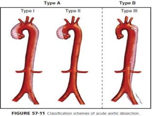
An aortic dissection is a tear in the inner layer of the aorta that allows blood to flow between the layers of the aortic wall, creating a false passageway. It is a medical emergency that requires prompt diagnosis and treatment. Type A dissections, which involve the ascending aorta, require emergency surgery while type B dissections, involving only the descending aorta, are generally treated medically with blood pressure control. Surgical treatment of type A dissections aims to remove the damaged aortic segment and restore blood flow through the aorta using a graft.
A lecture on the echocardiographic evaluation of hypertrophic cardiomyopathy. Starts with an overview of the topic then a systematic approach to diagnosis and then a differential diagnosis followed by take-home messages and conclusion.
Dr Vivek Baliga discusses left atrial myxoma for medical students. Lecture includes a link to MCQs in the video. For access to video, please copy and paste this link --> https://youtu.be/JtkWxbVklgA
Repolarization ST wave Abnormalities
Precise guide for Allied Health Science Students especially cardiac specialty students, DGNM, B.Sc Nursing & M.Sc Nursing Students regarding Repolarization ST wave Abnormalities.
What is a SCAD (spontaneous coronary artery dissection)?Laura Haywood-Cory
PPT presentation I created to educate people about how SCAD survivors use social media to support each other and organize. Katherine Leon had input on this as well, and the two of us presented a slightly different version of this in the WomenHeart "Champions Educating Champions" webinar series back in November of 2011.
Carotid artery disease is commonly seen in association with atherosclerosis and complicate the situation. clearcut guidelines with necessary surgical details are provided in presentations.
A circulatory condition in which narrowed blood vessels reduce blood flow to the limbs.
Peripheral vascular disease is a sign of fatty deposits and calcium building up in the walls of the arteries (atherosclerosis). Risk factors include ageing, diabetes and smoking.
Symptoms may include leg pain, particularly when walking.
Tobacco cessation, exercise and a healthy diet are often successful treatments. When these changes aren't enough, medication or surgery can help.
Tom Selleck Health: A Comprehensive Look at the Iconic Actor’s Wellness Journeygreendigital
Tom Selleck, an enduring figure in Hollywood. has captivated audiences for decades with his rugged charm, iconic moustache. and memorable roles in television and film. From his breakout role as Thomas Magnum in Magnum P.I. to his current portrayal of Frank Reagan in Blue Bloods. Selleck's career has spanned over 50 years. But beyond his professional achievements. fans have often been curious about Tom Selleck Health. especially as he has aged in the public eye.
Follow us on: Pinterest
Introduction
Many have been interested in Tom Selleck health. not only because of his enduring presence on screen but also because of the challenges. and lifestyle choices he has faced and made over the years. This article delves into the various aspects of Tom Selleck health. exploring his fitness regimen, diet, mental health. and the challenges he has encountered as he ages. We'll look at how he maintains his well-being. the health issues he has faced, and his approach to ageing .
Early Life and Career
Childhood and Athletic Beginnings
Tom Selleck was born on January 29, 1945, in Detroit, Michigan, and grew up in Sherman Oaks, California. From an early age, he was involved in sports, particularly basketball. which played a significant role in his physical development. His athletic pursuits continued into college. where he attended the University of Southern California (USC) on a basketball scholarship. This early involvement in sports laid a strong foundation for his physical health and disciplined lifestyle.
Transition to Acting
Selleck's transition from an athlete to an actor came with its physical demands. His first significant role in "Magnum P.I." required him to perform various stunts and maintain a fit appearance. This role, which he played from 1980 to 1988. necessitated a rigorous fitness routine to meet the show's demands. setting the stage for his long-term commitment to health and wellness.
Fitness Regimen
Workout Routine
Tom Selleck health and fitness regimen has evolved. adapting to his changing roles and age. During his "Magnum, P.I." days. Selleck's workouts were intense and focused on building and maintaining muscle mass. His routine included weightlifting, cardiovascular exercises. and specific training for the stunts he performed on the show.
Selleck adjusted his fitness routine as he aged to suit his body's needs. Today, his workouts focus on maintaining flexibility, strength, and cardiovascular health. He incorporates low-impact exercises such as swimming, walking, and light weightlifting. This balanced approach helps him stay fit without putting undue strain on his joints and muscles.
Importance of Flexibility and Mobility
In recent years, Selleck has emphasized the importance of flexibility and mobility in his fitness regimen. Understanding the natural decline in muscle mass and joint flexibility with age. he includes stretching and yoga in his routine. These practices help prevent injuries, improve posture, and maintain mobilit
New Drug Discovery and Development .....NEHA GUPTA
The "New Drug Discovery and Development" process involves the identification, design, testing, and manufacturing of novel pharmaceutical compounds with the aim of introducing new and improved treatments for various medical conditions. This comprehensive endeavor encompasses various stages, including target identification, preclinical studies, clinical trials, regulatory approval, and post-market surveillance. It involves multidisciplinary collaboration among scientists, researchers, clinicians, regulatory experts, and pharmaceutical companies to bring innovative therapies to market and address unmet medical needs.
263778731218 Abortion Clinic /Pills In Harare ,sisternakatoto
263778731218 Abortion Clinic /Pills In Harare ,ABORTION WOMEN’S CLINIC +27730423979 IN women clinic we believe that every woman should be able to make choices in her pregnancy. Our job is to provide compassionate care, safety,affordable and confidential services. That’s why we have won the trust from all generations of women all over the world. we use non surgical method(Abortion pills) to terminate…Dr.LISA +27730423979women Clinic is committed to providing the highest quality of obstetrical and gynecological care to women of all ages. Our dedicated staff aim to treat each patient and her health concerns with compassion and respect.Our dedicated group ABORTION WOMEN’S CLINIC +27730423979 IN women clinic we believe that every woman should be able to make choices in her pregnancy. Our job is to provide compassionate care, safety,affordable and confidential services. That’s why we have won the trust from all generations of women all over the world. we use non surgical method(Abortion pills) to terminate…Dr.LISA +27730423979women Clinic is committed to providing the highest quality of obstetrical and gynecological care to women of all ages. Our dedicated staff aim to treat each patient and her health concerns with compassion and respect.Our dedicated group of receptionists, nurses, and physicians have worked together as a teamof receptionists, nurses, and physicians have worked together as a team wwww.lisywomensclinic.co.za/
Knee anatomy and clinical tests 2024.pdfvimalpl1234
This includes all relevant anatomy and clinical tests compiled from standard textbooks, Campbell,netter etc..It is comprehensive and best suited for orthopaedicians and orthopaedic residents.
Muktapishti is a traditional Ayurvedic preparation made from Shoditha Mukta (Purified Pearl), is believed to help regulate thyroid function and reduce symptoms of hyperthyroidism due to its cooling and balancing properties. Clinical evidence on its efficacy remains limited, necessitating further research to validate its therapeutic benefits.
Flu Vaccine Alert in Bangalore Karnatakaaddon Scans
As flu season approaches, health officials in Bangalore, Karnataka, are urging residents to get their flu vaccinations. The seasonal flu, while common, can lead to severe health complications, particularly for vulnerable populations such as young children, the elderly, and those with underlying health conditions.
Dr. Vidisha Kumari, a leading epidemiologist in Bangalore, emphasizes the importance of getting vaccinated. "The flu vaccine is our best defense against the influenza virus. It not only protects individuals but also helps prevent the spread of the virus in our communities," he says.
This year, the flu season is expected to coincide with a potential increase in other respiratory illnesses. The Karnataka Health Department has launched an awareness campaign highlighting the significance of flu vaccinations. They have set up multiple vaccination centers across Bangalore, making it convenient for residents to receive their shots.
To encourage widespread vaccination, the government is also collaborating with local schools, workplaces, and community centers to facilitate vaccination drives. Special attention is being given to ensuring that the vaccine is accessible to all, including marginalized communities who may have limited access to healthcare.
Residents are reminded that the flu vaccine is safe and effective. Common side effects are mild and may include soreness at the injection site, mild fever, or muscle aches. These side effects are generally short-lived and far less severe than the flu itself.
Healthcare providers are also stressing the importance of continuing COVID-19 precautions. Wearing masks, practicing good hand hygiene, and maintaining social distancing are still crucial, especially in crowded places.
Protect yourself and your loved ones by getting vaccinated. Together, we can help keep Bangalore healthy and safe this flu season. For more information on vaccination centers and schedules, residents can visit the Karnataka Health Department’s official website or follow their social media pages.
Stay informed, stay safe, and get your flu shot today!
2. Aortic dissection
• An aortic dissection is a tearing of the layers within
the aortic wall, classically associated with sudden-
onset chest or back pain, a pulse deficit, and
mediastinal widening on a chest radiograph.
• Depending on size and degree of aortic
involvement, it may result in marked hemodynamic
instability and, often, a rapid death.
3.
4. • Prompt diagnosis and appropriate treatment are
critical to maximize the possibility of survival.
• Significant dissections are often fatal and rarely
survive to clinical attention;
• majority of dissections seen in the critical care
environment are either Subacute, contained, or
sparing the major aortic vessels.
5. • Anatomy of injury in aortic dissection
• The tear usually originates in the intima.
• It then propagates into the media creating a
false channel for blood to flow and hematoma
to form.
• The dissection process may alternatively
originate with hemorrhage in the media that
secondarily causes disruption of the intima
6.
7. • In 70% of patients, the intimal tear, which is the
beginning of the dissection, occurs in the
ascending aorta.
• In 20% of patients it occurs in the descending
thoracic aorta,
• in 10% of patients it occurs in the aortic arch.
• Only rarely is an intimal tear identified in the
abdominal aorta.
8. DeBakey classifications of aortic
dissection
• The two classification systems most commonly used
both have anatomic as well as management
implications.
9. • The DeBakey classification describes
• three types of dissection :
Type I: extends from aortic root to beyond the
ascending aorta
• Type 11: involves only the ascending aorta
• Type Ill: Begins distal to the takeoff of the left
subclavian artery and has two subtypes
• Type III A: limited to the thoracic aorta
• Type 111 B: extends below the diaphragm
10.
11. • The Stanford classification has two types of
dissection
• Type A: involves the ascending aorta
• TYPE B: involves the descending aorta, distal
to the left subclavian artery
12. • Approximately 75% of patients with ruptured
aortic aneurysm will reach an emergency
department alive.
• Whereas for aortic dissection 40% die
immediately.
• Furthermore, only 50% to 70% will be alive 5
years after surgery depending on age and
underlying cause.
13. • For untreated acute dissection of the ascending
aorta the mortality rate is 1 % to 2% per hour after
onset.
• For type A dissections treated medically it is 20%
within the first 24 hours and 50% by 1 month after
presentation.
• Even with surgical intervention the mortality rate
for type A dissection may be as high as 10% after
24 hours and nearly 20% 1 month after repair.
14. • type B dissection is less dangerous than type A, it is
still associated with an extremely high mortality.
• The 30-day mortality rate for an uncomplicated
type B dissection approaches 10%.
• patients with type B dissection who have
complications such as limb ischemia, renal failure,
or visceral ischemia have a 2-day mortality upwards
of 20% and may prompt the need for surgical
intervention
16. • Hypertension:
• Present in 70% to 90% of patients with acute
dissection.
• Advanced age:
• Mean of 63 years in the International Registry of
Acute Aortic Dissection (IRAD).
• Male sex:
• Represented by 65% of patients in the IRAD.
• Family history:
• Recently recognized is a genetic, nonsyndromic
familial form of thoracic aortic dissection
17. • Trauma (deceleration/torsional injury) .
• Congenital and inflammatory disorders:
• Present as Marfan syndrome in almost 5% of
total patients in the IRAD
• and half of those patients under age 40 years
19. • confirmed genetic mutations known to
predispose to dissections
• (TGFBRI, TGFBRP, FBNI, ACTAP, or MYHI I ) .
20. • Pregnancy
• Associated with 50% of dissections in women
under age 40 and most frequently occurring in
the third trimester.
• This might be attributable to elevations in
cardiac output during pregnancy that cause
increased wall stress.
21. Circadian and seasonal variations
• Producing a higher frequency of dissection in
the morning hours and in the winter months.
• Iatrogenic
• Occurring as a consequence of invasive
procedures or surgery, especially when the
aorta has been entered or its main branches
have been cannulated, such as for
cardiopulmonary bypass
22. • The common clinical signs and symptoms of
aortic dissection
23. Pain
• The most common presenting symptom is
chest pain, occurring in up to 90% of patients
with acute dissection.
• Classically, for type A dissections, sudden
onset of severe anterior chest pain with
extension to the back occurs that is described
as ripping or tearing in nature
24. • The pain is usually of maximal intensity from
its inception and is frequently unremitting.
• It may migrate along the path of the
dissection.
• The pain of aortic dissection may mimic that
of myocardial ischemia.
• Patients with type B dissections are more
likely to be seen with back pain ( 64%) alone.
25. Syncope
• Syncope is a well-recognized clinical feature of
dissection, occurring in up to 13% of cases.
• Impairments of cerebral blood flow can be due to
Acute hypovolemia, low cardiac output, or
dissection-involvement of the cerebral vessels.
26. • Patients with a presenting syncope were
significantly more likely to die than were those
without syncope (34% vs. 23%),
• likely because of the frequent correlation with
associated
• cardiac tamponade,
• stroke,
• decreased consciousness,
• and spinal cord ischemia.
27. Neurologic symptoms
• 17% of patients were seen initially with neurologic
symptoms, 53% of which represented ischemic
stroke.
• Neurologic complications may result from
hypotension, malperfusion, distal
thromboembolism, or nerve compression.
• Acute paraplegia as a result of spinal cord
malperfusion has been described as a primary
manifestation in 1% to 3% of patients.
• Up to 50% of neurologic symptoms may be
transient
28. Cardiovascular manifestations
• The heart is the most frequently involved end-
organ in acute proximal aortic dissections.
• Acute aortic regurgitation may be present in 41
% to 76% of patients with proximal dissection
caused by widening of the aortic annulus
resulting in incomplete valve closure
• actual disruption of the aortic valve leaflets
from the dissection flap
29. • Clinical manifestations of dissection- related
aortic regurgitation span from
• mere diastolic murmurs without clinical
significance
• to overt congestive heart failure
• and cardiogenic shock.
30.
31. Myocardial ischemia or infarction
• May result from compromised coronary artery flow
by an expanding false lumen that compresses the
proximal coronary or by extension of the dissection
flap into the coronary artery ostium.
• This occurs in 7% to 19% of patients with proximal
aortic dissections.
• Clinically, these present as electrocardiographic
changes consistent with primary myocardial
ischemia and/or infarction.
• MI IS DANGEROUS FOR A PATIENT OF AORTIC
DISSECTION
32. • Cardiac tamponade is diagnosed in 8% to 10% of
patients seen with acute type A dissections.
• It is associated with a high mortality and should
prompt consideration for emergent drainage and
aortic repair.
• Hypertension occurs in greater than 50% of
patients with dissection, more commonly with
distal disease.
Ongoing renal ischemia can produce severe
hypertension
33. • Hypotension/shock may present in up to 20% of
patients with dissection.
• This may be a result of cardiac tamponade from
aortic rupture into the pericardium,
• dissection, or compression of the coronary
arteries, acute aortic regurgitation,
• acute blood loss,
• true lumen compression by distended false lumen,
or an intra- abdominal catastrophe.
• Cardiogenic shock In approximately 6% of cases.
• This can be due to acute aortic regurgitation or
ongoing myocardial ischemia
34. • Peripheral vascular complications
• Can manifest as pulse and/or blood pressure
differentials or deficits and occur in approximately
one third to one half of patients with proximal
dissection.
• Etiology is partial compression, obstruction,
thrombosis, or embolism of the aortic branch
vessels, resulting in cerebral, renal, visceral, or limb
ischemia.
• Peripheral pulse deficits should alert the clinician
to possible ongoing renal or visceral ischemia
unable to be detected from physical examination
or laboratory values alone
35.
36. Pulmonary complications
• May manifest as pleural effusions, which occur
most frequently on the left.
• Causes include rupture of the dissection into the
pleural space or weeping of fluid from the aorta
as an inflammatory response to the dissection
37. Laboratory abnormalities associated
with aortic dissection
• Laboratory data are usually unrevealing, but anemia
from blood loss into the false lumen can occur.
• A moderate leukocytosis (10,000-14,000 white cells
per mL) is sometimes seen.
• Lactic acid dehydrogenase and bilirubin levels may
be elevated because of hemolysis within the false
lumen.
38. • Disseminated intravascular coagulation has been
reported.
• Currently, randomized controlled data do not
support the use of D-dimers or experimental
serum markers
• (plasma smooth muscle myosin heavy chain
protein, high-sensitivity C-reactive protein)
39. Imaging modalities used to diagnose
aortic dissection
• On the basis of clinical risk factors and conditions,
presentation, and associated examination findings,
patients are stratified into Low- intermediate- or
high- risk categories
40. Further work-up is dictated by this pretest
probability index.
• Some patients with acute dissection initially have
no high-risk features,
creating a diagnostic dilemma.
• According to most recent guidelines, if a clear
alternative diagnosis is not established after the
initial evaluation, then obtaining a diagnostic aortic
imaging study should be considered
41. • Although lacking specificity,
a chest radiograph should be obtained as part of
the initial diagnostic evaluation.
•
• A radiograph abnormality is seen in up to 90% of
patients with aortic dissection;
• most frequent is widening of the aorta and
mediastinum
42. • Other findings may include a
• localized hump on the aortic arch,
• displacement of calcification in the aortic knob,
and pleural effusions.
• However, approximately 40% of radiographs in
acute dissection lack a widened mediastinum, and
as many as 16% are normal.
• Thus a negative radiograph must not delay
definitive aortic imaging in patients deemed at high
risk for aortic dissection by initial screening
43.
44.
45. • (CT), (MRI), and (TEE) Are all highly accurate
imaging modalities that may be used to make the
diagnosis;
• all can provide acceptable diagnostic accuracy.
• Transthoracic echocardiography has limited
diagnostic accuracy
46.
47.
48.
49.
50. Aortography
• Which was once the test of choice, is no longer
used routinely because it is invasive and time-
consuming and involves exposure to intravenous
contrast dye.
• The most recent comparative study with nonhelical
CT, MRI, and TEE showed 100% sensitivity for all
modalities, with better specificity of CT (100%) as
compared with TEE or MRI
51. • A recent metaanalysis found that all three
imaging techniques provided equally reliable
results.
52. • It should be noted, however, that the diagnosis
of acute aortic dissection can be difficult and
occasionally cannot be absolutely excluded by a
single imaging study.
• If a high clinical suspicion exists despite initially
negative imaging, then consideration should be
given to a second imaging modality.
54. Differentiate between the
management of Stanford type A and
type B dissections
• An acute type A dissection is a surgical emergency
• medical management is critical to halt progression
of dissection while diagnostic work-up takes place
55. • Pain management
• gradual down-titration of blood pressure
• are critical to prevent extension of dissection
56. • blood products and intravascular access should be
available in event of aortic rupture.
• Patients with uncomplicated type B dissection are
preferably managed medically with B- blockers
and other antihypertensive agents.
Surgical intervention has no demonstrable
superiority
• except in cases of
• failed medical management manifesting as
malperfusion,
• aortic expansion with potential for imminent
rupture,
• or intractable pain
57. •
Ongoing advances with less invasive interventions
(endovascular stent grafts and endovascular
fenestration procedures) suggest an expanded role
for interventional management in treatment of
acute type B dissection, especially in experienced
centers.
58. The strategies for medical
management of dissection and
commonly used medications
The goals of medical therapy are
to treat pain,
to aggressively control blood pressure,
to determine need for surgical or
endovascular intervention.
59. • Patients who are seen with hypotension should receive
the following:
• Prompt but judicious volume resuscitation and
hemodynamic support with intravenous vasopressors
to maintain a goal mean arterial pressure of 70 mm Hg
.
• Rapid search for underlying etiology (tamponade,
myocardial dysfunction, acute hemorrhage)
• Emergent surgical consultation for operative
management
60. • In those who are seen initially with hypertension,
• the blood pressure should generally be lowered to a
systolic of 100 to 120 mm Hg,
• to a mean of 60 to 65 mm Hg,
• or to the lowest level that is compatible with
perfusion of the vital organs.
• The aortic wall stress is affected by
• the heart rate,
• blood pressure,
• velocity of ventricular contraction .
61. • The ideal antihypertensive regimen must decrease
blood pressure without increasing cardiac output
through peripheral vasodilatation.
• This is because an increased cardiac output can
increase flow rates producing higher aortic wall
stress and thus propagating dissection
62. • Intravenous B-blockers (commonly esmolol,
labetalol, propranolol, or metoprolol) are
considered the first-line medical stabilization
regimen because they affect all three parameters
without increases in cardiac output and aortic wall
stress.
• In patients who are unable to tolerate B- blockade,
nondihydropyridine calcium channel antagonists
(verapamil, diltiazem) offer an acceptable
alternative
63. • Often, single-drug therapy alone is inadequate
to optimize blood pressure management.
• Adequate pain control is essential not only for
patient comfort but also to decrease sympathetic
mediated increases in heart rate and blood
pressure.
• This may be accomplished with intravenous
opioid analgesics.
64. • If B-blockade and adequate pain control are
ineffective to control blood pressure,
• addition of a rapidly acting, easily titratable
intravenous vasodilator, such as
nitroprusside should be considered.
65. • Other agents, such as Nicardipine, nitroglycerin,
and fenoldopam are also acceptable.
Vasodilator therapy without prior B-blockade may
cause reflex tachycardia and increased force of
ventricular contraction leading to greater wall
stress and potentially causing false lumen
propagation;
therefore adequate B-blockade must be established
first, before the vasodilator is initiated.
66. The surgical approach for repair of
Stanford type A dissection.
• The purpose of surgery is to
• resect the aortic segment containing the proximal
intimal tear,
• to obliterate the false channel,
• and to restore aortic continuity with a graft or by
reapproximating the transected ends of aorta.
• For patients with aortic insufficiency, it may be
possible to resuspend the aortic valve, but in some
cases replacement of the aortic valve is necessary.
67. • In some cases of proximal dissection,
reimplantation of the coronary arteries is required.
• If a DeBakey type II dissection is present,
entire dissected aorta should be replaced.
• Surgery to repair an aortic dissection generally
requires cardiopulmonary bypass and, often, deep
hypothermic circulatory arrest.
68. Recent alternatives to surgical repair o
• An endovascular technique of stent-grafting
and/or balloon fenestration may be used for
initial surgical treatment of some dissections.
• Indications for open or endograft treatment
are based on anatomic features of the lesion,
clinical presentation and course, patient
comorbidities, and anatomic constraints
related to endograft technology
69. • Dissections pose a complex situation because
branches of aorta may be perfused from either
true or false lumen.
• Often, both true and false lumens are patent and
some of visceral, renal, or lower extremity vessels
are fed by one channel and remainder by other.
• Consideration must be given to how blood flow
reaches vital organs before considering treatment
of a dissection with an endovascular stent-graft
70.
71. • For type B dissection, an increasing number of
reports show better results with endovascular
repair versus open surgical repair.
72. • However, longer-term (5 year) data are
needed to fully assess potential impact of
stent- grafting for acute dissection,
• including Effects on survival,
• clinical outcomes,
• and long-term aortic remodeling.
73.
74. The use of fenestrated endografts
• A new era in treatment of aortic dissections.
• Unsuitable anatomy is a significant barrier to use of
endovascular stent-grafts for most forms of aortic
disease,
• where ostia of major vessels would otherwise be
partially or completed covered with deployment
of a stent-graft
75. The use of fenestrated endografts
• Using preoperative Three-dimensional CT aortic
reconstruction customized stents can be
constructed,
• featuring holes (fenestrations) or side- branches
matched to patient-specific anatomy to ensure
perfusion to major aortic branch vessels.
76. • The use of fenestrated endografts
• Current trials are underway in Europe and
United States for their use for complex
aneurysmal disease, and expectations are high
for similar application to aortic dissection