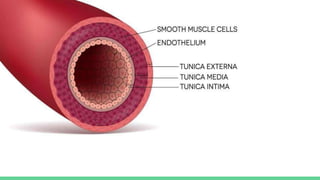
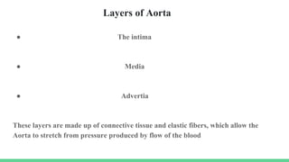
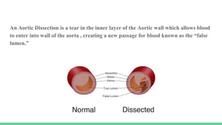
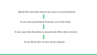
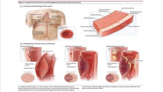
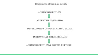
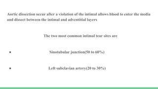
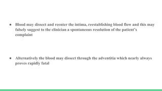
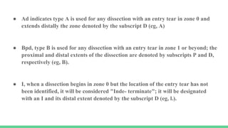
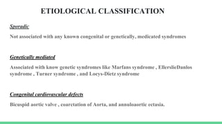
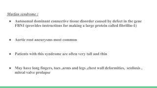
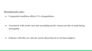
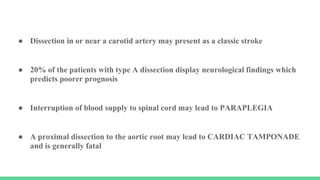
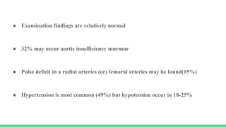
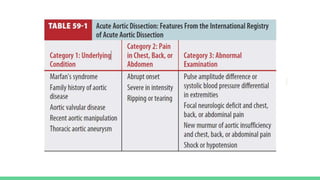
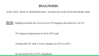
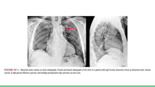
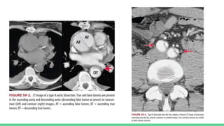
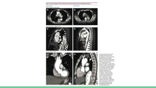
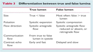
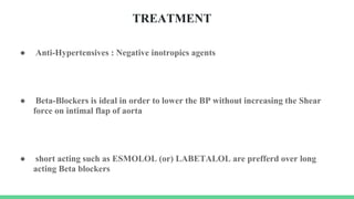
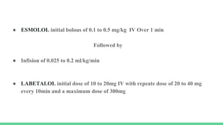
2. The Aorta is the largest artery in the body
It carries the blood from the heart to the branch arteries that supply the rest of the
body
The Aorta has the same dimensions as a garden hose and curves up from the heart
before extending down to the waist
3.
4. The Aorta is identified by 3 major sections
● The ascending Aorta
● The Arch of Aorta
● The descending aorta
5.
6. Layers of Aorta
● The intima
● Media
● Advertia
These layers are made up of connective tissue and elastic fibers, which allow the
Aorta to stretch from pressure produced by flow of the blood
8. An Aortic Dissection is a tear in the inner layer of the Aortic wall which allows blood
to enter into wall of the aorta , creating a new passage for blood known as the “false
lumen.”
9. Blood flow into false lumen can cause several problems
It can rob crucial blood from the rest of the body
It can cause the dissection to spread and affect other arteries
It can block flow in true aortic channel
10. These problems may cause decreased blood flow to vital organs
Aortic dissection also weakness the Aortic wall and may lead to rupture
Which may be fatal or to formation of a BALLOON like expansion of aorta
Known as aneurysm
11.
12. Aortic dissection are uncommon ,yet they are lethal
If untreated, can be fatal within the first 24 to 48 hours
Acute aortic syndrome occurs in the setting of chronic HTN and other factors
That leads to degeneration of the media of the Aortic wall
13. ● Bicuspid aortic valve, Marfan's syndrome , Ehlers-Danlos syndrome and family
history of aortic dissection
● Chronic cocaine or Amphetamine use accelerates atherosclerosis and increase
the risk of dissection
● All mechanism involve weakening of the medial layer and increasing intimal
wall stress
14. Response to stress may include
AORTIC DISSECTION
ANEURYSM FORMATION
DEVELOPMENT OF PENETRATING ULCER
INTRAMURAL HAEMORRHAGE
AORTIC DISSECTION & AORTIC RUPTURE
15. Aortic dissection occur after a violation of the intimal allows blood to enter the media
and dissect between the intimal and adventitial layers
The two most common intimal tear sites are
● Sinotubular junction(50 to 60%)
● Left subclavian artery(20 to 30%)
16.
17. ● Blood may dissect and reenter the intima, reestablishing blood flow and this may
falsely suggest to the clinician a spontaneous resolution of the patient’s
complaint
● Alternatively the blood may dissect through the adventitia which nearly always
proves rapidly fatal
18. ● Aorta dissection has a bimodal age
● The first peak involves younger patients condition due to connective tissue
disorder
● 2nd peak includes those age > 50 years with chronic HTN and/or Ischemic heart
disease
● Prior aortic disease/dissection is a risk factor for recurrent
19. CLASSIFIED INTO TWO SEPARATE
STANFORD
TYPE A DISSECTION: Involvement of
ascending aorta
TYPE B DISSECTION: Restricted to
only descending Aorta
DeBakey system
TYPE 1: Involves the ascending aorta, the arch
and descending aorta
TYPE 2: only the ascending aorta
TYPE 3: only to descending aorta
TYPE 3a : Tear confined only to descending
thoracic artery
TYPE 3b : Tear originates in the descending
thoracic aorta and extends below the
diaphragm
20.
21.
22. ● Ad indicates type A is used for any dissection with an entry tear in zone 0 and
extends distally the zone denoted by the subscript D (eg, A)
● Bpd, type B is used for any dissection with an entry tear in zone 1 or beyond; the
proximal and distal extents of the dissection are denoted by subscripts P and D,
respectively (eg, B).
● I, when a dissection begins in zone 0 but the location of the entry tear has not
been identified, it will be considered "Inde- terminate"; it will be designated
with an I and its distal extent denoted by the subscript D (eg, l.).
23. ETIOLOGICAL CLASSIFICATION
Sporadic
Not associated with any known congenital or genetically, medicated syndromes
Genetically mediated
Associated with know genetic syndromes like Marfans syndrome , EllerslieDanlos
syndrome , Turner syndrome , and Loeys-Dietz syndrome
Congenital cardiovascular defects
Bicuspid aortic valve , coarctation of Aorta, and annuloaortic ectasia.
24. Marfan syndrome :
● Autosomal dominant connective tissue disorder caused by defect in the gene
FBN1 (provides instructions for making a large protein called fibrillin-1)
● Aortic root aneurysms most common
● Patients with this syndrome are often very tall and thin
● May have long fingers, toes ,arms and legs ,chest wall deformities, scoliosis ,
mitral valve prolapse
25. Bicuspid aortic valve :
● Congenital condition affects 1% of population
● Associated with aortic root and ascending aortic Aneurysm due to underlying
aortopathy
● Patients with this are risk for aortic dissection (5 to 10 times higher)
26. Loeys Dietz Aneurysm syndrome :
● Autosomal dominate disorder caused by mutation in TGFBR1 or TGFBR2
(provides instructions for making a protein called transforming growth factor
Beta receptor
● Leading to arterial tortuosity ,wide set eye (hypertelorism) , bifid or broad uvula
, cleft palate and aortic dissection and aneurysm involving branch vessels
27. Familial thoracic aortic aneurysm and dissection :
● Caused by mutation in various genes including ACTA2 , MYH11 , TGFBR1 and
TGFBR2 , FBN1
● Autosomal dominate disorder with predominant features of thoracic aortic
aneurysm and dissection
28. Vascular Ehlers Danlos syndrome :
● Autosomal dominate genetic disorder that resulst from a mutation in gene
COL3A1 leading to abnormal collagen synthesis
● Features include hyperflexible fingers , hyperlucent skin with visible viens, easy
brusability and varicose viens
● Patients are at risk for spontaneous arterial dissection and rupture
29. Turner syndrome :
● Chromosomal disorder affecting 1 in 2,000 live born girls caused by the
complete or partial loss of second sex chromosome
● Features include short statue ,webbed neck and underlying cardiovascular
defects including Bicuspid aortic valve in 25% and Coarctation of Aorta in 12%
● Turners patients are at increased risk of aortic dissection
30. PREDISPOSING FACTORS
Factors increasing the risk of aortic dissection
It's A B C D
● A - Age & Sex / Aortic instrumentation or Previous heart diseases
● B - Bicuspid valve and Blunt chest trauma
● C - Coarctation of Aorta
● D - Pregnancy & Delivery ; Genetic disorders
● Inflammatory or Infectious conditions
31. ● As many as third of patients will subsequently requires surgery on the
remaining aorta because of late enlargement (Aneurysm formation).
● After aortic dissection, most patients require medications to control blood
pressure, which ensures that stress on the aorta will is minimized.
● Patents will typically be placed on BETA-BLOCKERS , angiotensin receptor
blockers , Calcium channel blockers and or other appropriate combinations
32. ● An Intramural hematoma may resolves spontaneously or may lead to dissection
● Penetrating atherosclerosis ulcer can lead to intramural hematoma aortic
dissection (or) perforation of the aorta
● Intramural hematoma affects female individuals in approximately 62% of cases
33. CLINICAL FEATURES
Classically presented with severe chest pain that radiates to an area between the
scapula and abdomen
64% describe sharp pain & 50% describe tearing or ripping pain
Others may note
● SHORTNESS OF BREATH
● PAIN IN ARMS OR LEG
● WEAKNESS or LOSS OF CONSCIOUSNESS
34. ● Dissection in or near a carotid artery may present as a classic stroke
● 20% of the patients with type A dissection display neurological findings which
predicts poorer prognosis
● Interruption of blood supply to spinal cord may lead to PARAPLEGIA
● A proximal dissection to the aortic root may lead to CARDIAC TAMPONADE
and is generally fatal
35. ● Examination findings are relatively normal
● 32% may occur aortic insufficiency murmur
● Pulse deficit in a radial arteries (or) femoral arteries may be found(15%)
● Hypertension is most common (49%) but hypotension occur in 18-25%
36.
37. DIAGNOSIS
Acute chest , Back or abdominal pain , syncope (or) acute focal neurologic signs
ECG - findings include new Q waves or ST Segment elevation in 3 to 4%
ST Segment depression in 15 to 22% and
Nonspecific ST and T waves changes in 41% to 62%
Its normal in 19 to 31% of patients
38. D-DIMER
● Levels rises in acute aortic dissection as they do in pulmonary embolism
● D-DIMER levels >1600ng/mL within first 6 hrs
● So this test may be useful in identifying patients
● However D-DIMER levels may not be elevated in dissection variants such as aortic
intramural hematoma or penetrating aortic ulcer
39. Smooth muscle myosine heavy-chain assay
● Performed in the first 24 hours
● Levels are higher in the first 3 hours
● 2.5 fold increase has a sensitivity of 91% and 98% for aortic dissection
40. Measurement of the degradation products of plasma fibrin and
fibrinogen
● Plasma fibrin degradation product level of 12.6 Micrograms/mL or higher is
suggestive of possibility of aortic dissection with false lumen in symptomatic
patients
● Plasma fibrin degradation product level of 5.6 microgram/mL or higher is
suggestive of the possibility of dissection with complete thrombosis of the false
lumen
41. ● CHEST X RAY - Most common radiographic abnormality is widened
mediastinum (or) abnormal aortic contour
Other possible findings include
pleural effusion
Displacement of aortic intimal calcification
Deviation of trachea , mainstream bronchi , or esophagus
IMAGING
42.
43. ECHOCARDIOGRAPHY
● Transesophageal echocardiography may be used as sensitive and specific as CT
● The procedure generally must be performed under moderate sedation or even
general anesthesia
● The accuracy and precision of transesophageal echocardiography are highly
operated dependent
44. ● Evaluation of the aorta is a routine part of the standard echocardiographic
examination.
● Transthoracic echocardiography (TTE) permits adequate assessment of several
aortic segments, particularly the aortic root and proximal ascending aorta
● TOE overcomes the limitations of TTE in thoracic aorta assessment
● TTE & TOE should be used in complementary manner.
45. ● Echocardiography is useful for assessing aortic size,biophysical properties and
atherosclerotic involvement of the thoracic aorta
● TTE may be used as the initial modality in emergency settings
● Intimal falps in proximal ascending aorta , pericardial effusion/Tamponade and
left ventricular function can be easily visualized by TTE
● TTE does not rule out aortic dissection and other imaging techniques must be
considered
46. ● TOE should defined entry tear location , a mechanisms and severity of aortic
regurgitation and true lumen compression
47. Transthoracic echocardiography
● Although TTE is not the technique of choice for overall assessment of Aorta .
● TTE is one of the technique most used to measure proximal aortic segments in
clinical practice .
● Using different windows , The Proximal ascending aorta is visualized in the left
and right parasternal long axis view.
● In all patients with suspected aortic disease the right parasternal view is
recommended for estimating the true size of ascending aorta
48. ● Modified subcostal views may in some cases (more in children) be helpful , but
here the ascending aorta is far from the transducer .
● All these views also permit assessment of the Aortic valve , which is often
involved in diseases of the ascending aorta
● From this windows aortic Coarctation can be visualized and functionally
evaluated by continous-wave by colour doppler.
● Dilatation and Aneurysm, plaque , calcification, thrombus or a dissection
membrane are detectable if image quality is sufficient
49.
50.
51. TRANSESOPHAGEAL ECHOCARDIOGRAPHY
● The most important TOE view of ascending aorta ,aortic root and aortic valve
are the high TOE Long-axisand short axis views
● TOE is the USG technique of choice in thoracic aorta assessment and provides
high resolution images of the entire thoracic aorta except for a small portion of
the distal ascending aorta near the innominate artery
● TOE overcomes limitations encountered by TTE
52.
53. TTE
Classically limited in the diagnoses
Sensitivity 78%-90% for ascending aorta
dissection
Specificity for Type A dissection ranges 87%-96%
and for Type B dissection 60-83%
TTE is used in emergency room
TOE
Advance in the diagnosis of dissection
Sensitivity 86-100%
Specificity 90%-100%
It's is just performed just prior to surgery to
confirm the diagnosis in suspected Type B
dissection
54. ● CT is the imaging modality of choice for diagnostic of dissection
● CT protocols should be both with or without IV contrast
● CT may also diagnose intramural hematoma and penetrating atherosclerotic
ulcer
COMPUTER TOMOGRAPHY
55. Aortography- The gold standard diagnostic modality for aortic dissection
Benefits includes accurate visualization of the true and false lumen , intimal flap,
aortic regurgitation arteries
56.
57.
58.
59. ● Coronary/Pulmonary/Aortic CT angiography or the triple rule out
which is used to differentiate acute coronary artery disease, pulmonary
embolism and acute aortic dissection has not been shown to improve diagnostic
yield .
● MRI has been used to evaluate stable patients with suspected aortic disease
60. TREATMENT
● Anti-Hypertensives : Negative inotropics agents
● Beta-Blockers is ideal in order to lower the BP without increasing the Shear
force on intimal flap of aorta
● short acting such as ESMOLOL (or) LABETALOL are prefferd over long
acting Beta blockers
61. ● ESMOLOL initial bolous of 0.1 to 0.5 mg/kg IV Over 1 min
Followed by
● Infision of 0.025 to 0.2 ml/kg/min
● LABETALOL initial dose of 10 to 20mg IV with repeate dose of 20 to 40 mg
every 10min and a maximum dose of 300mg
62. SURGICAL TREATMENT
● All the patients with acute aortic dissection should be evaluated by
cardiothoracic surgeon.
● For the patients whose dissection involves the ascending aorta (type A dissection)
, immediate surgery is indicated
● If the dissection involves only the descending Aorta (type B) Medical treatment
is indicated ad surgery usually not recommended
63. ● However if the dissection rapidly progresses the aorta ruptures , or vital organ
become threatened by lack of blood flow
● An interventional radiologist or surgeon may use catheter-based procedure to
improve vital organ arterial perfusion , or urgent aortic surgery may be
required
● This typically requires a Dacron graft (a synthetic material) to replace part of
the aorta to prevent blood flow into the false lumen .
● On average the risk of death from acute type A aortic dissection is
approximately 20%
64.
65. TREATING COMPLICATIONS
● Because may important arteries branch from the aorta , other arteries may be
affected when a dissection spreads.
● If the arteries that provide blood to the heart are compromised, they may need
to be repaired during surgery (which could require a coronary artery bypass).
● The Aortic valve, a 1-way valve to allow blood to flow out of the heart into the
aorta ,may need to be repaired or even replaced by a prosthetic valve if a
dissection causes severe leaking of the valve
66. ● After a dissection, patients will usually be required to stay in the intensive care
unit so that they can be continuously monitored.
● Recovery from surgery usually requires 7 to 10 days.
● Before a patient discharged, another CT SCAN or MRI is often obtained as a
baseline study and to ensure that the dissection has not progressed
67. LIVING WITH AORTIC DISSECTION
● Patients are at highest risk for complaints during the 2 years after an aortic
dissection.
● Complications may begin without symptoms, so Patients must be observed
closely.
● Optimal follow up typically consists of baseline CT SCAN or MRI within the
first 3 months of the acute dissection and repeat imaging of the aorta at every 6
months for the first 2 years
● Over long term, continued imaging of the aorta at least yearly is usually
recommended
68. ● After aortic dissection most patients require medications to control blood
pressure which ensures the stress on the Aortic wall is minimized .
● Patient will typically be placed on beta blockers, angiotensin-converting
inhibitors, angiotensin receptor blockers , Calcium channel blockers and or
other appropriate combinations
● Lifestyle modifications are necessary to reduce the risk of long-term
complications
● Patients are counseled to avoid strenuous physical activity and activities that
involve heavy lifting because such activity may dramatically increase the BP and
therefore stress on the Aortic wall
69. ● Normal daily activities such as cooking, bathing, driving and climbing stairs are
not restricted , although contact sports are not recommended.
● Many patients may require a change in occupation, as sedentary jobs are often
more appropriate for the patients who have suffered a dissection
● Lifestyle recommendations should be thoroughly discussed with cardiologist and
primary care physician
70. CONCLUSION
● Aortic dissection is a life threatening condition that must be diagnosed and
treated without delay.
● Dissection involving the beginning portion of the Aorta (Type A) are treated
with emergency surgery and those involving the descending Aorta (Type B) are
usually treated with medications.
● After successful surgery for Type A dissection, many patients may expect a
relatively good short-term outlook.
71. ● However, many patients require more surgery in the future and many with Type
B dissections will ultimately require surgery.
● Continues evaluation of the affected Aorta with CT or MRI imaging necessary.
● Because aortic dissection may be related to underlying genetic abnormality of
the aorta , first degree relatives should be screened for aortic enlargement.