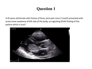
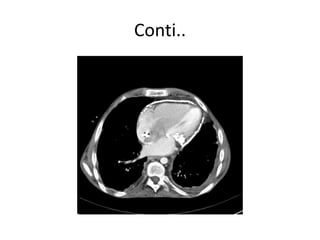
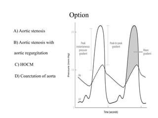
This document contains a 14 question cardiology MCQ exam based on chapters from Harrison's 18th Edition on noninvasive cardiac imaging modalities and diagnostic cardiac catheterization. Each question is multiple choice with 4 answer options and includes the reference used to write the question. The questions cover topics like echocardiography findings, fractional flow reserve measurement, intravascular ultrasound, stress myocardial perfusion imaging, positron emission tomography, computed tomography of the chest, coronary angiography, and hemodynamic measurements.

![Question-15
Normal Values for Hemodynamic Measurements include all of the following
except?
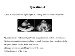
A) Systemic vascular resistance 900–1400 (dyn-s)/cm5
B) Pulmonary vascular resistance 40–120 (dyn-s)/cm5
C) Cardiac index [(L-min)/m2] 2.8–4.2
D) Pulmonary capillary wedge (mean) 4-6 mmhg](https://image.slidesharecdn.com/cardiologyexammcq-161223192050/85/Cardiology-exam-MCQ-37-320.jpg)