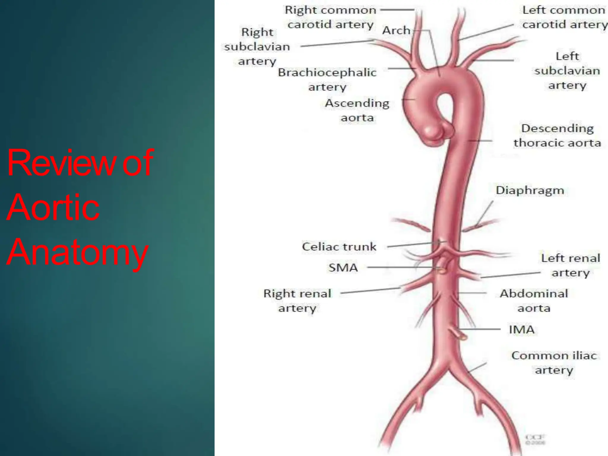
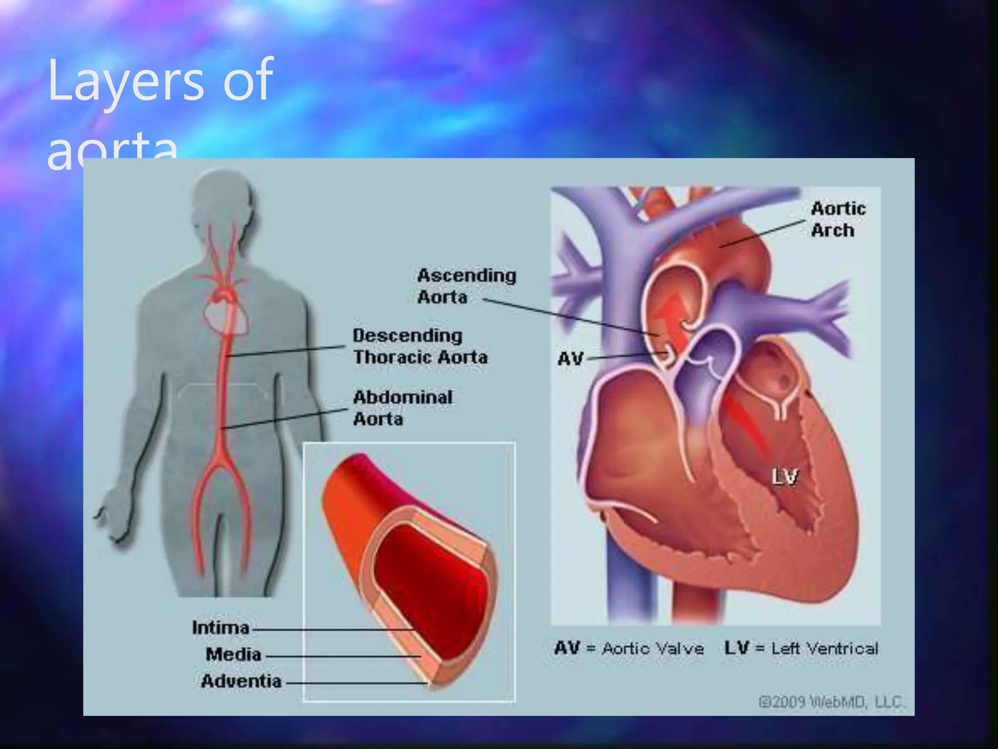
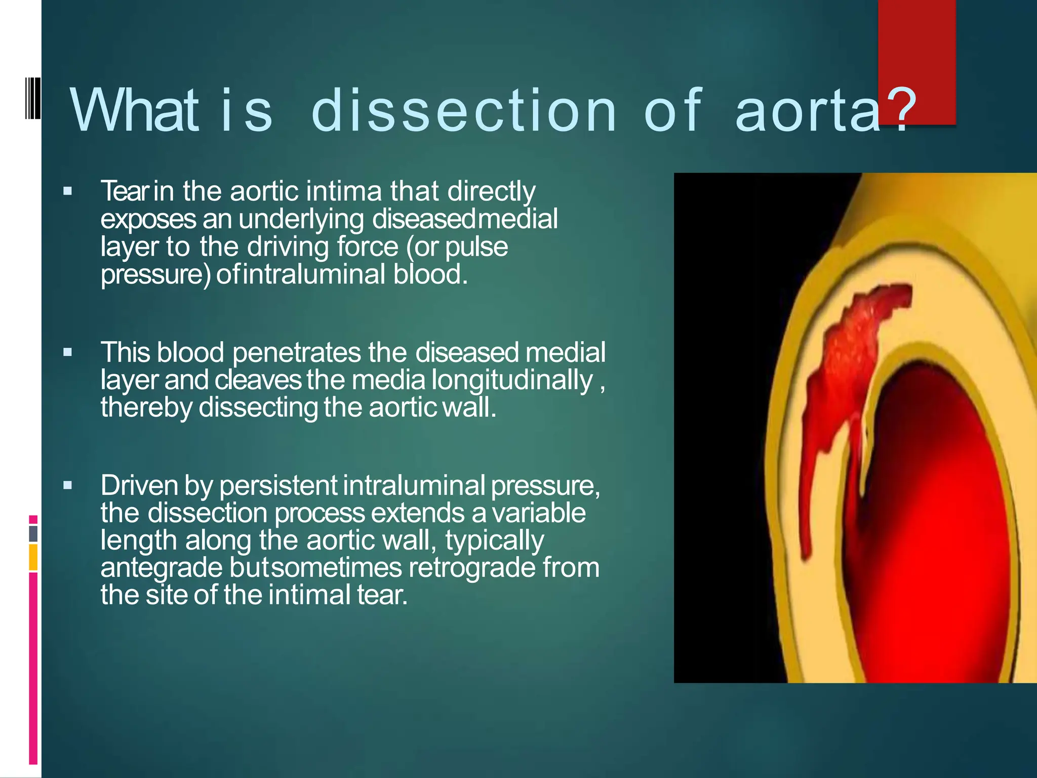
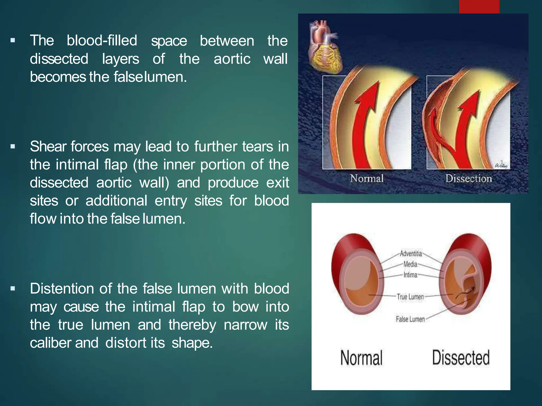
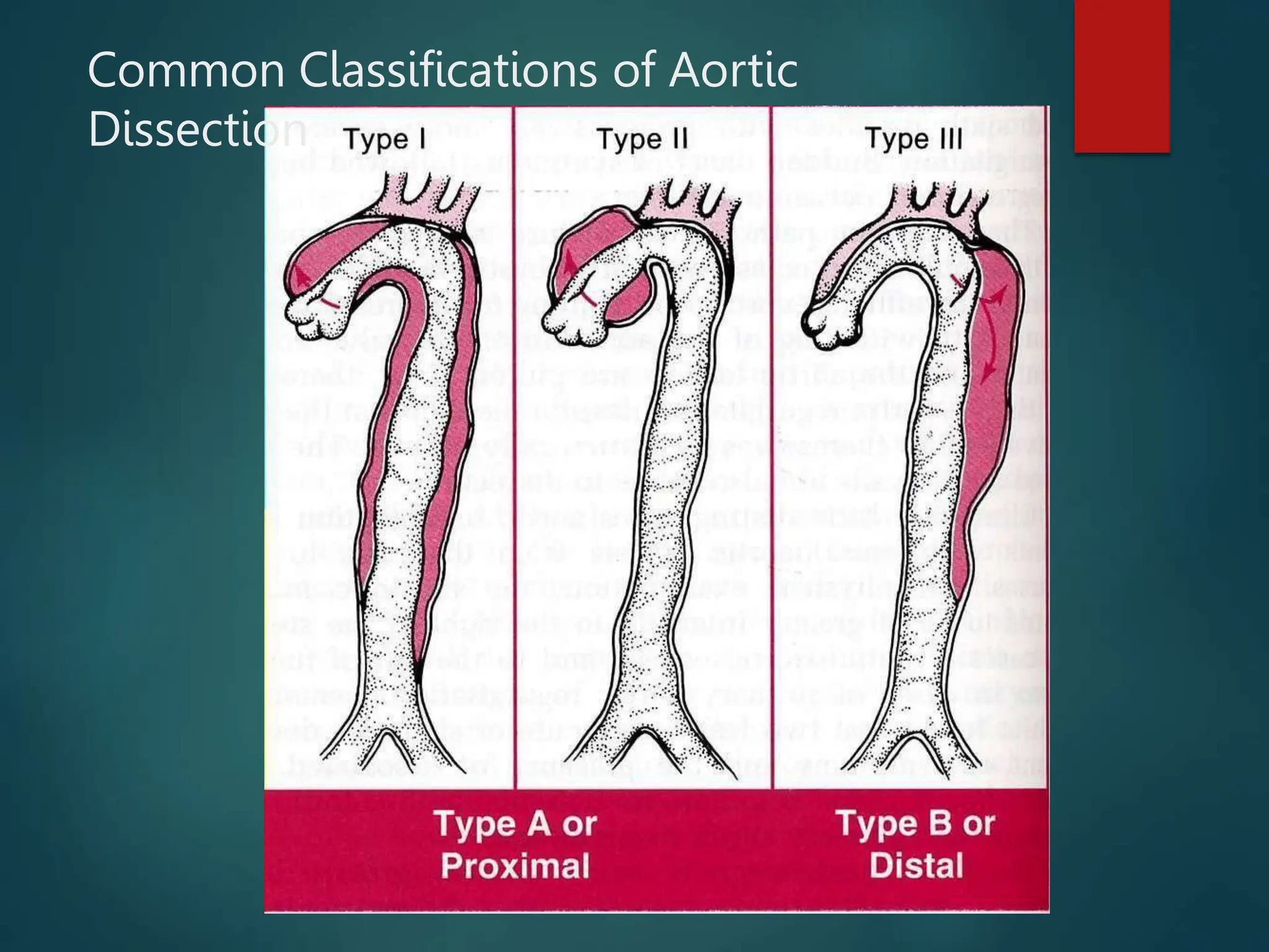
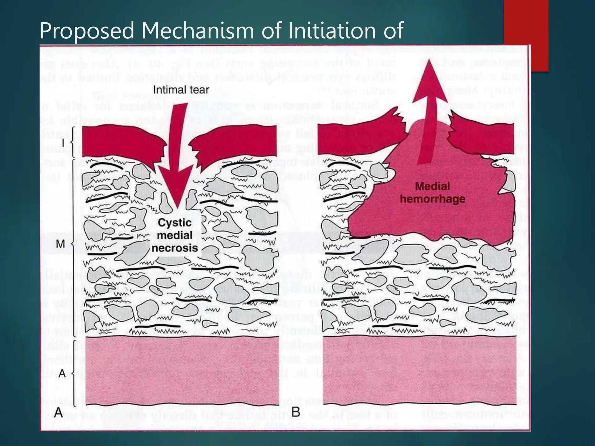
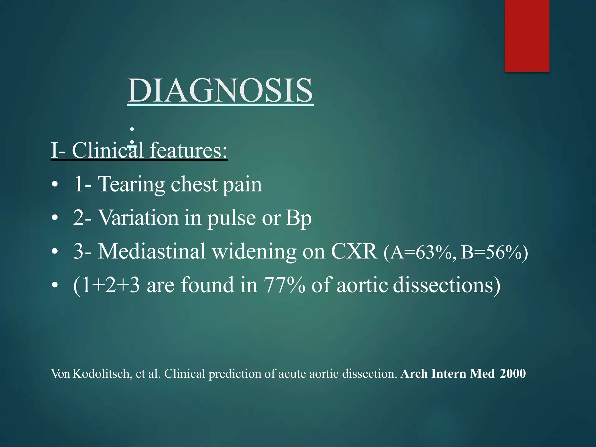
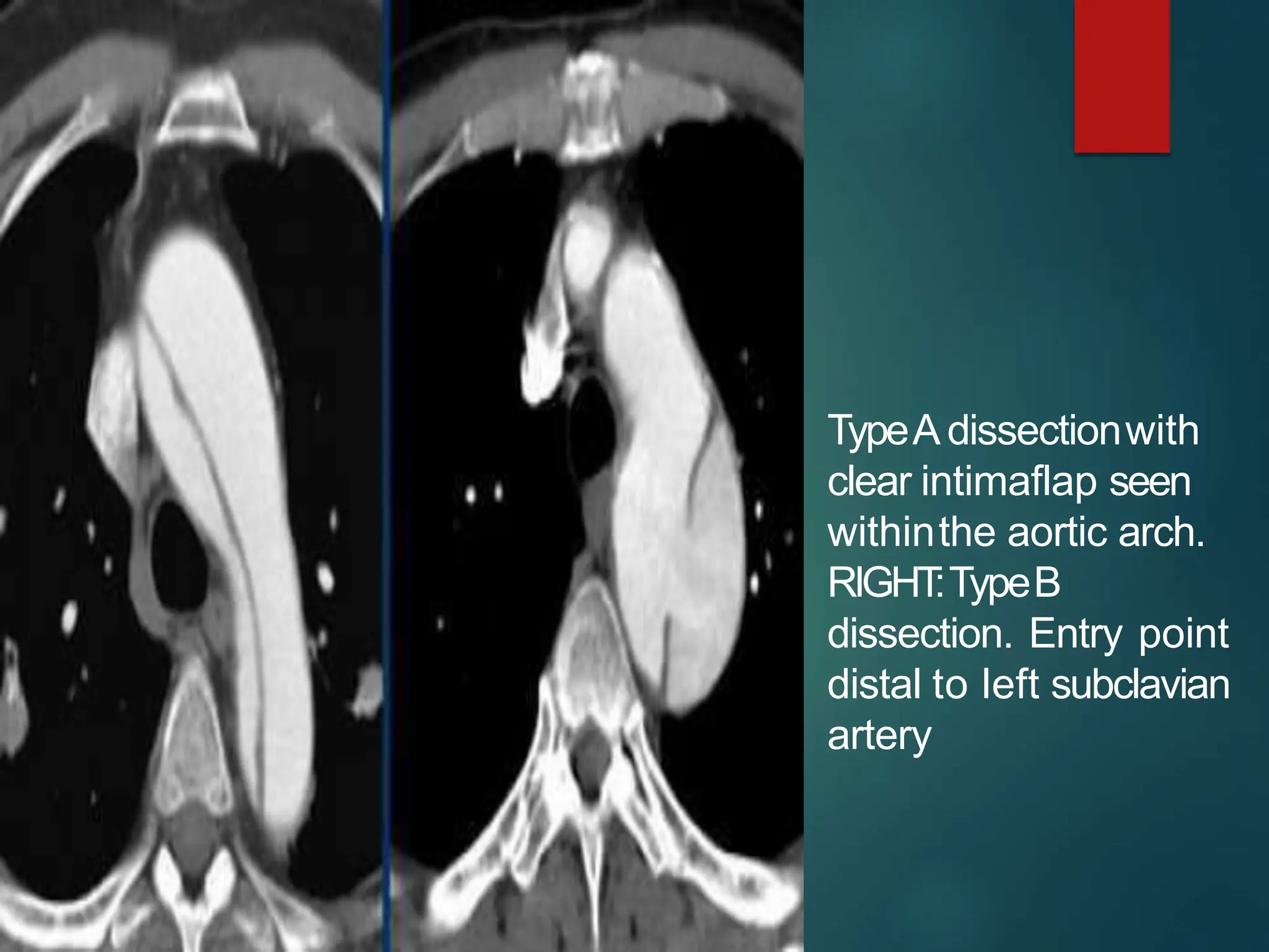
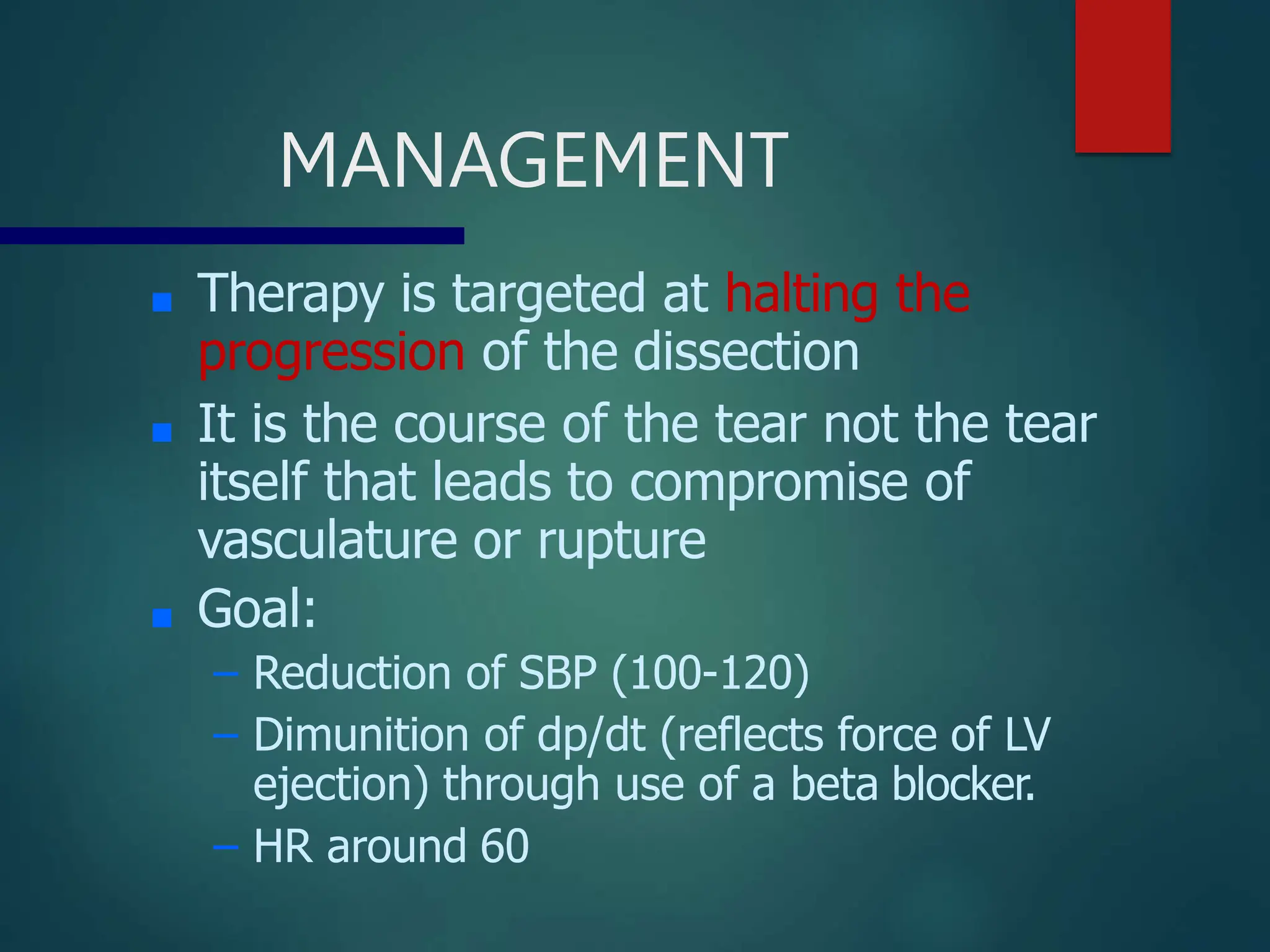
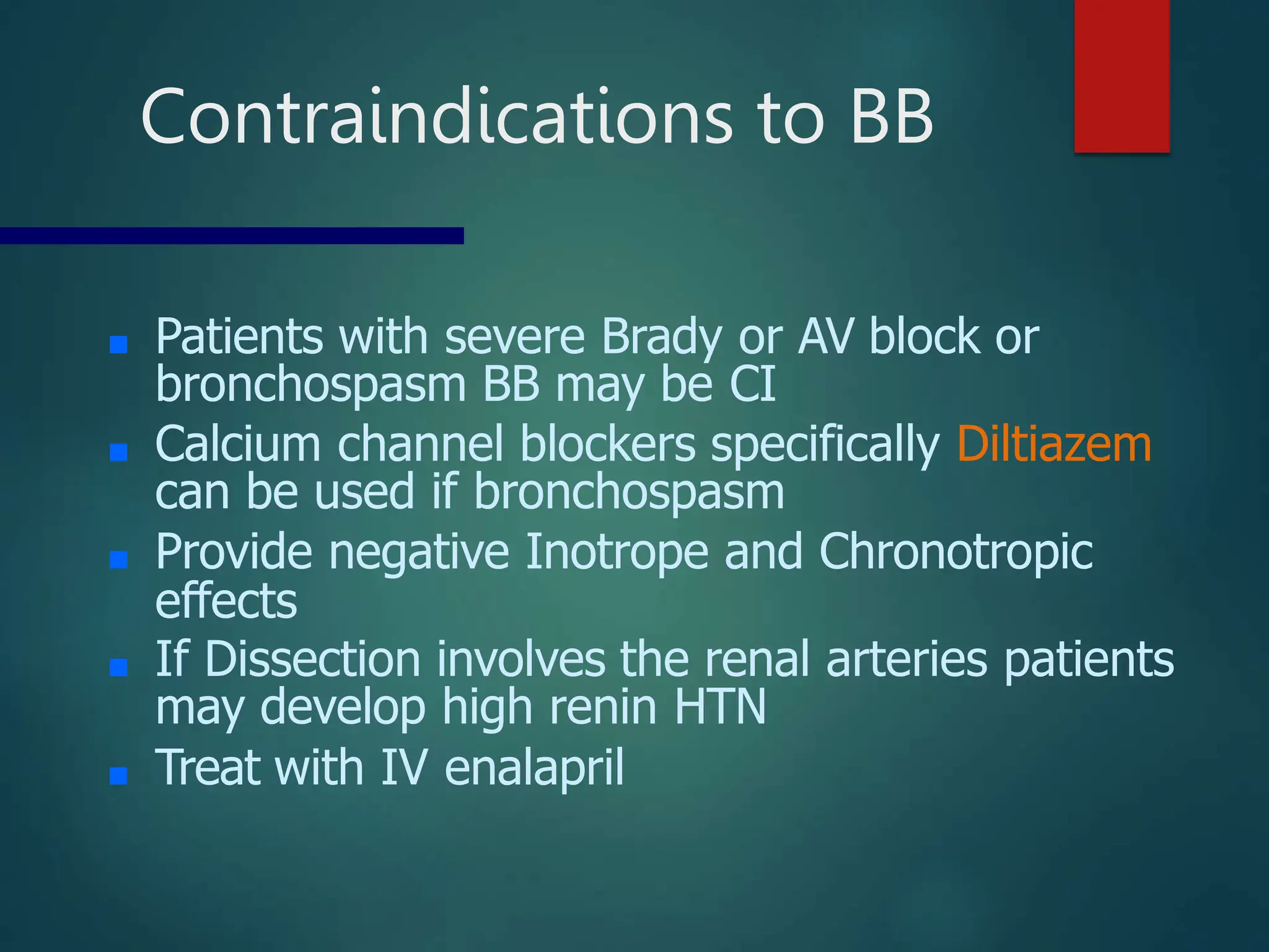
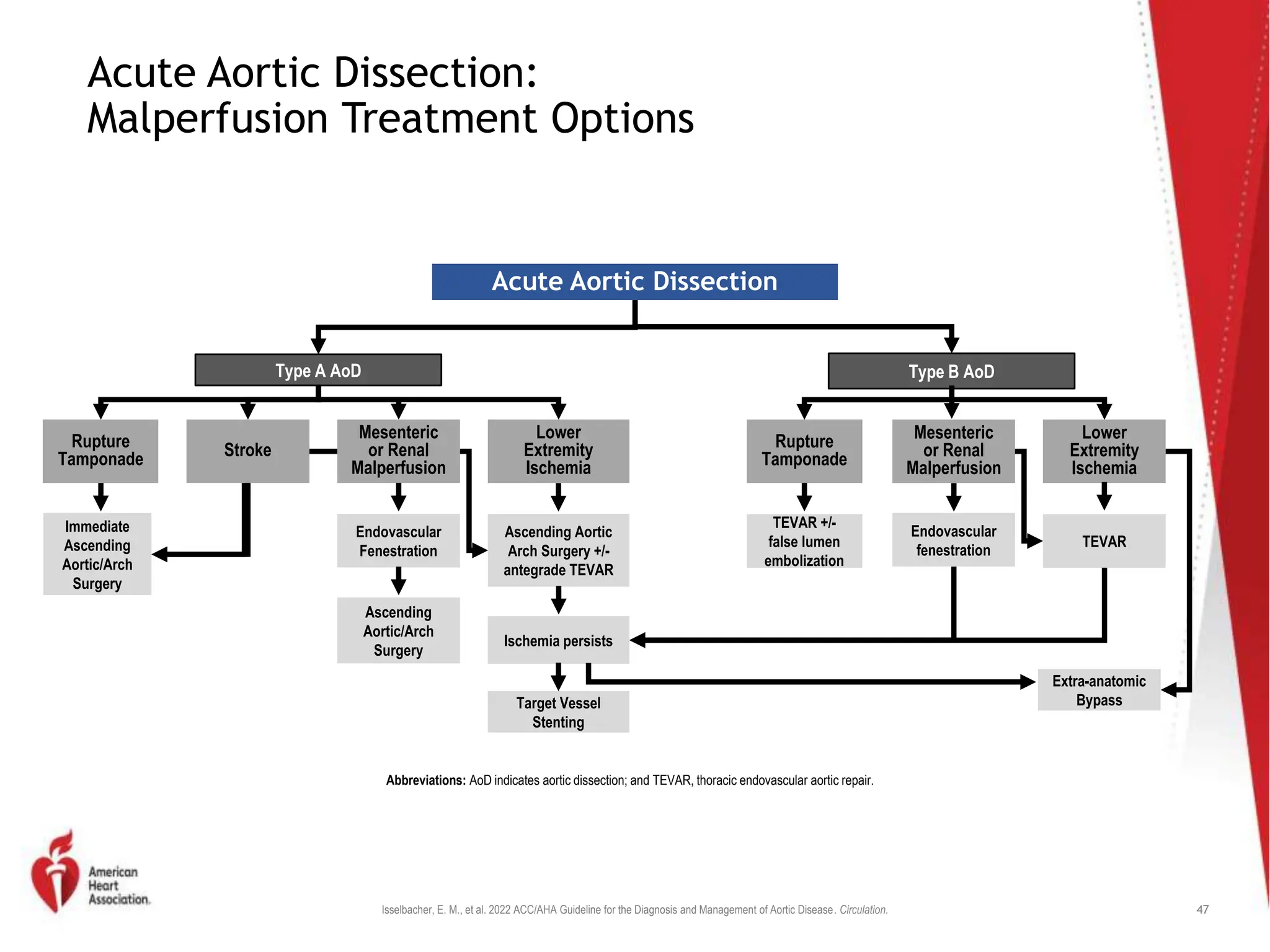
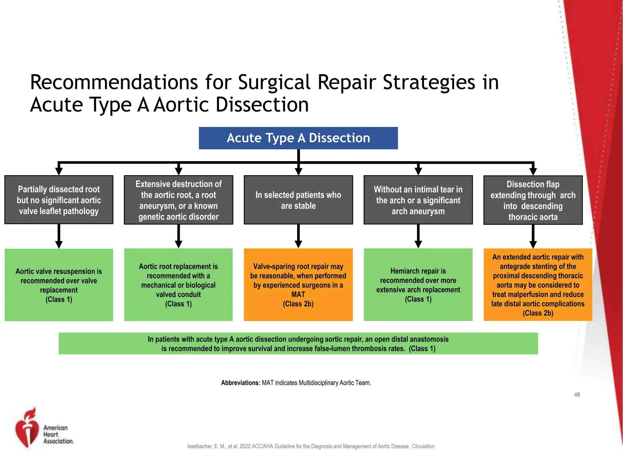
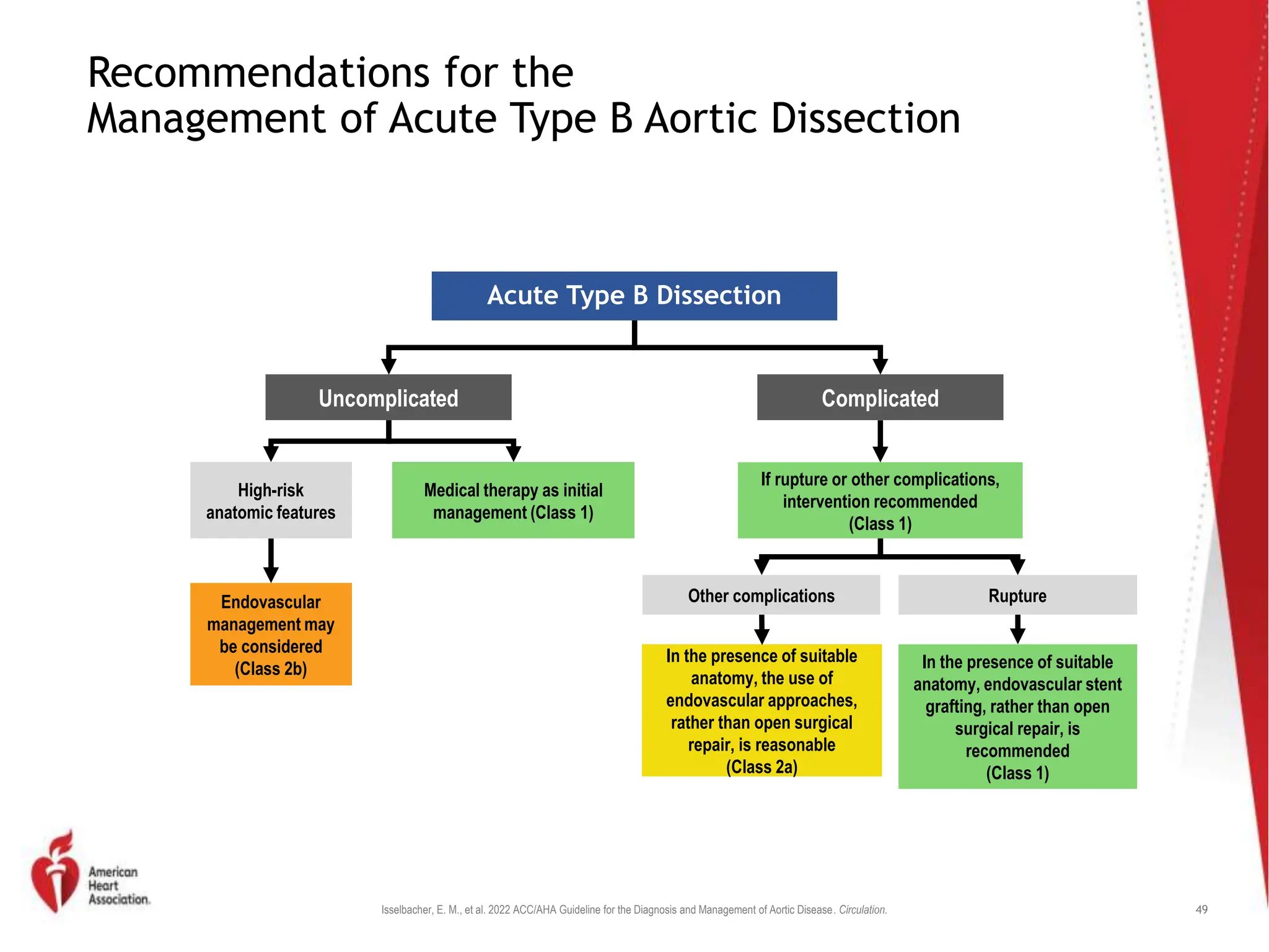
BP CONTROL IN
AORTIC
EMERGENCIES
DR.HANIFMOHDALI
12TH SEPTEMBER2023
What is aortic emergencies
??
 Aortic emergencies comprise different clinical entities
with only one thing in common : if not diagnosed and
managed in due time they are life threatening
Example of aortic
emergencies
 Aortic trauma
 Aneurysm rupture
 Acute aortic syndrome
 Aortic dissection
 Intramural haematoma
 Penetrating aortic ulcer
 Aorto-enteric fistula
 Aortitis
AORTIC DISSECTION
Outline
 Emergencies –aortic dissection
 Pathophysiology
 Sign and symptoms
 Modalities
 Diagnosis
 Management
Reviewof
Aortic
Anatomy
Layers of
aorta
What i s dissection of aorta?
 Tearin the aortic intima that directly
exposes an underlying diseasedmedial
layer to the driving force (or pulse
pressure) ofintraluminal blood.
 This blood penetrates the diseased medial
layer andcleavesthe media longitudinally ,
thereby dissectingthe aortic wall.
 Driven by persistent intraluminalpressure,
the dissection process extends avariable
length along the aortic wall, typically
antegrade butsometimes retrograde from
the site of the intimal tear.
 The blood-filled space between the
dissected layers of the
becomes the falselumen.
aortic wall
 Shear forces may lead to further tears in
the intimal flap (the inner portion of the
dissected aortic wall) and produce exit
sites or additional entry sites for blood
flow into the falselumen.
 Distention of the false lumen with blood
may cause the intimal flap to bow into
the true lumen and thereby narrow its
caliber and distort its shape.
EPIDEMIOLOGY :
 Uncommon but potentiallycatastrophic illness.
 Occurswith anincidenceof at least 2000cases
per year.
 Early mortality is ashigh as5%per hour if
untreated.
 Thepeakincidence–fifth and sixth decadesof
life,
 Male tofemale--- 3:1
Classifications
■ Acute: when diagnosis is made within
2 weeks of initial onset of symptoms
■ Chronic: >2 weeks of symptoms
■ One third of patients fall into the
chronic category.
Common Classifications of Aortic
Dissection
Clinical Presentation
:
 Sudden onset of sharp , tearing , intractable
chest pain, may radiate to the back, esp,
interscapular region.
 Asymmetrical peripheralpulse
 Diastolic murmur orbruit
 Pulmonary edema
 Previously hypertensive, now in shock
LOCATION
:
 Ascending aorta65%
 Descending aorta,20%
just distal to the origin of the left subclavian
artery at the site of the ligamentum
arteriosum.
 Aortic arch 10%and
 Abdominal aorta5%
Predisposing Factors
■ Chronic Systemic HTN (80%)
■ Proximal Dissection:
– Peak age 50-55 years
■ Distal Dissection:
– Peak Age 60-70 years
■ Direct Iatrogenic Trauma: 5% of cases
■ Indirect Trauma (eg sudden deceleration)
Predisposing Factors
■ Hereditary Connective Tissue Diseases
– Marfan Syndrome
– Ehler Danlos Syndrome
■ Chromosomal Aberrations
– Turners Syndrome
– Noonans Syndrome
■ Aortic Diseases
– Aortic Dilatation
– Aortic Aneurysm
– Aortic Arteritis
– Bicuspid Aortic Valve
– Coarctation oa aorta
Predisposing Factors
■ Females in 3rd Trimester Pregnancy or
1st Stage of Labor
■ Case Reports of:
– Cocaine (Perron et al. Am J Emerg Med 1992)
– Abrupt Discontinuation of Beta Blockers
al. Cardiology 1993; 83:128-131)
(Eber et
Cystic Medial Degeneration
■ Medial Degeneration predisposes dissection
by decreasing cohesiveness of layers of aortic
wall
– More extensive in patients with:
■ HTN
■ Marfan Syndrome
■ Bicuspid Aortic Valves
– But, even in other causes of dissection, medial
degeneration is much greater than expected with
normal aging.
Pathogenesis
■ Intimal tears occur in regions of aorta
results in separation of the intima
from media(90%)
■ Rapture of vas vasorum resulting
IMH(intramural haematoma)
■ Penetrating atherosclerotic ulcer
Proposed Mechanism of Initiation of
Dissection
Evolution of a Penetrating
atherosclerotic Ulcer of Aorta
Natural
History
■ Hydrodynamic forces propagate the
dissection until rupture occurs either:
– Back into the lumen of the aorta
– Through the adventitia (causing death)
■ Mortality Rates (if untreated)
– 5% per hour
– 25% die within 24 hr
– 50% die within a week
– 90% within a year
DIAGNOSIS
:
I- Clinical features:
• 1- Tearing chest pain
• 2- Variation in pulse or Bp
• 3- Mediastinal widening on CXR (A=63%, B=56%)
• (1+2+3 are found in 77% of aortic dissections)
Von Kodolitsch, et al. Clinical prediction of acute aortic dissection. Arch Intern Med 2000
Differential diagnosis
• Acute coronary syndrome
• Pericarditis
• Pulmonary embolus
• Aortic aneurysm without dissection
• Musculoskeletal pain
• Mediastinal tumors
• Pleuritis
• Cholecystitis
• Atherosclerotic or cholesterolembolism
• Peptic ulcer disease or perforatingulcer
ECG
■ ECG: 1/3 exhibit LVH
■ ECG is important to rule out any
ischemic changes or MI which would
lead to an alternate diagnosis
■ ECG may display infarction
■ ECG – usually shows nonspecific ST-T
wave changes.
CXR
■ Widened Mediastinum suggestive but
not diagnostic.
■ Seen Anywhere from 50% in most
reports.
■ And in one report of 236 cases as high
as: 90% (Spittell, Mayo Clinic Proc 68:642,1993.)
Normal Aorta in CXR 3 years
Prior
Enlargement of the Aortic
Knob
A Case of Proximal Aortic Dissection
Aortography
■ Sensitivity: 86-88%
■ Specificity: 75-94%
■ False negatives if intramural hematoma
or thrombosis of false lumen
■ Good at detecting branch vessel
involvement and Coronary Artery
invovlvement.
CT
■ Sensitivity 83-94%
■ Specificity of 100%
■ Spiral CT increased sensitivity to 96%
■ Non-invasive with rapid availability (MC initial imaging
modality in IRAD pts)
■ Needs contrast to be effective
■ Disadvantages:
– Cannot Detect AR
– Does not detect Site of Intimal Tear well
– Cannot detect Coronary Artery Involvement
Left Anterior Oblique View Contrast Enhanced CT
Intimal Flap originates beyond Left SC Artery
Intramural Hematoma
(IMH)
Crescenteric Hematoma that does not Enhance
confirming a intramural Hematoma that does
not communicate with the Aortic Lumen.
MRI
■ Gold Standard for Diagnosis
■ Sensitivity and Specificity of 98-100%
■ Disadvantages:
– Limited Availability
– Relatively CI in unstable patients.
– CI:
Pacemakers
Certain types of vascular clips
Older metallic heart valves
MRi
1.Intimal flap
2.Slowflow and clot in false lumen
Lumen Partition of athree-dimensional
contrast-enhanced MRAshows intimal flap
(arrows ) in the distal aortic arch and descending
aorta.
TypeA dissectionwith
clear intimaflap seen
withinthe aortic arch.
RIGHT
:TypeB
dissection. Entry point
distal to left subclavian
artery
MANAGEMENT
■ Therapy is targeted at halting the
progression of the dissection
■ It is the course of the tear not the tear
itself that leads to compromise of
vasculature or rupture
■ Goal:
– Reduction of SBP (100-120)
– Dimunition of dp/dt (reflects force of LV
ejection) through use of a beta blocker.
– HR around 60
The key for medical Rx:
Systolic pressure < 100-120mmhg
Shearing force Contractility
preventing propagation of the dissection
Labetolol
■ Effectively lowers dP/dT as well as reducing
arterial pressure
■ Initial dose is 20mg followed by 40 to 80 mg
– 15 minutes (max 200mg IV)
■ Once BP controlled maintenance by
continuous infusion
■ Infusion at 2mg/min titrating up to 5 –10
mg/min
Esmolol
■ Ultra short acting BB for those with labile
blood pressure or those that are surgical
candidates. (Long acting medications may affect intraoperative
bp management)
■ Load with 500 mcg/kg bolus
■ Infusion starts @ 50mcg/kg/min titrate to 200
mcg/kg/min for control
Contraindications to BB
■ Patients with severe Brady or AV block or
bronchospasm BB may be CI
■ Calcium channel blockers specifically Diltiazem
can be used if bronchospasm
■ Provide negative Inotrope and Chronotropic
effects
■ If Dissection involves the renal arteries patients
may develop high renin HTN
■ Treat with IV enalapril
Other
Considerations
■ Hypotension must ensure if its true or false
■ May be secondary to compromise of artery by
dissection (pseudohypotension) so check both
arms
■ If true hypotension may indicate rupture or
tamponade
■ Fluids first then use norepinephrineor
phenylephrine
■ Dopamine should be avoided since it can
raise dP/dT unless used at low doses for renal
perfusion
Indications forDefinitive
Surgical and Medical
Therapy in AD
Isselbacher, E. M., et al. 2022 ACC/AHA Guideline for the Diagnosis and Management of Aortic Disease. Circulation.
Recommendations for Surgery for Sporadic Aneurysms of
the Aortic Root and Ascending Aorta
Symptomatic Asymptomatic
Proceed to
surgical repair
(Class 1)
Aneurysms of the aortic root or ascending aorta
Diameter ≥5.5
cm
Diameter <5.5
cm
Maximum
diameter of
≥5.0 cm
Pt. height >1 SD
above or below the
mean and max.
cross-sectional
aortic area/height
ratio of ≥10 cm2/m
Aortic size index
of ≥3.08 cm/m2 or
aortic height index
of ≥3.21 cm/m
Confirmed rapid
growth rate (≥0.3
cm/y over 2y OR
≥0.5 cm in 1y)
Surgery is
reasonable by
experienced
surgeons in a
MAT (Class 2a)
Surgery may be
reasonable when
performed by
experienced
surgeons in a MAT
(Class 2b)
Pt. undergoing repair or replacement of tricuspid AV
with a concomitant aneurysm of the ascending aorta
with max. diameter of ≥4.5 cm
Ascending aortic replacement is reasonable
by experienced surgeons in MAT (Class 2a)
Pt. undergoing cardiac surgery for other than AV repair
or replacement with aneurysm of the aortic root or
ascending aorta and max. diameter of ≥5.0 cm
Ascending aortic replacement may be
reasonable (Class 2b)
Abbreviations: AV indicates aortic valve; cm, centimeter; CT, computed tomography; y, year; MAT, multidisciplinary aortic team; max,
maximal; pt, patient; SD, standard deviation; and y, year.
45
Isselbacher, E. M., et al. 2022 ACC/AHA Guideline for the Diagnosis and Management of Aortic Disease. Circulation.
Guidance for Repair of Intact Descending Thoracic
Aortic Aneurysms
Descending thoracic aortic aneurysm repair thresholds
<5.5cm
Patients with
average
operative risk and
elevated risk of
rupture
Risk factors for
increased
aneurysm rupture
• Growth ≥0.5cm/y
• Symptomatic
• Marfan or Loeys-
Dietz syndrome
• HTAD
• Saccular
aneurysm
• Female sex
• Mycotic aneurysm
>5.5cm
Patients with
average
operative risk and
average risk of
rupture
>>5.5cm
Patients with
elevated operative
risk and average
risk of rupture
Risk factors for
increased operative
morbidity
• Advanced age,
particularly ≥75 y
• CKD3 or greater
• COPD and FEV1
<50% predicted
• Prior stroke
• Functional
dependence
• Unfavorable
anatomy for TEVAR
Abbreviations: cm indicates centimeter; CKD, chronic kidney disease; COPD, chronic obstructive pulmonary disease; CT, computed tomography; FEV,
forced expiratory volume; HTAD, heritable thoracic aortic disease; TEVAR, thoracic endovascular aortic repair; and y, year.
46
Isselbacher, E. M., et al. 2022 ACC/AHA Guideline for the Diagnosis and Management of Aortic Disease. Circulation.
Acute Aortic Dissection:
Malperfusion Treatment Options
Acute Aortic Dissection
Type A AoD Type B AoD
Mesenteric
or Renal
Malperfusion
Lower
Extremity
Ischemia
Rupture
Tamponade Stroke
Mesenteric
or Renal
Malperfusion
Lower
Extremity
Ischemia
Rupture
Tamponade
Ascending Aortic
Arch Surgery +/-
antegrade TEVAR
Endovascular
fenestration
Target Vessel
Stenting
Ischemia persists
TEVAR +/-
false lumen
embolization
Immediate
Ascending
Aortic/Arch
Surgery
Endovascular
Fenestration
TEVAR
Ascending
Aortic/Arch
Surgery
Extra-anatomic
Bypass
Abbreviations: AoD indicates aortic dissection; and TEVAR, thoracic endovascular aortic repair.
47
Isselbacher, E. M., et al. 2022 ACC/AHA Guideline for the Diagnosis and Management of Aortic Disease. Circulation.
Recommendations for Surgical Repair Strategies in
Acute Type A Aortic Dissection
Acute Type A Dissection
Partially dissected root
but no significant aortic
valve leaflet pathology
Without an intimal tear in
the arch or a significant
arch aneurysm
Extensive destruction of
the aortic root, a root
aneurysm, or a known
genetic aortic disorder
In selected patients who
are stable
Dissection flap
extending through arch
into descending
thoracic aorta
Aortic valve resuspension is
recommended over valve
replacement
(Class 1)
Valve-sparing root repair may
be reasonable, when performed
by experienced surgeons in a
MAT
(Class 2b)
Hemiarch repair is
recommended over more
extensive arch replacement
(Class 1)
Aortic root replacement is
recommended with a
mechanical or biological
valved conduit
(Class 1)
An extended aortic repair with
antegrade stenting of the
proximal descending thoracic
aorta may be considered to
treat malperfusion and reduce
late distal aortic complications
(Class 2b)
In patients with acute type A aortic dissection undergoing aortic repair, an open distal anastomosis
is recommended to improve survival and increase false-lumen thrombosis rates. (Class 1)
Abbreviations: MAT indicates Multidisciplinary Aortic Team.
48
Isselbacher, E. M., et al. 2022 ACC/AHA Guideline for the Diagnosis and Management of Aortic Disease. Circulation.
Recommendations for the
Management of Acute Type B Aortic Dissection
Acute Type B Dissection
Uncomplicated Complicated
High-risk
anatomic features
Other complications Rupture
Endovascular
management may
be considered
(Class 2b)
Medical therapy as initial
management (Class 1)
If rupture or other complications,
intervention recommended
(Class 1)
In the presence of suitable
anatomy, the use of
endovascular approaches,
rather than open surgical
repair, is reasonable
(Class 2a)
In the presence of suitable
anatomy, endovascular stent
grafting, rather than open
surgical repair, is
recommended
(Class 1)
49
Take Home
Message
• Acute ascending aortic dissection is included in the
differential diagnosis of patients with acute chestpain.
• Aortic dissection is considered as the most common
disaster of aorta.
• Stanford type A dissection is a surgicalemergency that
requires urgent diagnosis and immediate surgical
intervention.
Reference
 ESC guideline 2014
 2022 ACC/AHA Guidelines for the diagnosis and
management of aortic disease
 National institute of health 2022
AORTIC DISSECTION and management of aortic dissection

AORTIC DISSECTION and management of aortic dissection

  • 1.
  • 2.
    What is aorticemergencies ??  Aortic emergencies comprise different clinical entities with only one thing in common : if not diagnosed and managed in due time they are life threatening
  • 3.
    Example of aortic emergencies Aortic trauma  Aneurysm rupture  Acute aortic syndrome  Aortic dissection  Intramural haematoma  Penetrating aortic ulcer  Aorto-enteric fistula  Aortitis
  • 4.
  • 5.
    Outline  Emergencies –aorticdissection  Pathophysiology  Sign and symptoms  Modalities  Diagnosis  Management
  • 6.
  • 7.
  • 8.
    What i sdissection of aorta?  Tearin the aortic intima that directly exposes an underlying diseasedmedial layer to the driving force (or pulse pressure) ofintraluminal blood.  This blood penetrates the diseased medial layer andcleavesthe media longitudinally , thereby dissectingthe aortic wall.  Driven by persistent intraluminalpressure, the dissection process extends avariable length along the aortic wall, typically antegrade butsometimes retrograde from the site of the intimal tear.
  • 9.
     The blood-filledspace between the dissected layers of the becomes the falselumen. aortic wall  Shear forces may lead to further tears in the intimal flap (the inner portion of the dissected aortic wall) and produce exit sites or additional entry sites for blood flow into the falselumen.  Distention of the false lumen with blood may cause the intimal flap to bow into the true lumen and thereby narrow its caliber and distort its shape.
  • 10.
    EPIDEMIOLOGY :  Uncommonbut potentiallycatastrophic illness.  Occurswith anincidenceof at least 2000cases per year.  Early mortality is ashigh as5%per hour if untreated.  Thepeakincidence–fifth and sixth decadesof life,  Male tofemale--- 3:1
  • 11.
    Classifications ■ Acute: whendiagnosis is made within 2 weeks of initial onset of symptoms ■ Chronic: >2 weeks of symptoms ■ One third of patients fall into the chronic category.
  • 12.
    Common Classifications ofAortic Dissection
  • 13.
    Clinical Presentation :  Suddenonset of sharp , tearing , intractable chest pain, may radiate to the back, esp, interscapular region.  Asymmetrical peripheralpulse  Diastolic murmur orbruit  Pulmonary edema  Previously hypertensive, now in shock
  • 14.
    LOCATION :  Ascending aorta65% Descending aorta,20% just distal to the origin of the left subclavian artery at the site of the ligamentum arteriosum.  Aortic arch 10%and  Abdominal aorta5%
  • 15.
    Predisposing Factors ■ ChronicSystemic HTN (80%) ■ Proximal Dissection: – Peak age 50-55 years ■ Distal Dissection: – Peak Age 60-70 years ■ Direct Iatrogenic Trauma: 5% of cases ■ Indirect Trauma (eg sudden deceleration)
  • 16.
    Predisposing Factors ■ HereditaryConnective Tissue Diseases – Marfan Syndrome – Ehler Danlos Syndrome ■ Chromosomal Aberrations – Turners Syndrome – Noonans Syndrome ■ Aortic Diseases – Aortic Dilatation – Aortic Aneurysm – Aortic Arteritis – Bicuspid Aortic Valve – Coarctation oa aorta
  • 17.
    Predisposing Factors ■ Femalesin 3rd Trimester Pregnancy or 1st Stage of Labor ■ Case Reports of: – Cocaine (Perron et al. Am J Emerg Med 1992) – Abrupt Discontinuation of Beta Blockers al. Cardiology 1993; 83:128-131) (Eber et
  • 18.
    Cystic Medial Degeneration ■Medial Degeneration predisposes dissection by decreasing cohesiveness of layers of aortic wall – More extensive in patients with: ■ HTN ■ Marfan Syndrome ■ Bicuspid Aortic Valves – But, even in other causes of dissection, medial degeneration is much greater than expected with normal aging.
  • 19.
    Pathogenesis ■ Intimal tearsoccur in regions of aorta results in separation of the intima from media(90%) ■ Rapture of vas vasorum resulting IMH(intramural haematoma) ■ Penetrating atherosclerotic ulcer
  • 20.
    Proposed Mechanism ofInitiation of Dissection
  • 21.
    Evolution of aPenetrating atherosclerotic Ulcer of Aorta
  • 22.
    Natural History ■ Hydrodynamic forcespropagate the dissection until rupture occurs either: – Back into the lumen of the aorta – Through the adventitia (causing death) ■ Mortality Rates (if untreated) – 5% per hour – 25% die within 24 hr – 50% die within a week – 90% within a year
  • 23.
    DIAGNOSIS : I- Clinical features: •1- Tearing chest pain • 2- Variation in pulse or Bp • 3- Mediastinal widening on CXR (A=63%, B=56%) • (1+2+3 are found in 77% of aortic dissections) Von Kodolitsch, et al. Clinical prediction of acute aortic dissection. Arch Intern Med 2000
  • 24.
    Differential diagnosis • Acutecoronary syndrome • Pericarditis • Pulmonary embolus • Aortic aneurysm without dissection • Musculoskeletal pain • Mediastinal tumors • Pleuritis • Cholecystitis • Atherosclerotic or cholesterolembolism • Peptic ulcer disease or perforatingulcer
  • 25.
    ECG ■ ECG: 1/3exhibit LVH ■ ECG is important to rule out any ischemic changes or MI which would lead to an alternate diagnosis ■ ECG may display infarction ■ ECG – usually shows nonspecific ST-T wave changes.
  • 26.
    CXR ■ Widened Mediastinumsuggestive but not diagnostic. ■ Seen Anywhere from 50% in most reports. ■ And in one report of 236 cases as high as: 90% (Spittell, Mayo Clinic Proc 68:642,1993.)
  • 27.
    Normal Aorta inCXR 3 years Prior
  • 28.
    Enlargement of theAortic Knob A Case of Proximal Aortic Dissection
  • 29.
    Aortography ■ Sensitivity: 86-88% ■Specificity: 75-94% ■ False negatives if intramural hematoma or thrombosis of false lumen ■ Good at detecting branch vessel involvement and Coronary Artery invovlvement.
  • 30.
    CT ■ Sensitivity 83-94% ■Specificity of 100% ■ Spiral CT increased sensitivity to 96% ■ Non-invasive with rapid availability (MC initial imaging modality in IRAD pts) ■ Needs contrast to be effective ■ Disadvantages: – Cannot Detect AR – Does not detect Site of Intimal Tear well – Cannot detect Coronary Artery Involvement
  • 31.
    Left Anterior ObliqueView Contrast Enhanced CT Intimal Flap originates beyond Left SC Artery
  • 32.
  • 33.
    Crescenteric Hematoma thatdoes not Enhance confirming a intramural Hematoma that does not communicate with the Aortic Lumen.
  • 35.
    MRI ■ Gold Standardfor Diagnosis ■ Sensitivity and Specificity of 98-100% ■ Disadvantages: – Limited Availability – Relatively CI in unstable patients. – CI: Pacemakers Certain types of vascular clips Older metallic heart valves
  • 36.
    MRi 1.Intimal flap 2.Slowflow andclot in false lumen Lumen Partition of athree-dimensional contrast-enhanced MRAshows intimal flap (arrows ) in the distal aortic arch and descending aorta.
  • 37.
    TypeA dissectionwith clear intimaflapseen withinthe aortic arch. RIGHT :TypeB dissection. Entry point distal to left subclavian artery
  • 38.
    MANAGEMENT ■ Therapy istargeted at halting the progression of the dissection ■ It is the course of the tear not the tear itself that leads to compromise of vasculature or rupture ■ Goal: – Reduction of SBP (100-120) – Dimunition of dp/dt (reflects force of LV ejection) through use of a beta blocker. – HR around 60
  • 39.
    The key formedical Rx: Systolic pressure < 100-120mmhg Shearing force Contractility preventing propagation of the dissection
  • 40.
    Labetolol ■ Effectively lowersdP/dT as well as reducing arterial pressure ■ Initial dose is 20mg followed by 40 to 80 mg – 15 minutes (max 200mg IV) ■ Once BP controlled maintenance by continuous infusion ■ Infusion at 2mg/min titrating up to 5 –10 mg/min
  • 41.
    Esmolol ■ Ultra shortacting BB for those with labile blood pressure or those that are surgical candidates. (Long acting medications may affect intraoperative bp management) ■ Load with 500 mcg/kg bolus ■ Infusion starts @ 50mcg/kg/min titrate to 200 mcg/kg/min for control
  • 42.
    Contraindications to BB ■Patients with severe Brady or AV block or bronchospasm BB may be CI ■ Calcium channel blockers specifically Diltiazem can be used if bronchospasm ■ Provide negative Inotrope and Chronotropic effects ■ If Dissection involves the renal arteries patients may develop high renin HTN ■ Treat with IV enalapril
  • 43.
    Other Considerations ■ Hypotension mustensure if its true or false ■ May be secondary to compromise of artery by dissection (pseudohypotension) so check both arms ■ If true hypotension may indicate rupture or tamponade ■ Fluids first then use norepinephrineor phenylephrine ■ Dopamine should be avoided since it can raise dP/dT unless used at low doses for renal perfusion
  • 44.
  • 45.
    Isselbacher, E. M.,et al. 2022 ACC/AHA Guideline for the Diagnosis and Management of Aortic Disease. Circulation. Recommendations for Surgery for Sporadic Aneurysms of the Aortic Root and Ascending Aorta Symptomatic Asymptomatic Proceed to surgical repair (Class 1) Aneurysms of the aortic root or ascending aorta Diameter ≥5.5 cm Diameter <5.5 cm Maximum diameter of ≥5.0 cm Pt. height >1 SD above or below the mean and max. cross-sectional aortic area/height ratio of ≥10 cm2/m Aortic size index of ≥3.08 cm/m2 or aortic height index of ≥3.21 cm/m Confirmed rapid growth rate (≥0.3 cm/y over 2y OR ≥0.5 cm in 1y) Surgery is reasonable by experienced surgeons in a MAT (Class 2a) Surgery may be reasonable when performed by experienced surgeons in a MAT (Class 2b) Pt. undergoing repair or replacement of tricuspid AV with a concomitant aneurysm of the ascending aorta with max. diameter of ≥4.5 cm Ascending aortic replacement is reasonable by experienced surgeons in MAT (Class 2a) Pt. undergoing cardiac surgery for other than AV repair or replacement with aneurysm of the aortic root or ascending aorta and max. diameter of ≥5.0 cm Ascending aortic replacement may be reasonable (Class 2b) Abbreviations: AV indicates aortic valve; cm, centimeter; CT, computed tomography; y, year; MAT, multidisciplinary aortic team; max, maximal; pt, patient; SD, standard deviation; and y, year. 45
  • 46.
    Isselbacher, E. M.,et al. 2022 ACC/AHA Guideline for the Diagnosis and Management of Aortic Disease. Circulation. Guidance for Repair of Intact Descending Thoracic Aortic Aneurysms Descending thoracic aortic aneurysm repair thresholds <5.5cm Patients with average operative risk and elevated risk of rupture Risk factors for increased aneurysm rupture • Growth ≥0.5cm/y • Symptomatic • Marfan or Loeys- Dietz syndrome • HTAD • Saccular aneurysm • Female sex • Mycotic aneurysm >5.5cm Patients with average operative risk and average risk of rupture >>5.5cm Patients with elevated operative risk and average risk of rupture Risk factors for increased operative morbidity • Advanced age, particularly ≥75 y • CKD3 or greater • COPD and FEV1 <50% predicted • Prior stroke • Functional dependence • Unfavorable anatomy for TEVAR Abbreviations: cm indicates centimeter; CKD, chronic kidney disease; COPD, chronic obstructive pulmonary disease; CT, computed tomography; FEV, forced expiratory volume; HTAD, heritable thoracic aortic disease; TEVAR, thoracic endovascular aortic repair; and y, year. 46
  • 47.
    Isselbacher, E. M.,et al. 2022 ACC/AHA Guideline for the Diagnosis and Management of Aortic Disease. Circulation. Acute Aortic Dissection: Malperfusion Treatment Options Acute Aortic Dissection Type A AoD Type B AoD Mesenteric or Renal Malperfusion Lower Extremity Ischemia Rupture Tamponade Stroke Mesenteric or Renal Malperfusion Lower Extremity Ischemia Rupture Tamponade Ascending Aortic Arch Surgery +/- antegrade TEVAR Endovascular fenestration Target Vessel Stenting Ischemia persists TEVAR +/- false lumen embolization Immediate Ascending Aortic/Arch Surgery Endovascular Fenestration TEVAR Ascending Aortic/Arch Surgery Extra-anatomic Bypass Abbreviations: AoD indicates aortic dissection; and TEVAR, thoracic endovascular aortic repair. 47
  • 48.
    Isselbacher, E. M.,et al. 2022 ACC/AHA Guideline for the Diagnosis and Management of Aortic Disease. Circulation. Recommendations for Surgical Repair Strategies in Acute Type A Aortic Dissection Acute Type A Dissection Partially dissected root but no significant aortic valve leaflet pathology Without an intimal tear in the arch or a significant arch aneurysm Extensive destruction of the aortic root, a root aneurysm, or a known genetic aortic disorder In selected patients who are stable Dissection flap extending through arch into descending thoracic aorta Aortic valve resuspension is recommended over valve replacement (Class 1) Valve-sparing root repair may be reasonable, when performed by experienced surgeons in a MAT (Class 2b) Hemiarch repair is recommended over more extensive arch replacement (Class 1) Aortic root replacement is recommended with a mechanical or biological valved conduit (Class 1) An extended aortic repair with antegrade stenting of the proximal descending thoracic aorta may be considered to treat malperfusion and reduce late distal aortic complications (Class 2b) In patients with acute type A aortic dissection undergoing aortic repair, an open distal anastomosis is recommended to improve survival and increase false-lumen thrombosis rates. (Class 1) Abbreviations: MAT indicates Multidisciplinary Aortic Team. 48
  • 49.
    Isselbacher, E. M.,et al. 2022 ACC/AHA Guideline for the Diagnosis and Management of Aortic Disease. Circulation. Recommendations for the Management of Acute Type B Aortic Dissection Acute Type B Dissection Uncomplicated Complicated High-risk anatomic features Other complications Rupture Endovascular management may be considered (Class 2b) Medical therapy as initial management (Class 1) If rupture or other complications, intervention recommended (Class 1) In the presence of suitable anatomy, the use of endovascular approaches, rather than open surgical repair, is reasonable (Class 2a) In the presence of suitable anatomy, endovascular stent grafting, rather than open surgical repair, is recommended (Class 1) 49
  • 50.
    Take Home Message • Acuteascending aortic dissection is included in the differential diagnosis of patients with acute chestpain. • Aortic dissection is considered as the most common disaster of aorta. • Stanford type A dissection is a surgicalemergency that requires urgent diagnosis and immediate surgical intervention.
  • 51.
    Reference  ESC guideline2014  2022 ACC/AHA Guidelines for the diagnosis and management of aortic disease  National institute of health 2022