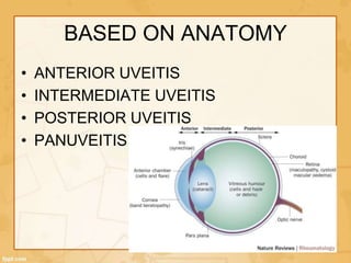
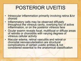
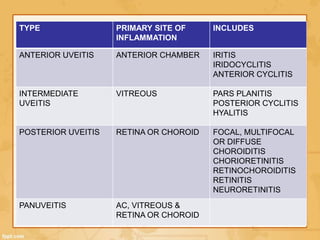
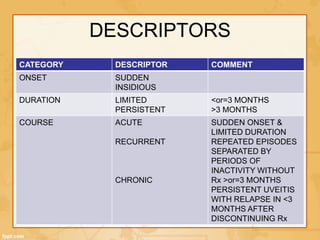
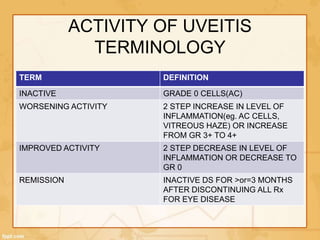
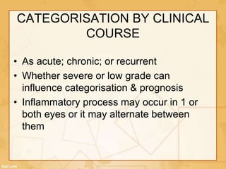
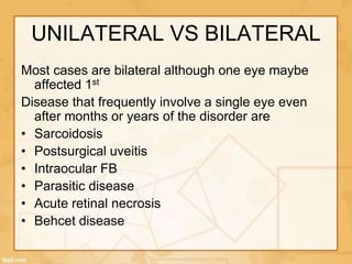
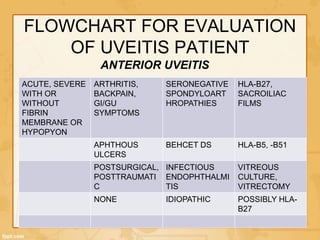
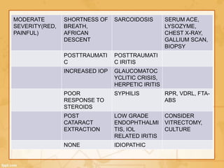
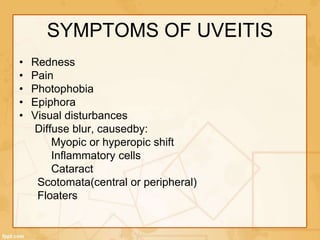
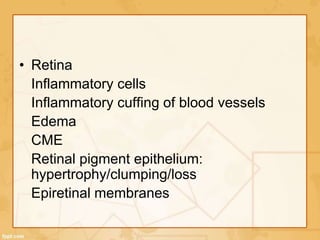
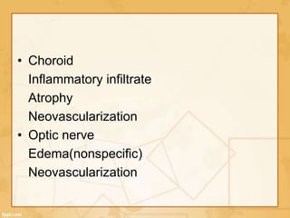
2. • Uvea consists of the middle, pigmented
vascular structures of the eye & includes
the iris, ciliary body & choroid
• Uveitis is broadly defined as inflammation
of the uvea
• Since uveitis is frequently associated with
systemic disease, a thorough history,
physical examination & workup is needed
to categorize it
3. CLASSIFICATION
• BASED ON ANATOMY
• CLINICAL COURSE
• ETIOLOGY
• HISTOLOGY
• STANDARDIZATION OF UVEITIS
NOMENCLATURE (SUN) WORKING GROUP IN
2005 DEVELOPED ANATOMICAL
CLASSIFICATION, DESCRIPTORS,
STANDARDISED GRADING SYSTEM AND
TERMINOLOGY
4. BASED ON ANATOMY
• ANTERIOR UVEITIS
• INTERMEDIATE UVEITIS
• POSTERIOR UVEITIS
• PANUVEITIS
5. ANTERIOR UVEITIS
• AC is the primary site of inflammation
• Inflammation confined to the anterior
chamber is called iritis
• If it spills over to retrolental space, it is
called iridocyclitis
• If it involves cornea, its called
keratouveitis
• If it involves sclera and uveal tract, its
called sclerouveitis
6. INTERMEDIATE UVEITIS
• The major site of inflammation is the
vitreous
• Inflammation of the middle
portion(posterior ciliary body, pars plana)
of the eye manifests primarily as floaters
affecting vision; the eye frequently
appears quiet externally
• Visual loss is primarily a result of CSME or
less commonly cataract formation
7. POSTERIOR UVEITIS
• Intraocular inflammation primarily involving retina &/or
choroid
• Inflammatory cells may be observed diffusely
throughout the vitreous cavity, overlying foci of active
inflammation, or on the posterior vitreous face
• Ocular examn reveals focal, multifocal or diffuse areas
of retinitis or choroiditis with varying degrees of
vitreous cellular activity
• Macular edema, retinal vasculitis and retinal or
choroidal neovascularisation are structural
complications of certain uveitic entities & not
considered essential to the anatomical classification
8. PANUVEITIS
• Primary sites are AC, vitreous & retina or
choroid
• Many systemic infectious & noninfectious
diseases associated with uveitis may
produce diffuse intraocular inflammation
with concomitant iridocyclitis & posterior
uveitis
9. TYPE PRIMARY SITE OF
INFLAMMATION
INCLUDES
ANTERIOR UVEITIS ANTERIOR CHAMBER IRITIS
IRIDOCYCLITIS
ANTERIOR CYCLITIS
INTERMEDIATE
UVEITIS
VITREOUS PARS PLANITIS
POSTERIOR CYCLITIS
HYALITIS
POSTERIOR UVEITIS RETINA OR CHOROID FOCAL, MULTIFOCAL
OR DIFFUSE
CHOROIDITIS
CHORIORETINITIS
RETINOCHOROIDITIS
RETINITIS
NEURORETINITIS
PANUVEITIS AC, VITREOUS &
RETINA OR CHOROID
10. DESCRIPTORS
CATEGORY DESCRIPTOR COMMENT
ONSET SUDDEN
INSIDIOUS
DURATION LIMITED
PERSISTENT
<or=3 MONTHS
>3 MONTHS
COURSE ACUTE
RECURRENT
CHRONIC
SUDDEN ONSET &
LIMITED DURATION
REPEATED EPISODES
SEPARATED BY
PERIODS OF
INACTIVITY WITHOUT
Rx >or=3 MONTHS
PERSISTENT UVEITIS
WITH RELAPSE IN <3
MONTHS AFTER
DISCONTINUING Rx
11. ACTIVITY OF UVEITIS
TERMINOLOGY
TERM DEFINITION
INACTIVE GRADE 0 CELLS(AC)
WORSENING ACTIVITY 2 STEP INCREASE IN LEVEL OF
INFLAMMATION(eg. AC CELLS,
VITREOUS HAZE) OR INCREASE
FROM GR 3+ TO 4+
IMPROVED ACTIVITY 2 STEP DECREASE IN LEVEL OF
INFLAMMATION OR DECREASE TO
GR 0
REMISSION INACTIVE DS FOR >or=3 MONTHS
AFTER DISCONTINUING ALL Rx
FOR EYE DISEASE
12. CATEGORISATION BY CLINICAL
COURSE
• As acute; chronic; or recurrent
• Whether severe or low grade can
influence categorisation & prognosis
• Inflammatory process may occur in 1 or
both eyes or it may alternate between
them
13. HISTOLOGY-GRANULOMATOUS OR
NONGRANULOMATOUS
• Ocular examination offers a unique opportunity to determine the
type of infiltrating inflammatory cells involved without taking a biopsy
• In anterior uveitis, inflammatory cells attach to the corneal
endothelium in conglomerates called keratic precipitates(KPs)
• Appearance of KPs can be used to classify it as granulomatous &
non granulomatous
• Non granulomatous characterised by fine white collections of
lymphocytes, plasma cells & pigment. These can form in any
disease. It only alerts that anterior inflammatory disease has
occurred
• Granulomatous KPs are large greasy appearing collections of
lymphocytes, plasma cells & giant cells called ‘mutton fat’ KPs is a
useful diagnostic clue
• Other ocular findings suggestive of granulomatous inflammation are
iris nodules & choroidal granulomas
15. UNILATERAL VS BILATERAL
Most cases are bilateral although one eye maybe
affected 1st
Disease that frequently involve a single eye even
after months or years of the disorder are
• Sarcoidosis
• Postsurgical uveitis
• Intraocular FB
• Parasitic disease
• Acute retinal necrosis
• Behcet disease
16. FLOWCHART FOR EVALUATION
OF UVEITIS PATIENT
ACUTE, SEVERE
WITH OR
WITHOUT
FIBRIN
MEMBRANE OR
HYPOPYON
ARTHRITIS,
BACKPAIN,
GI/GU
SYMPTOMS
SERONEGATIVE
SPONDYLOART
HROPATHIES
HLA-B27,
SACROILIAC
FILMS
APHTHOUS
ULCERS
BEHCET DS HLA-B5, -B51
POSTSURGICAL,
POSTTRAUMATI
C
INFECTIOUS
ENDOPHTHALMI
TIS
VITREOUS
CULTURE,
VITRECTOMY
NONE IDIOPATHIC POSSIBLY HLA-
B27
ANTERIOR UVEITIS
17. MODERATE
SEVERITY(RED,
PAINFUL)
SHORTNESS OF
BREATH,
AFRICAN
DESCENT
SARCOIDOSIS SERUM ACE,
LYSOZYME,
CHEST X-RAY,
GALLIUM SCAN,
BIOPSY
POSTTRAUMATI
C
POSTTRAUMATI
C IRITIS
INCREASED IOP GLAUCOMATOC
YCLITIC CRISIS,
HERPETIC IRITIS
POOR
RESPONSE TO
STEROIDS
SYPHILIS RPR, VDRL, FTA-
ABS
POST
CATARACT
EXTRACTION
LOW GRADE
ENDOPHTHALMI
TIS, IOL
RELATED IRITIS
CONSIDER
VITRECTOMY,
CULTURE
NONE IDIOPATHIC
18. Chronic, minimal
redness pain
Child especially
with arthritis
JIA-related
iridocyclitis
ANA, ESR,
rheumatoid factor
Heterochromia,
diffuse kp, u/l
Fuchs
heterochromic
iridocyclitis
None
Postsurgical Low grade
endophthalmitis(e
g.
Propionibacterium
acnes); iol related
Consider
vitrectomy,
capsulectomy,
with culture
None Idiopathic
19. INTERMEDIATE UVEITIS
Mild to moderate Shortness of
breath, african
descent
Sarcoidosis As above
Tick exposure,
erythema
chronicum
migrans rash
Lyme disease Elisa
Neurologic
symptoms
Multiple sclerosis MRI brain
Over age 50 Intraocular
lymphoma
Vitrectomy,
cytology
None Pars planitis
23. Diffuse Dermatological/
CNS symptoms
VKH syndrome FA, LP to
document CSF
pleocytosis
Postsurgical/trau
matic; bilateral
Sympathetic
ophthalmia
FA
U/L Infectious
endophthalmitis
As above
Child; History of
geophagia
Toxocariasis As above
24. CHORIORETINITIS WITHOUT
VITRITIS
Focal None, history of
carcinoma
Neoplastic Metastatic workup
Multifocal Ohio/Missisippi
Valley
Ocular
Histoplasmosis
FA if macula
involved
Lesions confined
to posterior pole
White dot
syndrome(eg.
APMPPE,
MEWDS, PIC)
FA
Geographic
pattern of scars
Serpiginous
choroiditis
FA
Diffuse From Africa,
Central/South
America
Onchocerciasis
25. VASCULITIS
Aphthous ulcers,
hypopyon
Behcet disease As above
Malar rash, Female,
arthralgias
SLE ANA
c/c sinusitis with
hemorrhagic
rhinorrhoea, dyspnoea,
renal insufficiency,
purpura
Wegeners
granulomatosis
c-ANCA
31. SYMPTOMS
• Depends on which part is inflammed, rapidity of
onset, duration of disease & course of the disease
• Acute onset anterior uveitis causes pain,
photophobia, redness & blurred vision.
• Pain results due to acute onset inflammation of iris
or from secondary glaucoma
• Pain associated with ciliary spasm in iritis may be
a reffered pain that seems to radiate over areas
severed by trigeminal nerve
• Epiphora, redness & photophobia are usually
present when inflammation involves the iris,
cornea or iris-ciliary body
32.
33. • Chronic iridocyclitis in patients with juvenile
idiopathic arthritis maynot be associated with any
symptoms at all
• But with c/c iridocyclitis blurred vision may result
as a result of calcific band keratopathy, cataract or
CME
• Intermediate uveitis produces symptoms of
floaters & blurred vision
• Floaters result from shadows cast by vitreous cells
on the retina
• Blurred vision may be due to CME or vitreous
opacities in the visual axis
34. • Presenting symptoms in posterior uveitis include painless
decreased visual aquity, floaters, photopsia,
metamorphopsia, scotoma, nyctalopia, or a combinaton of
these
• This blurred vision maybe due to the primary effects of
uveitis such as retinitis &/or choroiditis directly affecting
macular functions, or to the complication of inflammation
such as CME, epiretinal membrane, retinal ischemia &
choroidal neovascularisation
• It may also result from refractive error such as myopic or
hyperopic shift associated with macular edema, hypotony
or a change in lens position
• Other causes are opacities in visual axis from inflammatory
cells, fibrin or protein in the anterior chamber; KPs;
secondary cataract; vitreous debris; macular edema; &
retinal atrophy
35. SYMPTOMS OF UVEITIS
• Redness
• Pain
• Photophobia
• Epiphora
• Visual disturbances
Diffuse blur, causedby:
Myopic or hyperopic shift
Inflammatory cells
Cataract
Scotomata(central or peripheral)
Floaters
42. • Chemical mediators of acute stage of inflammation
include serotonin, complement & plasmin
• Leukotrienes, kinins & prostaglandins modify 2nd
phase of acute response
• Polymorphonuclear leukocytes, eosinophils &
mast cells contribute to inflammatory signs, but
lymphocyte is the predominant inflammatory cell in
the innner eye in uveitis
• These mediators result in vascular dilatation(ciliary
flush), increased vascular permeability(aqueous
flare), & chemotaxis of inflammatory cells into the
eye( aqueous & vitreous cellular reaction)
43. • Perilimbal vascular engorgement(ciliary flush) or
diffuse injection of the conjunctiva, episclera or
both is typical with acute anterior uveitis
• The AC reaction can be described as
• Serous(aqueous flare caused by protein influx)
• Purulent(PML & necrotic debris causing
hypopyon)
• Fibrinous(plasmoid or intense fibrinous exudate)
• Sanguinoid(inflammatory cells with erythrocytes
manifested by hypopyon mixed with hyphema)
44. • SUN grading of Cells & Flare
• AC cells
• Graded according to number of cells seen
in a 1mm*1mm high-powered beam at full
intensity at 45deg to 60 deg
• Flare may be graded similarly
45. GRADE CELLS CELLS IN FIELD
0 <1
0.5+ 1-5
1+ 6-15
2+ 16-25
3+ 26-50
4+ >50
47. • Iris involvement may manifest as either
anterior or posterior synechiae, iris
nodules(Koeppe nodules at the pupillary
border, Busaca nodules within the iris
stroma & Berlin nodules in the angle), iris
granulomas, heterochromia(eg, Fuchs
heterochromic iridocyclitis) or stromal
atrophy(eg, herpetic uveitis)
48. • With involvement of ciliary body & trabecular
meshwork, IOP is often low secondary to
decreased aqueous production or increased
alternative outflow
• But IOP may increase if meshwork becomes
clogged by cells or debris or if trab meshwork
itself is the site of inflammation(trabeculitis)
• Pupillary block with iris bombe & secondary
angle closure may also lead to an acute rise
in IOP
49. INTERMEDIATE SEGMENT
• Vitreal inflammatory cells graded as
• NIH grading for vitreous haze has been adopted by SUN
group. With this standardized photos are used for comparison
GRADE NO. OF CELLS
0 NO CELLS
0.5+ 1-10
1+ 11-20
2+ 21-30
3+ 31-100
4+ >100
50. • Other changes are
• Snowball opacities- common with sarcoidosis or
intermediate uveitis
• Exudates over pars plana(snowbank). Active
snowbanks have a fluffy or shaggy appearance; if
it becomes inactive the pars plana appears gliotic
or fibrotic & smooth(not reffered as snowbanks)
• Vitreal strands
• Chronic uveitis may be associated with cyclitic
membrane formation, secondary ciliary body
detachment & hypotony
51. POSTERIOR SEGMENT
• Retinal or choroidal inflammatory infiltrates
• Inflammatory sheathing of arteries or veins
• Exudative, tractional or rhegmatogenous
retinal detachment
• RPE hypertrophy or atrophy
• Atrophy or swelling of retina, choroid or optic
nerve head
• Preretinal or subretinal fibrosis
• Retinal or choroidal neovascularisation