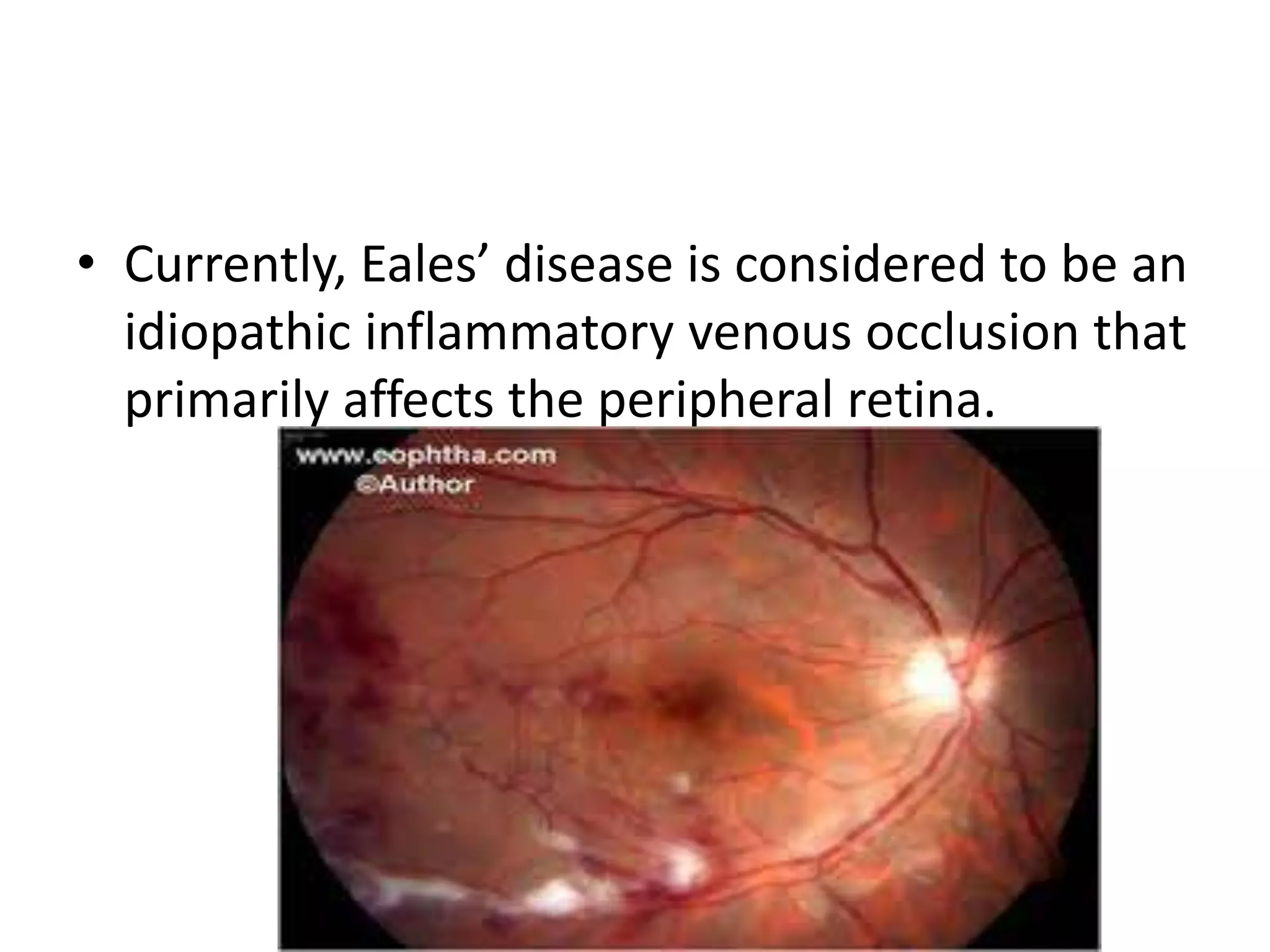
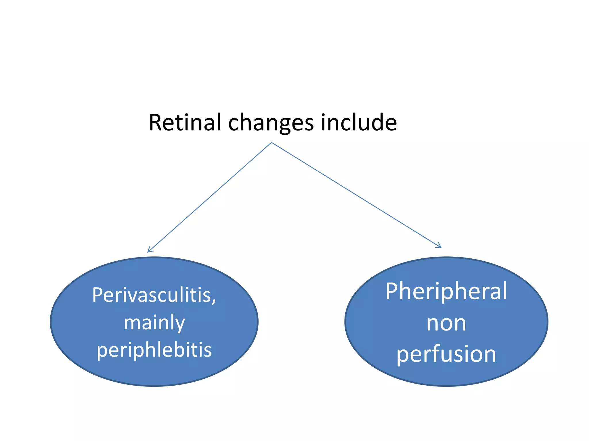
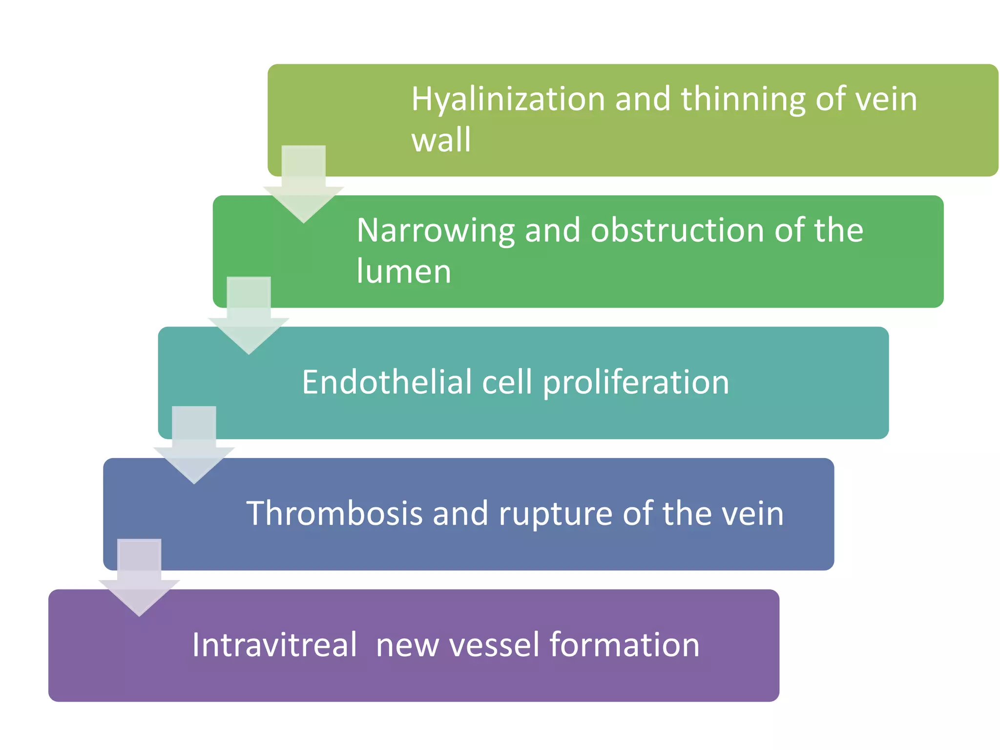
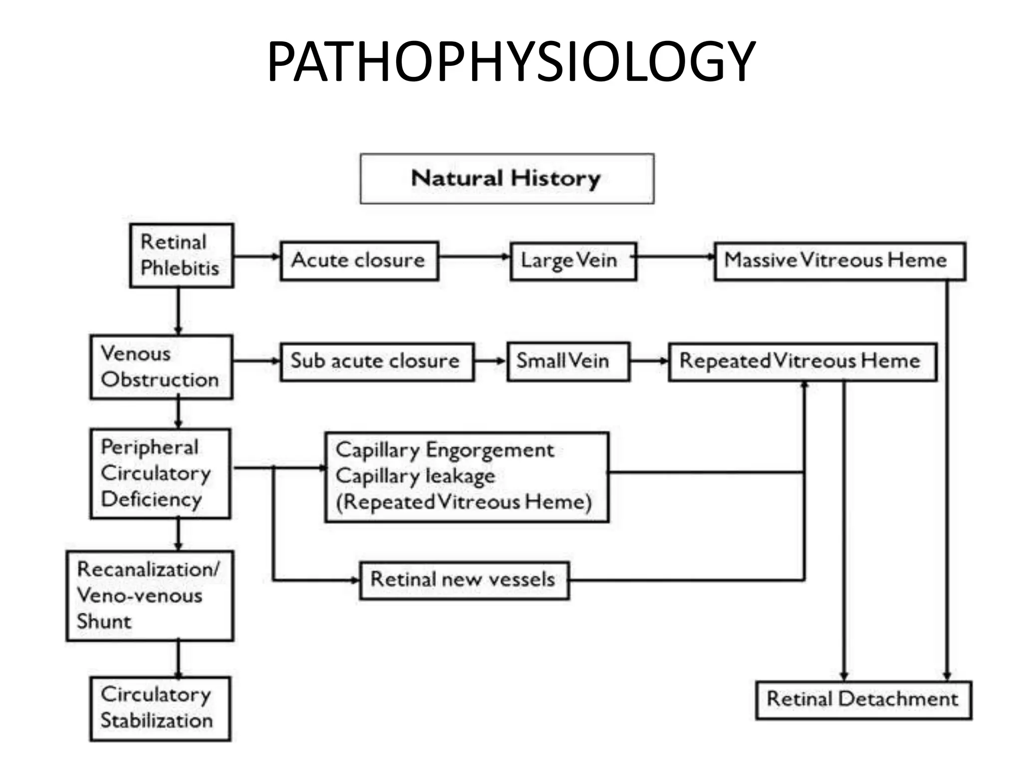
EALES DISEASE
dr.nirav b. Dadia
fellow
nandadeep eye hospital
In 1880 and 1882, Henry Eales - “primary recurrent
retinal hemorrhage”.
Similar conditions of retinal and vitreous hemorrhage
were described under the name of Eales’ Disease.
Eales didn’t mention any inflammatory signs
preceding or accompanying the hemorrhages.
• In 1887 Wadsworth reported on signs of
inflammation of the retinal vasculature - Eales’
disease and periphlebitis
• Elliot initially suggested that the disease be
called “periphlebitis retinae”.
• Currently, Eales’ disease is considered to be an
idiopathic inflammatory venous occlusion that
primarily affects the peripheral retina.
Perivasculitis,
mainly
periphlebitis
Retinal changes include
Pheripheral
non
perfusion
• This inflammation induced vascular occlusion can
lead to a proliferative vascular retinopathy, with
sequelae such as
recurrent vitreous hemorrhage and
traction retinal detachment.
PATHOPHYSIOLOGY
• Patchy perivascular or intramural infiltration of
lymphocytes or granulation tissue sometimes with or
without giant cells
• Plasma cells are occasionally present.
• Veins are primarily affected
• The vascular changes are usually seen on retinal periphery
Hyalinization and thinning of vein
wall
Narrowing and obstruction of the
lumen
Endothelial cell proliferation
Thrombosis and rupture of the vein
Intravitreal new vessel formation
PATHOPHYSIOLOGY
Systemic disease associated with Eales’ disease:
• Tuberculosis
• Hypersensitivity to tuberculoprotein
• Thromboangitis obliterans
• Neurologic disease
• Hematological abnormalities
• The assumption of tubercular aetiology is
based on active or healed tuberculosis in
some patient with Eales’ disease.
• Ophthalmoscopic evaluation in patient with
active or healed TB showed 1.3% had Eales’
disease .
Hypersensitivity to tuberculoprotein:
Allergic reaction to tuberculosis has been
reported by many authors till date.
Positive Mantoux reaction which is as high as
90% in some series.
Systemic disease:
• Several studies have shown association between
neurological and hematological disease.
• bilateral hearing loss 48% (Renie et al) , 25% (William
et al).
• 2 pt with Eales’ disease had progressive worsening of
neurological deficit (Rodier G).
• Myelopathy with Eales’ disease has been described by
many.
Immunological studies in Eales’
disease:
Immune mediated mechanism has been suggested by
many authors as a possible cause of Eales’ disease.
• Acute onset, steroid responsiveness, lymphocytic
infiltration and abnormal immunological parameters all
indicate an immunological basis of disease.
Immunological studies in Eales’
disease: cont…d
Altered immune response of type III and/or IV
reaction to an infectious agent (Muthukaruppan
et al).
• Raised IgG and IgA levels (Johnson et al) ,
elevated levels of circulating immune complexes
and antiretinal antibody (Kasp et al) ,
immunophenotyping predominant T cell CD4
• Higher frequencies of HLA B5(B51), DR1 and DR4
(Biswas et al)
Biochemical studies in Eales’ disease:
• Raised alpha-globulins and reduced albumin
levels in the serum samples.
• PDGF, IGF1, TGFa and TGFb play a key role in
neovascularisation.
• Raised serum alpha1 acid glycoproteins in 27
patients of Eales’ disease
Stages of Eales’ disease
Stage I: (Inflammatory stage)
• Localized areas of peripheral retinal edema with
sheathing of the smaller caliber vascular branches.
• Minute retinal hemorrhages as well as minute
vascular brackets or hooklets connecting two
adjoining vessels.
• Active periphlebitis with subhyaloid
hemorrhage over the macula.
Stage II (ischemic stage)
• Involvement of larger vessels and extend more
posteriorly .
• Veins as well as arterioles may be sheathed
• Widespread retinal hemorrhages and vitreous
looks hazy .
• Stage III (stage of neovascularisation)
• Peripheral new blood vessels with numerous
vitreous and retinal hemorrhages.
• The hemorrhages frequently recurs.
Sea- fan like neovascularization
• Stage IV (complicated stage)
• Massive retinal proliferans associated retinal
and massive vitreous hemorrhage.
• With this advanced disease the
neovascularization can cause tractional
rhegmatogenous retinal detachment.
Differential diagnosis:
• Vasculitis mimicking Eales’ disease
Systemic Ocular
Behcet’s disease Birdshot retinochoroidopathy
Lyme Borreliosis Coat’s disease
Leukemia Pars planitis
Multiple sclerosis Viral retinitis
Systemic lupus erythematosus
Toxocariasis
Toxoplasmosis
Tuberculosis
Wegener’s granulomatosis
• Proliferative vascular retinopathy mimicking
Eales’ disease:
Systemic Ocular
Diabetes mellitus BRVO
Sarcoidosis CRVO
Sickle cell disease ROP
Pars planitis
Coats’ disease
Clinical features
• Usually occurs in young , healthy people, with a peak
incidence between the ages of 30 and 40 years.
•
• It occurs more frequently in males 80-90%.
•
• 75% cases it presents before 49 years.
• Can be unilateral or bilateral.90% bilateral (Duke Elder)
retinal vasculitis
Vitreous floaters or blurring of vision,
symptoms attributable to recurrent vitreous
hemorrhages.
80% between the age of 20-40 years and 95%
were male (O.K Malla and co workers)
54.34% between 20-30 years and 94.73% male
Rare in more developed countries.
• More commonly reported from Indian
subcontinent. The reported incidence in India is 1 in
200-250 patient
• Anterior uveitis/Vitritis.
• Active perivasculitis with exudates around the veins
in one or more quadrants. Arterioles may be
affected.
Healed perivasculitis as
sheathing of the veins
Macular changes uncommon
Peripheral retinal
neovascularisation reported
in 36-84% of cases
Recurrent vitreous hemorrhages,
the hall mark of the disease
Some vitreous hemorrhages resolve,
some do not ( organize with multiple
VR adhesions & RRD/TRD
Some patient specially with multiple
sclerosis are asymptomatic.
Fundus fluorescein angiography
• To delineate areas of capillary nonperfusion, peripheral
retinal nonperfusion is present in all patients with
Eales’ disease.
• Retinal or disc neovascularisation
•
• Macular edema
• Helps in monitoring the regression and disappearance
of new vessels during treatment and follow up.
TREATMENT
• Symptomatic treatment.
• Treatment aim :
reducing retinal perivasculitis and associated vitritis ;
reducing risk of vitreous hemorrhage from new vessels
by retinal ablation and
surgical removal of non resolving vitreous hemorrhage
and/or vitreous membranes.
Treatment of Eales’ disease:
• Observation.
• Medical
Corticosteroids
Antituberculosis drugs
Immunosuppressive drugs.
• Retinal ablation
Photocoagulation
cryotherapy
• Surgical
vitrectomy
Observation:
• Patient with inactive retinal vasculitis
• Follow up 6 months to 1 year interval.
• Patient with fresh vitreous hemorrhage if retina is
found to be attached.
• Such vitreous hemorrhage usually clears by 6 to 8
weeks.
Medical therapy
• Corticosteroids are mainstay of therapy in active
perivasculitis stage of Eales’ disease.
• Majority of cases 1mg/kg body weight, tapered to
10mg/week over 6 to 8 weeks.
• Maintenance 15 to 20mg/day for 1 to 2 months.
• Periocular depot steroid injection may be added for
associated macular edema.
• Systemic and Periocular steroid useful in patient
having 3 quadrants involvement with macular
edema.
• Systemic steroid only if less than 3 quadrant
involvement.
• No difference in response between Mantoux
positive and negative cases.
• Immunosuppressive therapy in patient
unresponsive or have unacceptable side effects.
(Azathioprine and cyclosporine)
• Some investigators have recommended ATT
(Rifampicin and Isoniazid) for 9 months.
Photocoagulation
• Mainstay of therapy in proliferative stage of Eales’
disease.
• The aim
Regulate the circulation
To obliterate surface neovascularisation and
Close leaking intraretinal microvascular
abnormalities.
• Sectoral laser for capillary non
perfusion and PRP for
neovascularisation of disc.
• Occasional massive hemorrhage can
occur.
• After laser, regressing
neovascularisation can cause
macular distortion and retinal tear.
• Laser not advised in active
inflammatory stage
Vitreoretinal surgery
• Vitrectomy alone or combined with other
vitreoretinal surgical procedures is often
required.
• Nonresolving vitreous hemorrhage with
obscuration of central vision of 3 months
duration may be subjected to vitrectomy.
• Vitrectomy done between 3 to 6 months has
better results than done after 6 months (Kumar
et al).
• Early vitrectomy in patient with TRD, extensive
vitreous membranes or epimacular membranes.
• Endolaser can be given along with vitrectomy.
Tractional retinal fold after vitrectomy
Summary and conclusions:
• Characteristic clinical findings and angiographic
pattern.
• Mimic several ocular or systemic disease presenting
as retinal vasculitis or proliferative retinal
vasculopathy.
• Hypersensitivity to tubercular protein has been
considered a prime cause of Eales’ disease .
• Probable multifactorial etiology.
• HLA, retinal autoimmunity, mycobacterium
genome, free radical mediated damage.
• Corticosteroids in active disease and laser
photocoagulation in ischemic and proliferative
stage.
• Results of vitrectomy in non resolving vitreous
hemorrhage with or without retinal detachment
are satisfactory.
• Thank u for listening

Eales disease

  • 1.
    EALES DISEASE dr.nirav b.Dadia fellow nandadeep eye hospital
  • 2.
    In 1880 and1882, Henry Eales - “primary recurrent retinal hemorrhage”. Similar conditions of retinal and vitreous hemorrhage were described under the name of Eales’ Disease. Eales didn’t mention any inflammatory signs preceding or accompanying the hemorrhages.
  • 3.
    • In 1887Wadsworth reported on signs of inflammation of the retinal vasculature - Eales’ disease and periphlebitis • Elliot initially suggested that the disease be called “periphlebitis retinae”.
  • 4.
    • Currently, Eales’disease is considered to be an idiopathic inflammatory venous occlusion that primarily affects the peripheral retina.
  • 5.
  • 6.
    • This inflammationinduced vascular occlusion can lead to a proliferative vascular retinopathy, with sequelae such as recurrent vitreous hemorrhage and traction retinal detachment.
  • 7.
    PATHOPHYSIOLOGY • Patchy perivascularor intramural infiltration of lymphocytes or granulation tissue sometimes with or without giant cells • Plasma cells are occasionally present. • Veins are primarily affected • The vascular changes are usually seen on retinal periphery
  • 8.
    Hyalinization and thinningof vein wall Narrowing and obstruction of the lumen Endothelial cell proliferation Thrombosis and rupture of the vein Intravitreal new vessel formation
  • 9.
  • 10.
    Systemic disease associatedwith Eales’ disease: • Tuberculosis • Hypersensitivity to tuberculoprotein • Thromboangitis obliterans • Neurologic disease • Hematological abnormalities
  • 11.
    • The assumptionof tubercular aetiology is based on active or healed tuberculosis in some patient with Eales’ disease. • Ophthalmoscopic evaluation in patient with active or healed TB showed 1.3% had Eales’ disease .
  • 12.
    Hypersensitivity to tuberculoprotein: Allergicreaction to tuberculosis has been reported by many authors till date. Positive Mantoux reaction which is as high as 90% in some series.
  • 13.
    Systemic disease: • Severalstudies have shown association between neurological and hematological disease. • bilateral hearing loss 48% (Renie et al) , 25% (William et al). • 2 pt with Eales’ disease had progressive worsening of neurological deficit (Rodier G). • Myelopathy with Eales’ disease has been described by many.
  • 14.
    Immunological studies inEales’ disease: Immune mediated mechanism has been suggested by many authors as a possible cause of Eales’ disease. • Acute onset, steroid responsiveness, lymphocytic infiltration and abnormal immunological parameters all indicate an immunological basis of disease.
  • 15.
    Immunological studies inEales’ disease: cont…d Altered immune response of type III and/or IV reaction to an infectious agent (Muthukaruppan et al). • Raised IgG and IgA levels (Johnson et al) , elevated levels of circulating immune complexes and antiretinal antibody (Kasp et al) , immunophenotyping predominant T cell CD4 • Higher frequencies of HLA B5(B51), DR1 and DR4 (Biswas et al)
  • 16.
    Biochemical studies inEales’ disease: • Raised alpha-globulins and reduced albumin levels in the serum samples. • PDGF, IGF1, TGFa and TGFb play a key role in neovascularisation. • Raised serum alpha1 acid glycoproteins in 27 patients of Eales’ disease
  • 17.
    Stages of Eales’disease Stage I: (Inflammatory stage) • Localized areas of peripheral retinal edema with sheathing of the smaller caliber vascular branches. • Minute retinal hemorrhages as well as minute vascular brackets or hooklets connecting two adjoining vessels.
  • 19.
    • Active periphlebitiswith subhyaloid hemorrhage over the macula.
  • 20.
    Stage II (ischemicstage) • Involvement of larger vessels and extend more posteriorly . • Veins as well as arterioles may be sheathed • Widespread retinal hemorrhages and vitreous looks hazy .
  • 22.
    • Stage III(stage of neovascularisation) • Peripheral new blood vessels with numerous vitreous and retinal hemorrhages. • The hemorrhages frequently recurs.
  • 24.
    Sea- fan likeneovascularization
  • 25.
    • Stage IV(complicated stage) • Massive retinal proliferans associated retinal and massive vitreous hemorrhage. • With this advanced disease the neovascularization can cause tractional rhegmatogenous retinal detachment.
  • 27.
    Differential diagnosis: • Vasculitismimicking Eales’ disease Systemic Ocular Behcet’s disease Birdshot retinochoroidopathy Lyme Borreliosis Coat’s disease Leukemia Pars planitis Multiple sclerosis Viral retinitis Systemic lupus erythematosus Toxocariasis Toxoplasmosis Tuberculosis Wegener’s granulomatosis
  • 28.
    • Proliferative vascularretinopathy mimicking Eales’ disease: Systemic Ocular Diabetes mellitus BRVO Sarcoidosis CRVO Sickle cell disease ROP Pars planitis Coats’ disease
  • 29.
    Clinical features • Usuallyoccurs in young , healthy people, with a peak incidence between the ages of 30 and 40 years. • • It occurs more frequently in males 80-90%. • • 75% cases it presents before 49 years. • Can be unilateral or bilateral.90% bilateral (Duke Elder) retinal vasculitis
  • 30.
    Vitreous floaters orblurring of vision, symptoms attributable to recurrent vitreous hemorrhages. 80% between the age of 20-40 years and 95% were male (O.K Malla and co workers) 54.34% between 20-30 years and 94.73% male Rare in more developed countries.
  • 31.
    • More commonlyreported from Indian subcontinent. The reported incidence in India is 1 in 200-250 patient • Anterior uveitis/Vitritis. • Active perivasculitis with exudates around the veins in one or more quadrants. Arterioles may be affected.
  • 33.
    Healed perivasculitis as sheathingof the veins Macular changes uncommon Peripheral retinal neovascularisation reported in 36-84% of cases
  • 34.
    Recurrent vitreous hemorrhages, thehall mark of the disease Some vitreous hemorrhages resolve, some do not ( organize with multiple VR adhesions & RRD/TRD Some patient specially with multiple sclerosis are asymptomatic.
  • 35.
    Fundus fluorescein angiography •To delineate areas of capillary nonperfusion, peripheral retinal nonperfusion is present in all patients with Eales’ disease. • Retinal or disc neovascularisation • • Macular edema • Helps in monitoring the regression and disappearance of new vessels during treatment and follow up.
  • 38.
  • 39.
    • Symptomatic treatment. •Treatment aim : reducing retinal perivasculitis and associated vitritis ; reducing risk of vitreous hemorrhage from new vessels by retinal ablation and surgical removal of non resolving vitreous hemorrhage and/or vitreous membranes.
  • 40.
    Treatment of Eales’disease: • Observation. • Medical Corticosteroids Antituberculosis drugs Immunosuppressive drugs. • Retinal ablation Photocoagulation cryotherapy • Surgical vitrectomy
  • 41.
    Observation: • Patient withinactive retinal vasculitis • Follow up 6 months to 1 year interval. • Patient with fresh vitreous hemorrhage if retina is found to be attached. • Such vitreous hemorrhage usually clears by 6 to 8 weeks.
  • 42.
    Medical therapy • Corticosteroidsare mainstay of therapy in active perivasculitis stage of Eales’ disease. • Majority of cases 1mg/kg body weight, tapered to 10mg/week over 6 to 8 weeks. • Maintenance 15 to 20mg/day for 1 to 2 months. • Periocular depot steroid injection may be added for associated macular edema.
  • 43.
    • Systemic andPeriocular steroid useful in patient having 3 quadrants involvement with macular edema. • Systemic steroid only if less than 3 quadrant involvement. • No difference in response between Mantoux positive and negative cases.
  • 44.
    • Immunosuppressive therapyin patient unresponsive or have unacceptable side effects. (Azathioprine and cyclosporine) • Some investigators have recommended ATT (Rifampicin and Isoniazid) for 9 months.
  • 45.
    Photocoagulation • Mainstay oftherapy in proliferative stage of Eales’ disease. • The aim Regulate the circulation To obliterate surface neovascularisation and Close leaking intraretinal microvascular abnormalities.
  • 46.
    • Sectoral laserfor capillary non perfusion and PRP for neovascularisation of disc. • Occasional massive hemorrhage can occur. • After laser, regressing neovascularisation can cause macular distortion and retinal tear. • Laser not advised in active inflammatory stage
  • 48.
    Vitreoretinal surgery • Vitrectomyalone or combined with other vitreoretinal surgical procedures is often required. • Nonresolving vitreous hemorrhage with obscuration of central vision of 3 months duration may be subjected to vitrectomy.
  • 49.
    • Vitrectomy donebetween 3 to 6 months has better results than done after 6 months (Kumar et al). • Early vitrectomy in patient with TRD, extensive vitreous membranes or epimacular membranes. • Endolaser can be given along with vitrectomy.
  • 50.
    Tractional retinal foldafter vitrectomy
  • 51.
    Summary and conclusions: •Characteristic clinical findings and angiographic pattern. • Mimic several ocular or systemic disease presenting as retinal vasculitis or proliferative retinal vasculopathy. • Hypersensitivity to tubercular protein has been considered a prime cause of Eales’ disease .
  • 52.
    • Probable multifactorialetiology. • HLA, retinal autoimmunity, mycobacterium genome, free radical mediated damage. • Corticosteroids in active disease and laser photocoagulation in ischemic and proliferative stage. • Results of vitrectomy in non resolving vitreous hemorrhage with or without retinal detachment are satisfactory.
  • 53.
    • Thank ufor listening