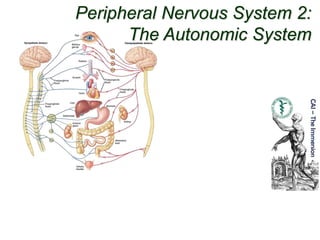
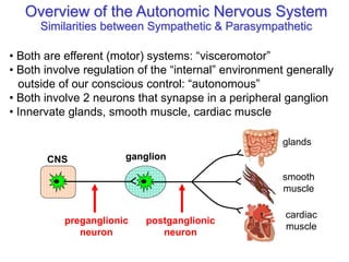
Peripheral Nervous System 2:
The Autonomic System
Overview of the Autonomic Nervous System
Similarities between Sympathetic & Parasympathetic
• Both are efferent (motor) systems: “visceromotor”
• Both involve regulation of the “internal” environment generally
outside of our conscious control: “autonomous”
• Both involve 2 neurons that synapse in a peripheral ganglion
• Innervate glands, smooth muscle, cardiac muscle
CNS ganglion
preganglionic
neuron
postganglionic
neuron
glands
smooth
muscle
cardiac
muscle
Overview of the Autonomic Nervous System
Differences between Sympathetic & Parasympathetic
Location of Preganglionic Cell Bodies
Thoracolumbar
T1 – L2/L3 levels
of the spinal cord
Craniosacral
Brain: CN III, VII, IX, X
Spinal cord: S2 – S4
Sympathetic Parasympathetic
Sympathetic
CNS ganglion
short preganglionic
neuron
long postganglionic
neuron
target
Parasympathetic
CNS ganglion
long preganglionic
neuron
target
short postganglionic
neuron
Overview of the Autonomic Nervous System
Differences between Sympathetic & Parasympathetic
Relative Lengths of Neurons
Overview of the Autonomic Nervous System
Differences between Sympathetic & Parasympathetic
Target Tissues
ParasympatheticSympathetic
• Organs of head, neck,
trunk, & external genitalia
• Organs of head, neck,
trunk, & external genitalia
• Adrenal medulla
• Sweat glands in skin
• Arrector muscles of hair
• ALL vascular smooth muscle
» Sympathetic system is distributed to essentially all
tissues (because of vascular smooth muscle)
» Parasympathetic system never reaches limbs or
body wall (except for external genitalia)
Overview of ANS
Functional Differences
Sympathetic
• “Fight or flight”
• Catabolic (expend energy)
Parasympathetic
• “Feed & breed”, “rest &
digest”
• Homeostasis
» Dual innervation of many
organs — having a brake
and an accelerator provides
more control
Structure of spinal nerves: Somatic pathways
dorsal root
dorsal root
ganglion
ventral root
spinal
nerve
dorsal
ramus
ventral
ramus
dorsal
horn
ventral
horn
somatic
sensory
nerve
(GSA)
somatic
motor
nerve
(GSE)
CNS
inter-
neuron
Mixed Spinal
Nerve
gray ramus
communicans white ramus
communicans
sympathetic
ganglion
spinal
nerve
dorsal
ramus
ventral
ramus
gray ramus
communicans white ramus
communicans
sympathetic
ganglion
intermediolateral
gray column
Structure of spinal nerves: Sympathetic pathways
Moore’s COA6 2010
somatic tissues
(body wall, limbs)
visceral tissues
(organs)
Sympathetic System: Preganglionic Cell Bodies
• Preganglionic cell bodies in
intermediolateral gray
• T1 – L2/L3
• Somatotopic organization
intermediolateral
gray columns
lateral
horn
T1 –
L2/L3
Clinical Relevance
» dysfunction due to cord injury
» spinal nerve impingement & OMM
» referred pain
Sympathetic System: Postganglionic Cell Bodies
Paravertebral
ganglia
Prevertebral
ganglia
• celiac ganglion
• sup. mesent. g.
• inf. mesent. g.
aorta
sympathetic
trunk (chain)
1. Paravertebral ganglia
• Located along sides of vertebrae
• United by preganglionics into Sympathetic Trunk
• Preganglionic neurons are thoracolumbar (T1–L2/L3)
but postganglionic neurons are cervical to coccyx
• Some preganglionics ascend or descend in trunk
synapse at
same level
ascend to
synapse at
higher level
descend to
synapse at
lower level
Moore’s COA6 2010
Sympathetic System: Postganglionic Cell Bodies
Paravertebral
ganglia
Prevertebral
ganglia
• celiac ganglion
• sup. mesent. g.
• inf. mesent. g.
aorta
sympathetic
trunk (chain)
2. Prevertebral (preaortic) ganglia
• Located anterior to abdominal aorta, in plexuses
surrounding its major branches
• Preganglionics reach prevertebral ganglia via
abdominopelvic splanchnic nerves
Moore’s COA6 2010
abdominopelvic
splanchnic
nerve
Sympathetic System: Summary
Moore’s COA6 2010
T1
L2
somatic tissues
(body wall, limbs)
visceral tissues
(organs)
postganglionics
via 31 spinal
nerves
to somatic tissues
of neck, body wall,
and limbs
sympathetic
trunk
prevertebral
ganglia
Cardiopulmonary Splanchnics:
postganglionic fibers to thoracic
viscera
Abdominopelvic Splanchnics:
preganglionic fibers to
prevertebral ganglia,
postganglionic fibers to
abdominopelvic viscera
Parasympathetic
Pathways
Moore’s COA6 2010
Cranial outflow
• CN III, VII, IX, X
• Four ganglia in head
• Vagus nerve (CN X) is major
preganglionic parasymp.
supply to thorax & abdomen
• Synapse in ganglia within
wall of the target organs (e.g.,
enteric plexus of GI tract)
Sacral outflow
• S2–S4 via pelvic splanchnics
• Hindgut, pelvic viscera, and
external genitalia
Clinical Relevance
» Surgery for colorectal cancer
puts pelvic splanchnics at risk
» Damage causes bladder &
sexual dysfunction
14
Central control of the
Autonomic NS
Amygdala: main limbic
region for emotions
-Stimulates sympathetic activity,
especially previously learned
fear-related behavior
-Can be voluntary when decide to
recall frightful experience -
cerebral cortex acts through
amygdala
-Some people can regulate some
autonomic activities by gaining
extraordinary control over their
emotions
Hypothalamus: main
integration center
Reticular formation: most
direct influence over
autonomic function
Parasympathetic
Overview of the Autonomic Nervous System
Differences between Sympathetic & Parasympathetic
Neurotransmitters
ACh, +
NE (ACh at sweat glands),
+ / -, α & ß receptors
ACh, + / -
muscarinic receptors
• All preganglionics release acetylcholine (ACh) & are excitatory (+)
• Symp. postgangl. — norepinephrine (NE) & are excitatory (+) or inhibitory (-)
• Parasymp. postgangl. — ACh & are excitatory (+) or inhibitory (-)
Sympathetic
• Excitation or inhibition is a receptor-dependent & receptor-mediated response
Potential for pharmacologic
modulation of autonomic responses
ACh, +

Ans 1

  • 1.
    Peripheral Nervous System2: The Autonomic System
  • 2.
    Overview of theAutonomic Nervous System Similarities between Sympathetic & Parasympathetic • Both are efferent (motor) systems: “visceromotor” • Both involve regulation of the “internal” environment generally outside of our conscious control: “autonomous” • Both involve 2 neurons that synapse in a peripheral ganglion • Innervate glands, smooth muscle, cardiac muscle CNS ganglion preganglionic neuron postganglionic neuron glands smooth muscle cardiac muscle
  • 3.
    Overview of theAutonomic Nervous System Differences between Sympathetic & Parasympathetic Location of Preganglionic Cell Bodies Thoracolumbar T1 – L2/L3 levels of the spinal cord Craniosacral Brain: CN III, VII, IX, X Spinal cord: S2 – S4 Sympathetic Parasympathetic
  • 4.
    Sympathetic CNS ganglion short preganglionic neuron longpostganglionic neuron target Parasympathetic CNS ganglion long preganglionic neuron target short postganglionic neuron Overview of the Autonomic Nervous System Differences between Sympathetic & Parasympathetic Relative Lengths of Neurons
  • 5.
    Overview of theAutonomic Nervous System Differences between Sympathetic & Parasympathetic Target Tissues ParasympatheticSympathetic • Organs of head, neck, trunk, & external genitalia • Organs of head, neck, trunk, & external genitalia • Adrenal medulla • Sweat glands in skin • Arrector muscles of hair • ALL vascular smooth muscle » Sympathetic system is distributed to essentially all tissues (because of vascular smooth muscle) » Parasympathetic system never reaches limbs or body wall (except for external genitalia)
  • 6.
    Overview of ANS FunctionalDifferences Sympathetic • “Fight or flight” • Catabolic (expend energy) Parasympathetic • “Feed & breed”, “rest & digest” • Homeostasis » Dual innervation of many organs — having a brake and an accelerator provides more control
  • 7.
    Structure of spinalnerves: Somatic pathways dorsal root dorsal root ganglion ventral root spinal nerve dorsal ramus ventral ramus dorsal horn ventral horn somatic sensory nerve (GSA) somatic motor nerve (GSE) CNS inter- neuron Mixed Spinal Nerve gray ramus communicans white ramus communicans sympathetic ganglion
  • 8.
    spinal nerve dorsal ramus ventral ramus gray ramus communicans whiteramus communicans sympathetic ganglion intermediolateral gray column Structure of spinal nerves: Sympathetic pathways
  • 9.
    Moore’s COA6 2010 somatictissues (body wall, limbs) visceral tissues (organs) Sympathetic System: Preganglionic Cell Bodies • Preganglionic cell bodies in intermediolateral gray • T1 – L2/L3 • Somatotopic organization intermediolateral gray columns lateral horn T1 – L2/L3 Clinical Relevance » dysfunction due to cord injury » spinal nerve impingement & OMM » referred pain
  • 10.
    Sympathetic System: PostganglionicCell Bodies Paravertebral ganglia Prevertebral ganglia • celiac ganglion • sup. mesent. g. • inf. mesent. g. aorta sympathetic trunk (chain) 1. Paravertebral ganglia • Located along sides of vertebrae • United by preganglionics into Sympathetic Trunk • Preganglionic neurons are thoracolumbar (T1–L2/L3) but postganglionic neurons are cervical to coccyx • Some preganglionics ascend or descend in trunk synapse at same level ascend to synapse at higher level descend to synapse at lower level Moore’s COA6 2010
  • 11.
    Sympathetic System: PostganglionicCell Bodies Paravertebral ganglia Prevertebral ganglia • celiac ganglion • sup. mesent. g. • inf. mesent. g. aorta sympathetic trunk (chain) 2. Prevertebral (preaortic) ganglia • Located anterior to abdominal aorta, in plexuses surrounding its major branches • Preganglionics reach prevertebral ganglia via abdominopelvic splanchnic nerves Moore’s COA6 2010 abdominopelvic splanchnic nerve
  • 12.
    Sympathetic System: Summary Moore’sCOA6 2010 T1 L2 somatic tissues (body wall, limbs) visceral tissues (organs) postganglionics via 31 spinal nerves to somatic tissues of neck, body wall, and limbs sympathetic trunk prevertebral ganglia Cardiopulmonary Splanchnics: postganglionic fibers to thoracic viscera Abdominopelvic Splanchnics: preganglionic fibers to prevertebral ganglia, postganglionic fibers to abdominopelvic viscera
  • 13.
    Parasympathetic Pathways Moore’s COA6 2010 Cranialoutflow • CN III, VII, IX, X • Four ganglia in head • Vagus nerve (CN X) is major preganglionic parasymp. supply to thorax & abdomen • Synapse in ganglia within wall of the target organs (e.g., enteric plexus of GI tract) Sacral outflow • S2–S4 via pelvic splanchnics • Hindgut, pelvic viscera, and external genitalia Clinical Relevance » Surgery for colorectal cancer puts pelvic splanchnics at risk » Damage causes bladder & sexual dysfunction
  • 14.
    14 Central control ofthe Autonomic NS Amygdala: main limbic region for emotions -Stimulates sympathetic activity, especially previously learned fear-related behavior -Can be voluntary when decide to recall frightful experience - cerebral cortex acts through amygdala -Some people can regulate some autonomic activities by gaining extraordinary control over their emotions Hypothalamus: main integration center Reticular formation: most direct influence over autonomic function
  • 15.
    Parasympathetic Overview of theAutonomic Nervous System Differences between Sympathetic & Parasympathetic Neurotransmitters ACh, + NE (ACh at sweat glands), + / -, α & ß receptors ACh, + / - muscarinic receptors • All preganglionics release acetylcholine (ACh) & are excitatory (+) • Symp. postgangl. — norepinephrine (NE) & are excitatory (+) or inhibitory (-) • Parasymp. postgangl. — ACh & are excitatory (+) or inhibitory (-) Sympathetic • Excitation or inhibition is a receptor-dependent & receptor-mediated response Potential for pharmacologic modulation of autonomic responses ACh, +