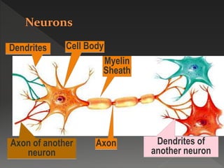
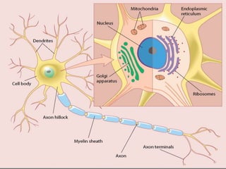
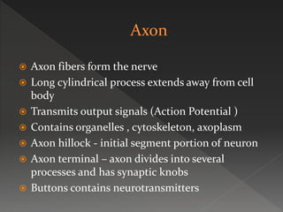
 Cell body (soma)
 Axon
 Dendrites
Axon of another
neuron
Cell BodyDendrites
Axon
Myelin
Sheath
Dendrites of
another neuron
 Soma or prikariyon
 Nucleus and cytoplamic organelles
 Nissels granules = RER
 Cytoskeletal proteins like neurofilaments
,microtubules , actin microfilaments
 Nucleus contain one or two nucleolus ,and no
centriole
 Terminally differentiated cells
 Soma extend into dendrites on one end and
axon on the other
 Membrane called plasmalemma
 Numerous short extensions from cell body
 Increasing surface area
 Dendritic spines
 Receive incoming signals and transmits them
 Helps in protein synthesis
 Helps in generating ACTION POTENTIAL
 Axon fibers form the nerve
 Long cylindrical process extends away from cell
body
 Transmits output signals (Action Potential )
 Contains organelles , cytoskeleton, axoplasm
 Axon hillock - initial segment portion of neuron
 Axon terminal – axon divides into several
processes and has synaptic knobs
 Buttons contains neurotransmitters
 It’s a protective insulator covering of the axon
 Formed by schwann cells
 Double layer membrane of a single schwann
cell wraps itself several times around axon
 1 mm in length , 8 -10 micrometer in
thickness
 Layers stick to each other due to protein p0
 Schwann cell nucleus lies in the outermost
layer
 Nodes of Ranvier
 Internodal distance is 1-2mm and each node is
0.5-1 micrometer
 Increases speed of conduction
 It reduces energy expenditure by the cell
 Provides protective covering of the axon
 Myleniated Axons
 Unmyelinated Axons
 Different neurons in body shows diff time of
myelination
 Starts early as 4th month of fetal life and
completes by 2 years
 Transfer of substances between cell body and
axon terminal
 For the development of neuron
 Bi-directional transport
 Anterograde transport
 Retrograde transport
 Transneuronal transport
 From ell body to axon terminal
 Mainly for neurotransmitters
 Two types – fast and slow
 Fast – 400mm/day by kinesin
 Slow- 0.5 – 2 mm/day
 Slow transport helps in nerve repair
 From axon terminal to cell body
 200mm /day done by dynin
 Keeps soma informed about synaptic
environment
 Transports viruses such as varicella Zoster ,
rabies
 Transport toxins
 Transports nerve growth factor
 Voltage gated Na channels are mainly
concentrated on nodes of ranvier
 Voltage gated Ca channels are mainly present
at the axon terminal for neurotransmitter
secretion
 Based on function
 Myelination
 Erlanger and gasser classification
 Demyelinating diseases
1) multiple sclerosis
2) Gullian barre syndrome
 Wallerian degeneration
 5 grades
 1st-transient loss of function
 2nd- severe injury due to prolonged pressure,
intact endoneurium complete recovery but
delayed
 3rd- severe damage to endoneurial sheath
 4th- disruption of nerve fasciculi
 5th- complete transection
 Neurotrophins
 Other growth factors ( FGF,IGF-1, PDGF ,
CNTF,GDNF)
 Nerve growth factor
 Brain derived neurotrophic factor
 Neurotrophin-3
 Neurotrophin 4/5
Neuron physiology

Neuron physiology

  • 3.
     Cell body(soma)  Axon  Dendrites
  • 4.
    Axon of another neuron CellBodyDendrites Axon Myelin Sheath Dendrites of another neuron
  • 6.
     Soma orprikariyon  Nucleus and cytoplamic organelles  Nissels granules = RER  Cytoskeletal proteins like neurofilaments ,microtubules , actin microfilaments  Nucleus contain one or two nucleolus ,and no centriole  Terminally differentiated cells
  • 7.
     Soma extendinto dendrites on one end and axon on the other  Membrane called plasmalemma
  • 8.
     Numerous shortextensions from cell body  Increasing surface area  Dendritic spines  Receive incoming signals and transmits them  Helps in protein synthesis  Helps in generating ACTION POTENTIAL
  • 10.
     Axon fibersform the nerve  Long cylindrical process extends away from cell body  Transmits output signals (Action Potential )  Contains organelles , cytoskeleton, axoplasm  Axon hillock - initial segment portion of neuron  Axon terminal – axon divides into several processes and has synaptic knobs  Buttons contains neurotransmitters
  • 12.
     It’s aprotective insulator covering of the axon  Formed by schwann cells  Double layer membrane of a single schwann cell wraps itself several times around axon  1 mm in length , 8 -10 micrometer in thickness  Layers stick to each other due to protein p0  Schwann cell nucleus lies in the outermost layer
  • 16.
     Nodes ofRanvier  Internodal distance is 1-2mm and each node is 0.5-1 micrometer
  • 17.
     Increases speedof conduction  It reduces energy expenditure by the cell  Provides protective covering of the axon
  • 18.
     Myleniated Axons Unmyelinated Axons
  • 19.
     Different neuronsin body shows diff time of myelination  Starts early as 4th month of fetal life and completes by 2 years
  • 20.
     Transfer ofsubstances between cell body and axon terminal  For the development of neuron  Bi-directional transport
  • 21.
     Anterograde transport Retrograde transport  Transneuronal transport
  • 22.
     From ellbody to axon terminal  Mainly for neurotransmitters  Two types – fast and slow  Fast – 400mm/day by kinesin  Slow- 0.5 – 2 mm/day  Slow transport helps in nerve repair
  • 23.
     From axonterminal to cell body  200mm /day done by dynin  Keeps soma informed about synaptic environment  Transports viruses such as varicella Zoster , rabies  Transport toxins  Transports nerve growth factor
  • 24.
     Voltage gatedNa channels are mainly concentrated on nodes of ranvier  Voltage gated Ca channels are mainly present at the axon terminal for neurotransmitter secretion
  • 25.
     Based onfunction  Myelination  Erlanger and gasser classification
  • 27.
     Demyelinating diseases 1)multiple sclerosis 2) Gullian barre syndrome
  • 28.
  • 29.
     5 grades 1st-transient loss of function  2nd- severe injury due to prolonged pressure, intact endoneurium complete recovery but delayed  3rd- severe damage to endoneurial sheath  4th- disruption of nerve fasciculi  5th- complete transection
  • 31.
     Neurotrophins  Othergrowth factors ( FGF,IGF-1, PDGF , CNTF,GDNF)
  • 32.
     Nerve growthfactor  Brain derived neurotrophic factor  Neurotrophin-3  Neurotrophin 4/5