Download as PPSX, PPTX


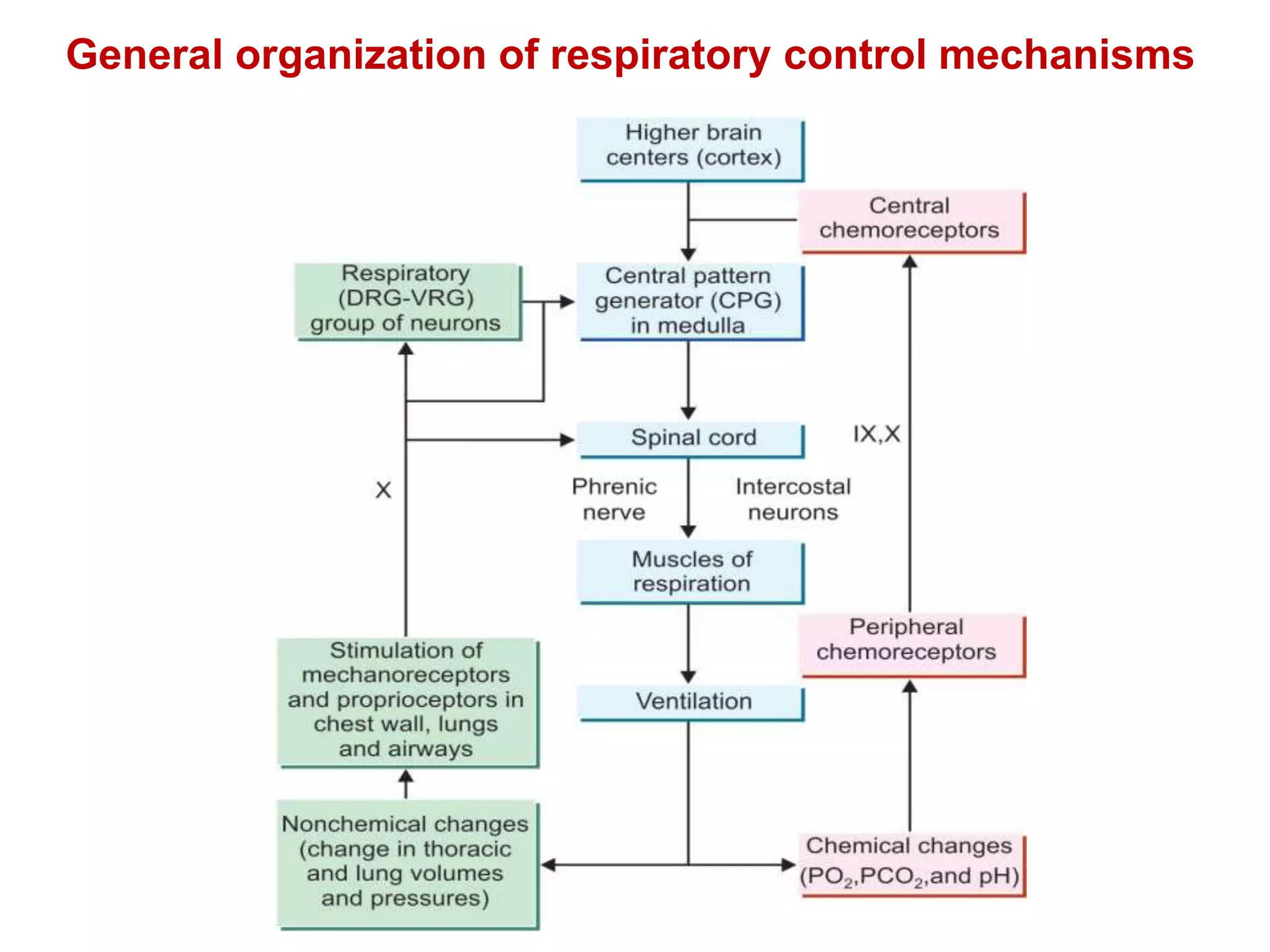


The document summarizes the neural and chemical regulation of respiration. It describes the key respiratory centers in the medulla and pons that control breathing. These include the dorsal and ventral respiratory groups in the medulla and the apneustic and pneumotaxic centers in the pons. Peripheral chemoreceptors in the carotid body and aortic body and central chemoreceptors in the medulla detect changes in blood gases like CO2 and pH to modulate breathing. Increased CO2 and H+ stimulate these chemoreceptors to enhance the activity of the respiratory centers and increase ventilation.
Introduction to respiratory regulation and learning objectives covering neural and chemical mechanisms.
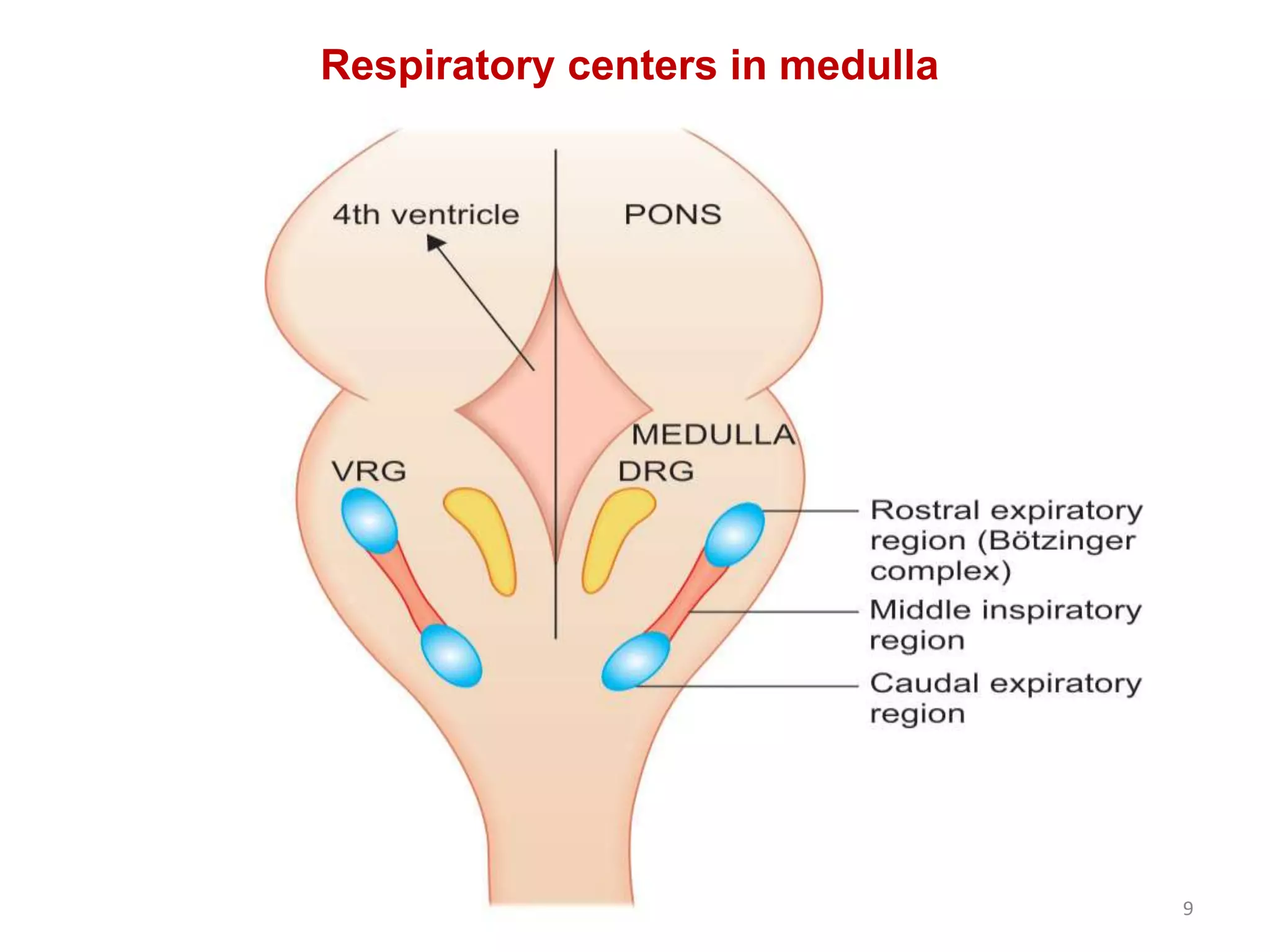
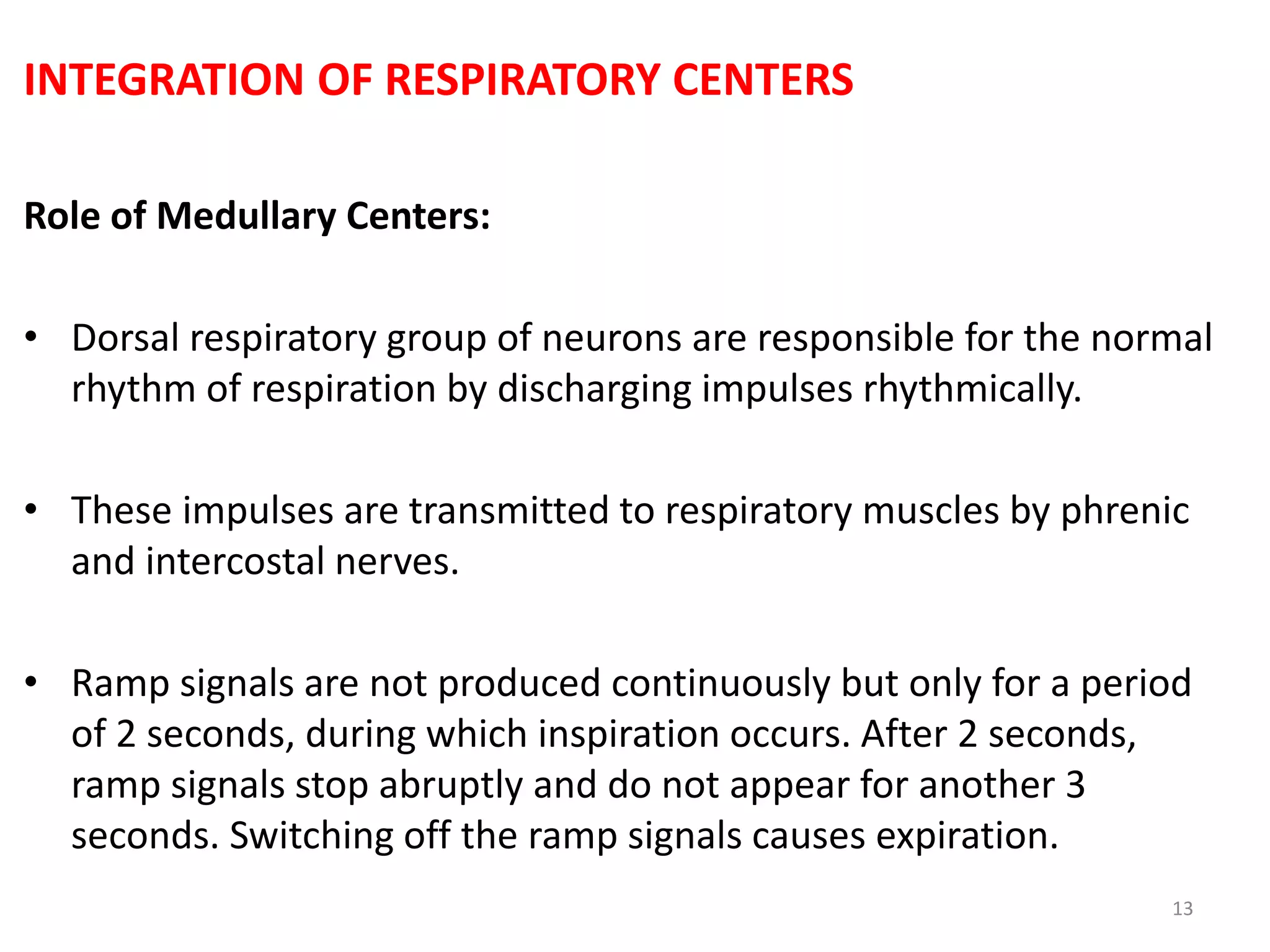
In-depth discussion on the neural regulation of respiration, including roles of medullary and pontine centers.
Details on the interaction and functions of medullary and pontine respiratory centers in controlling breathing.
Explanation of the pre-BÖtzinger complex as a rhythm generator and its influence on respiration.
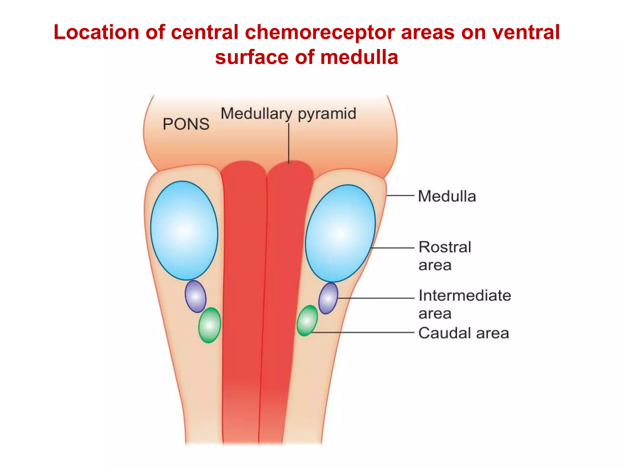
Mechanisms of respiration regulation through chemoreceptors sensitive to blood gas changes.
Summary of key points, references for further reading, and closing remarks.


