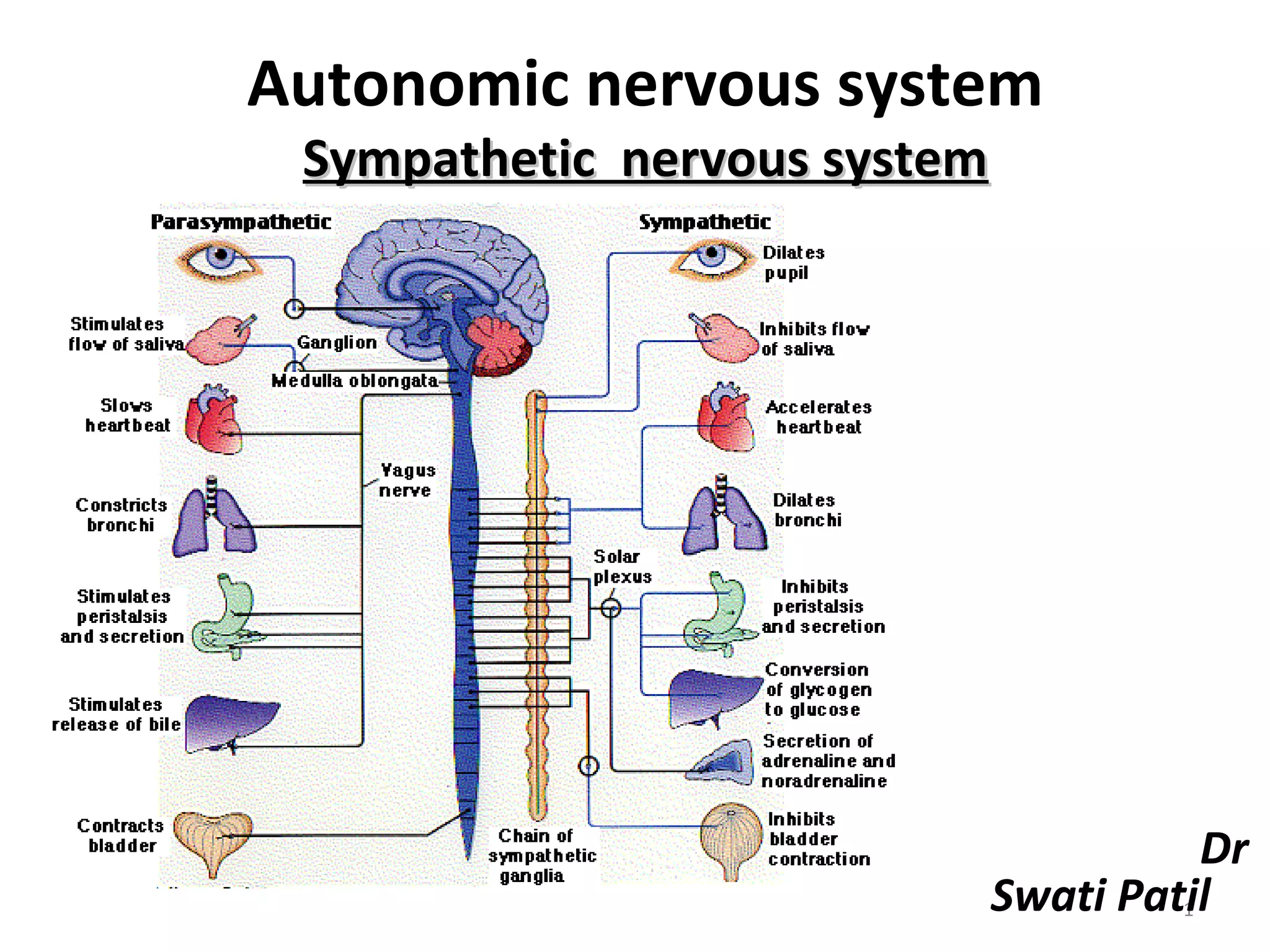
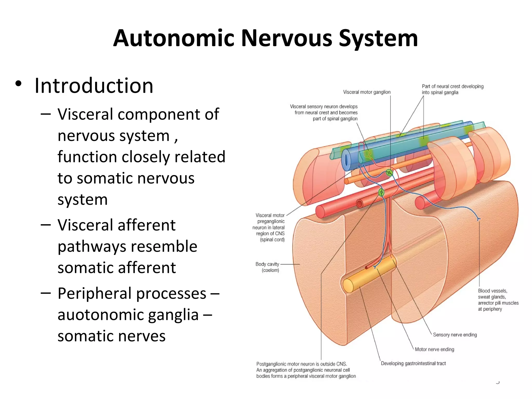
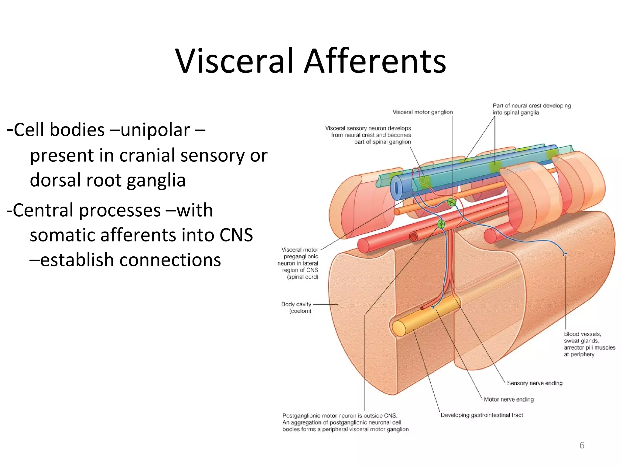
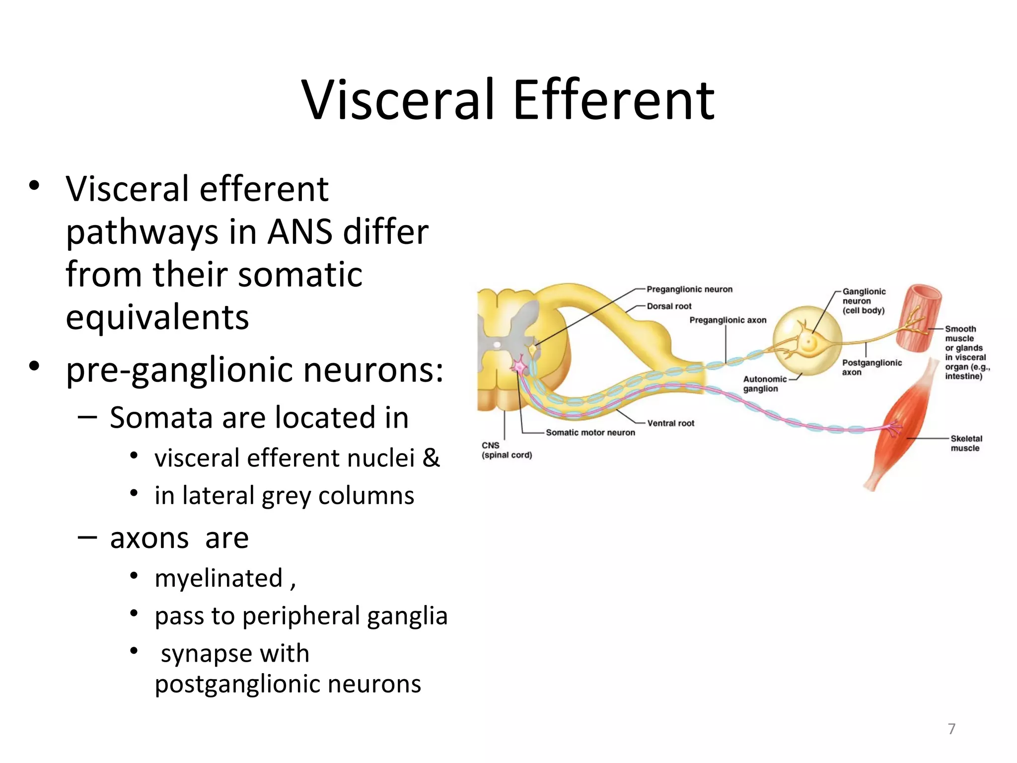
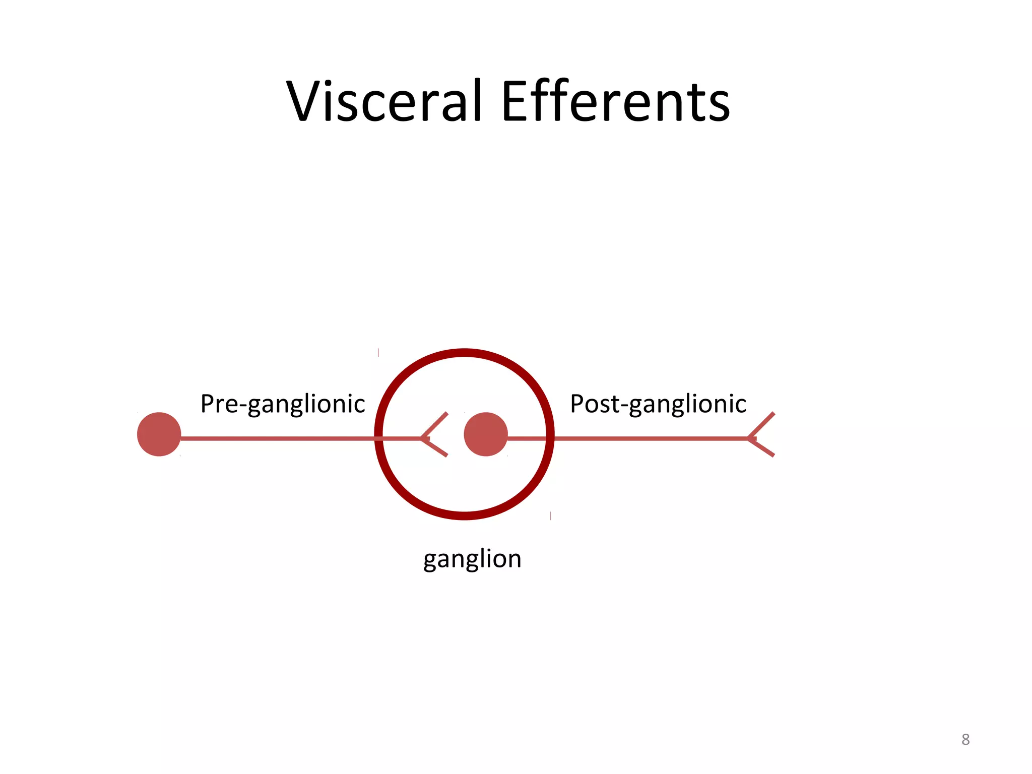
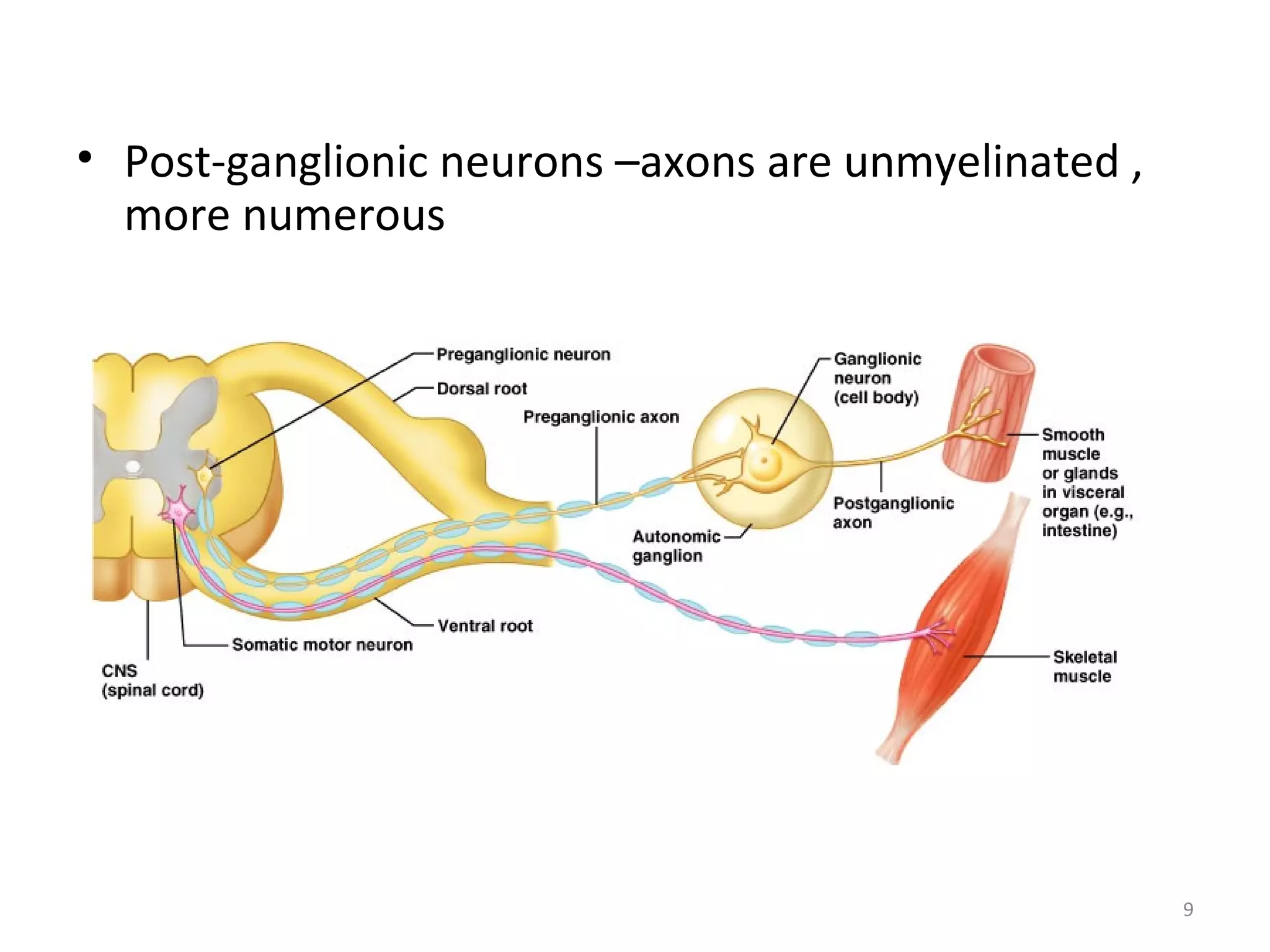
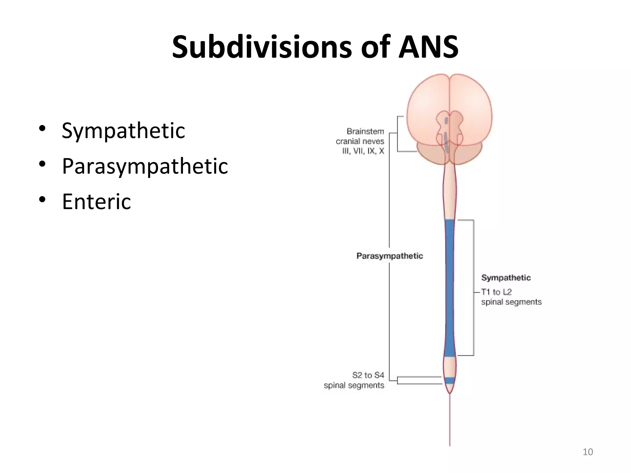
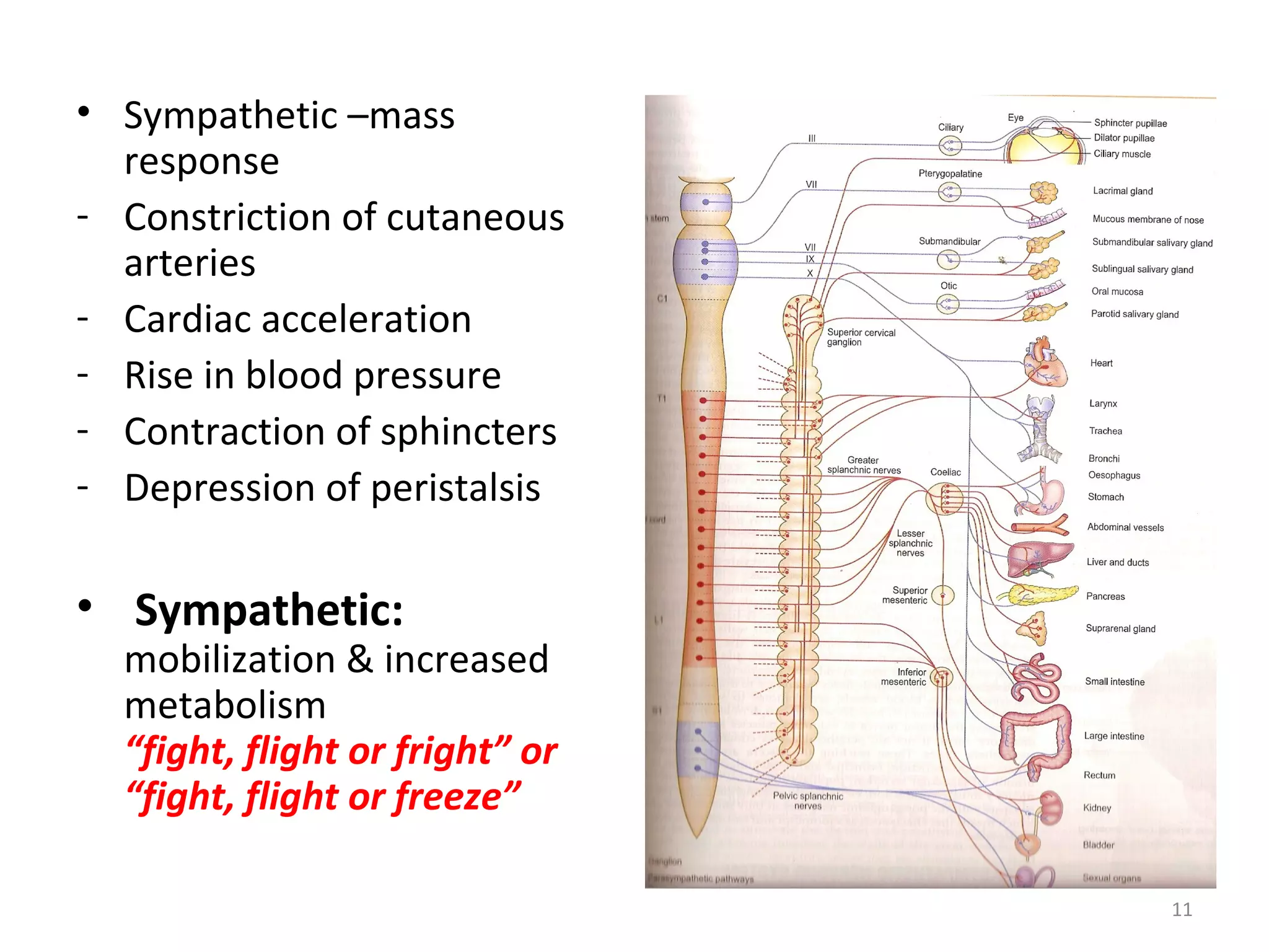
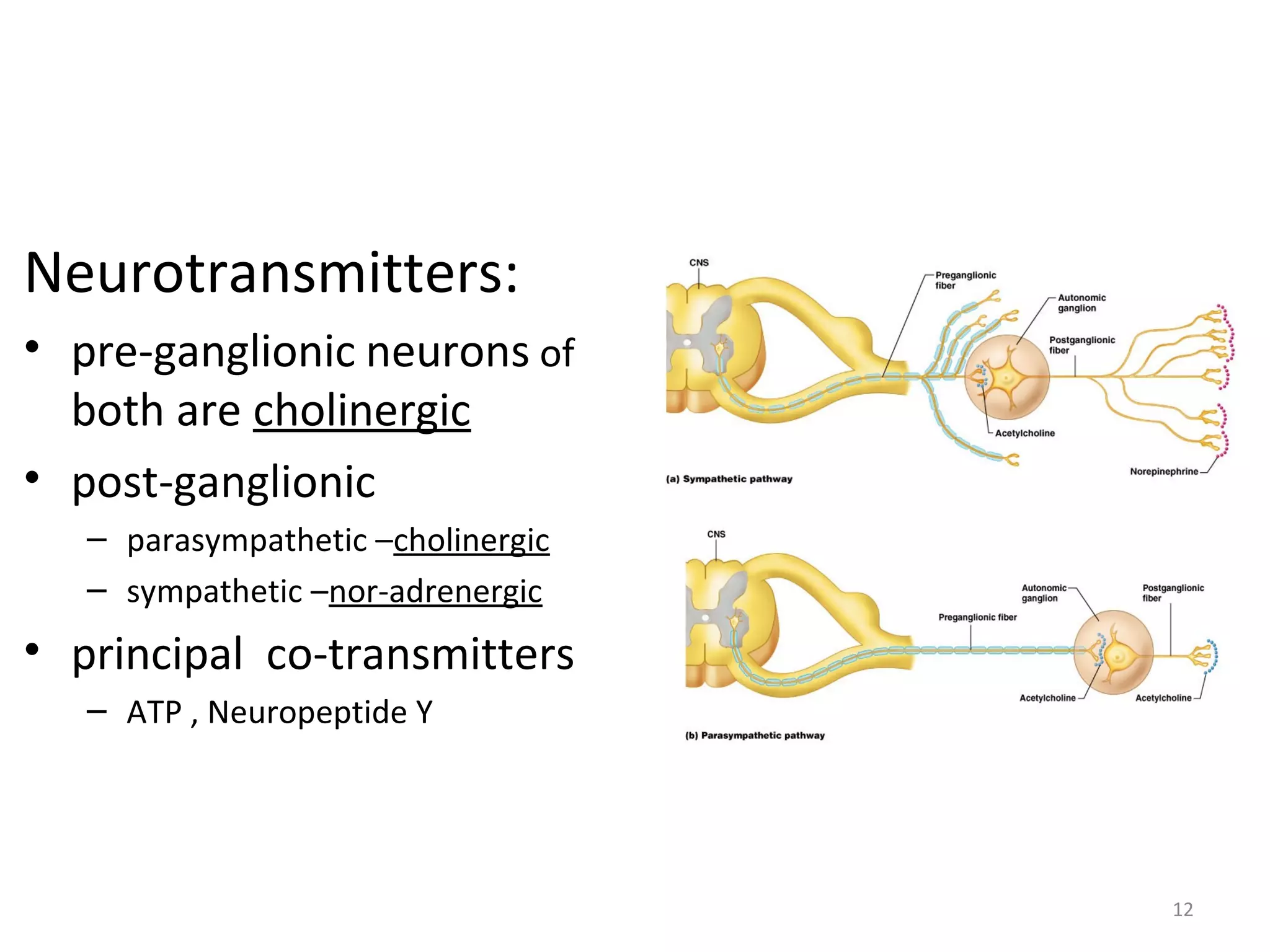
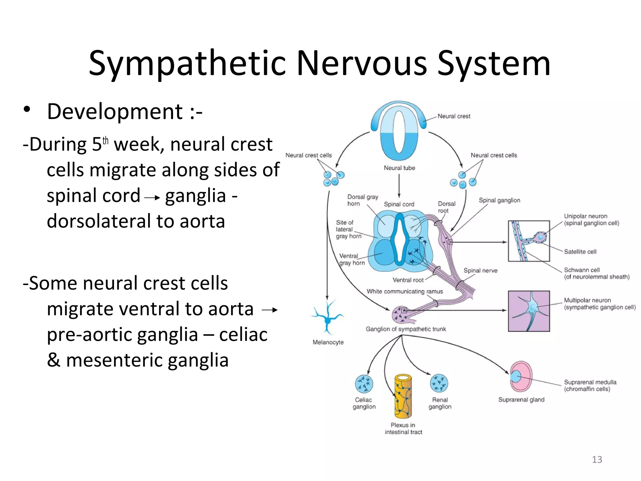
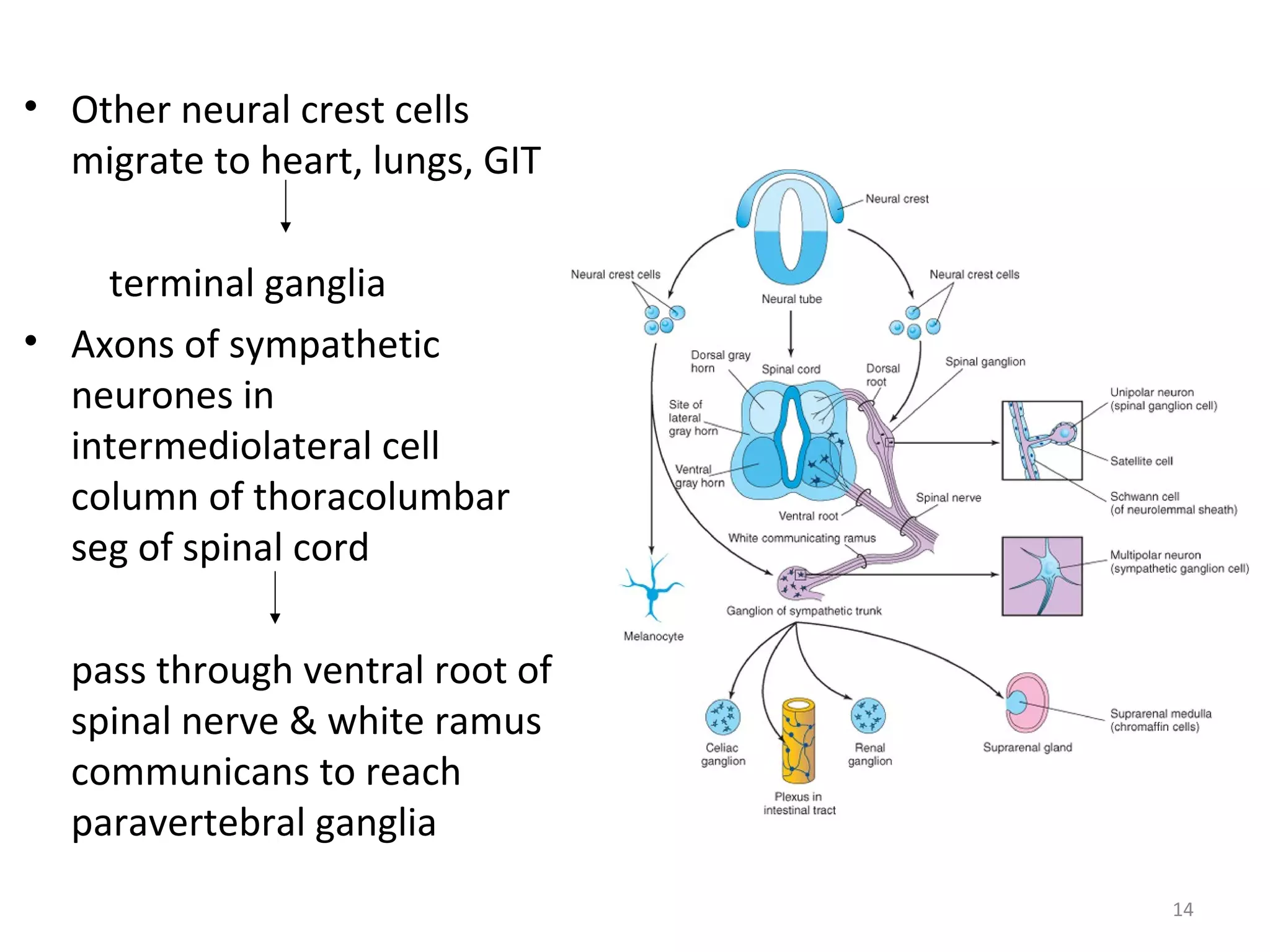
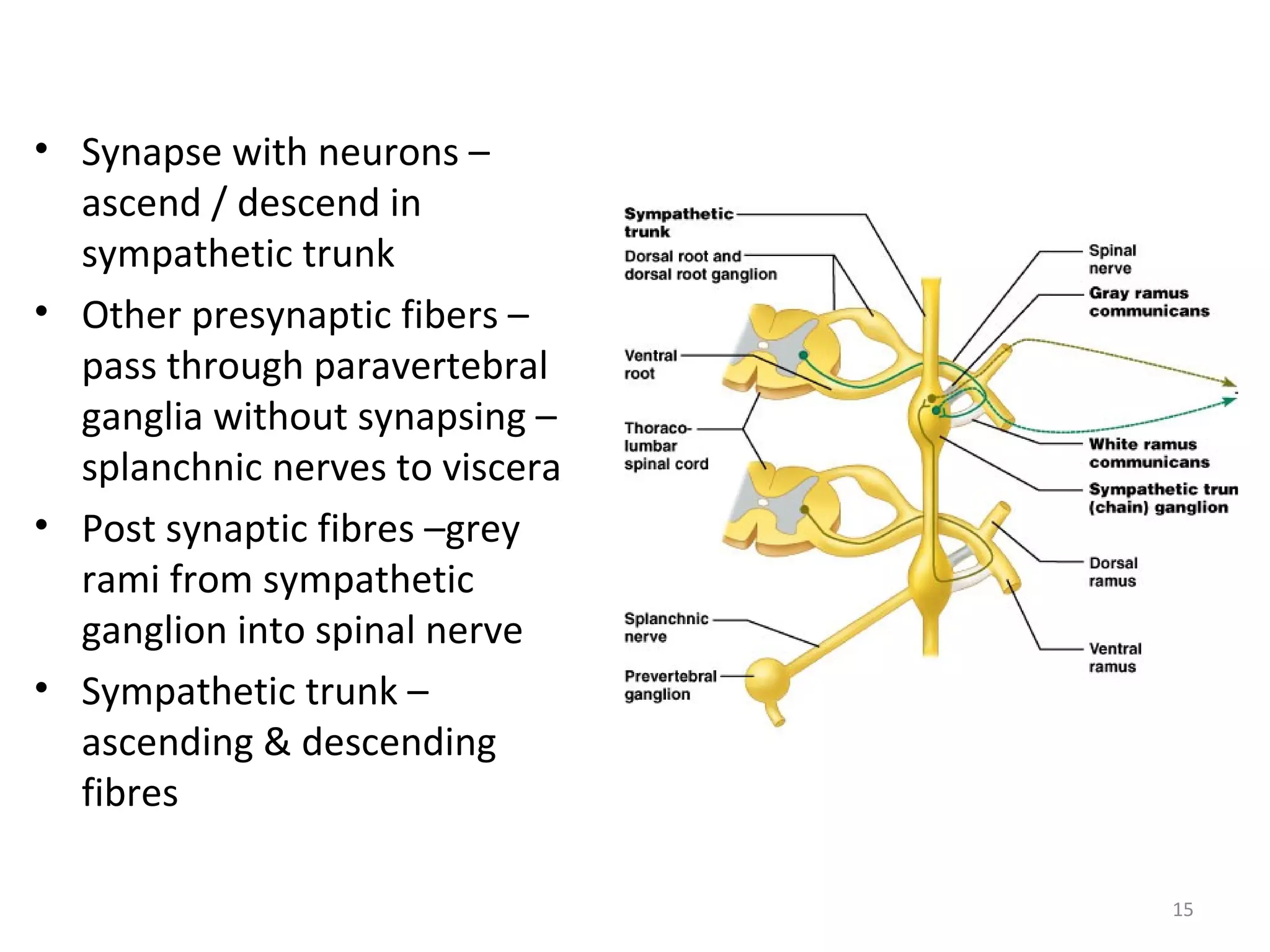
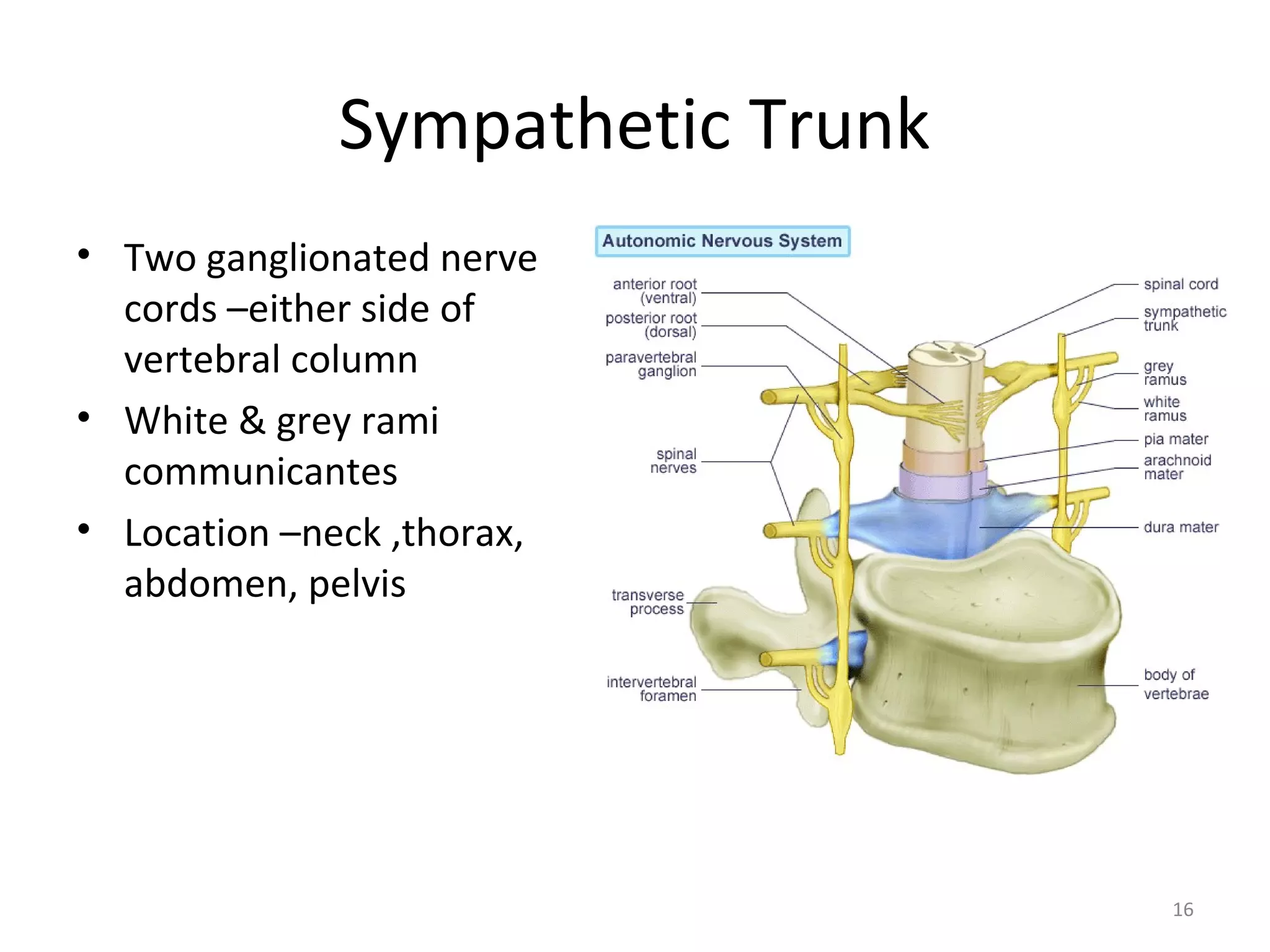
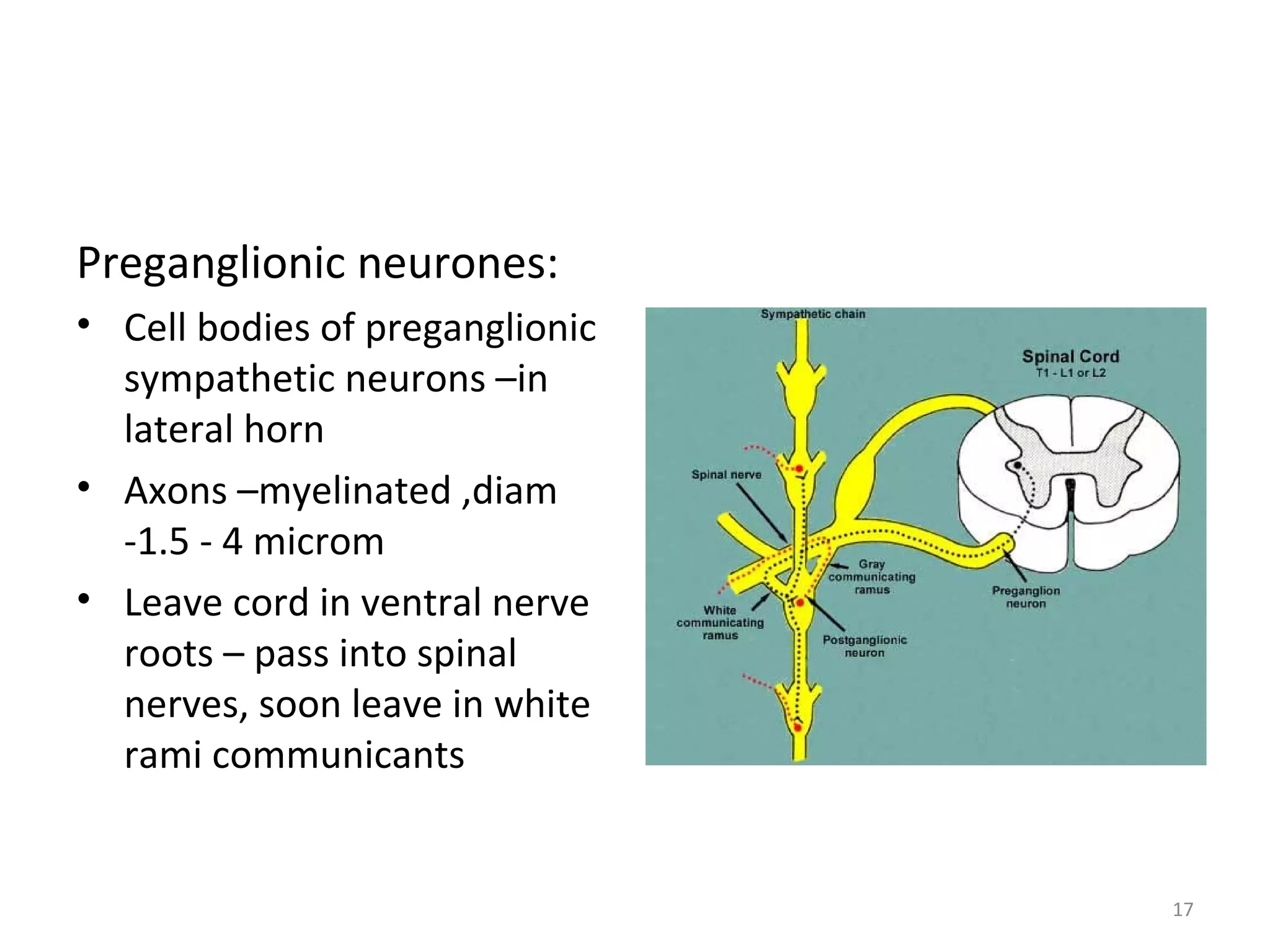
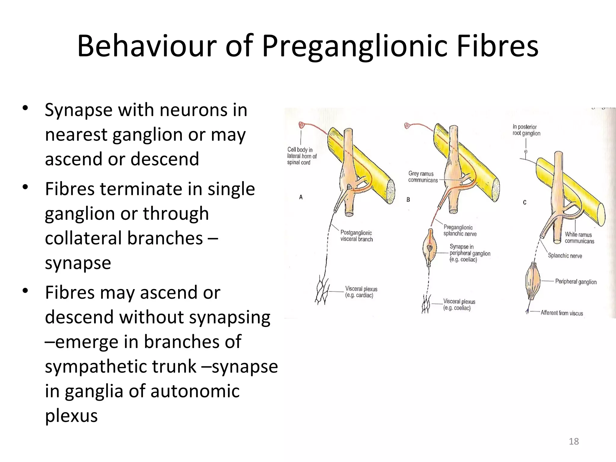
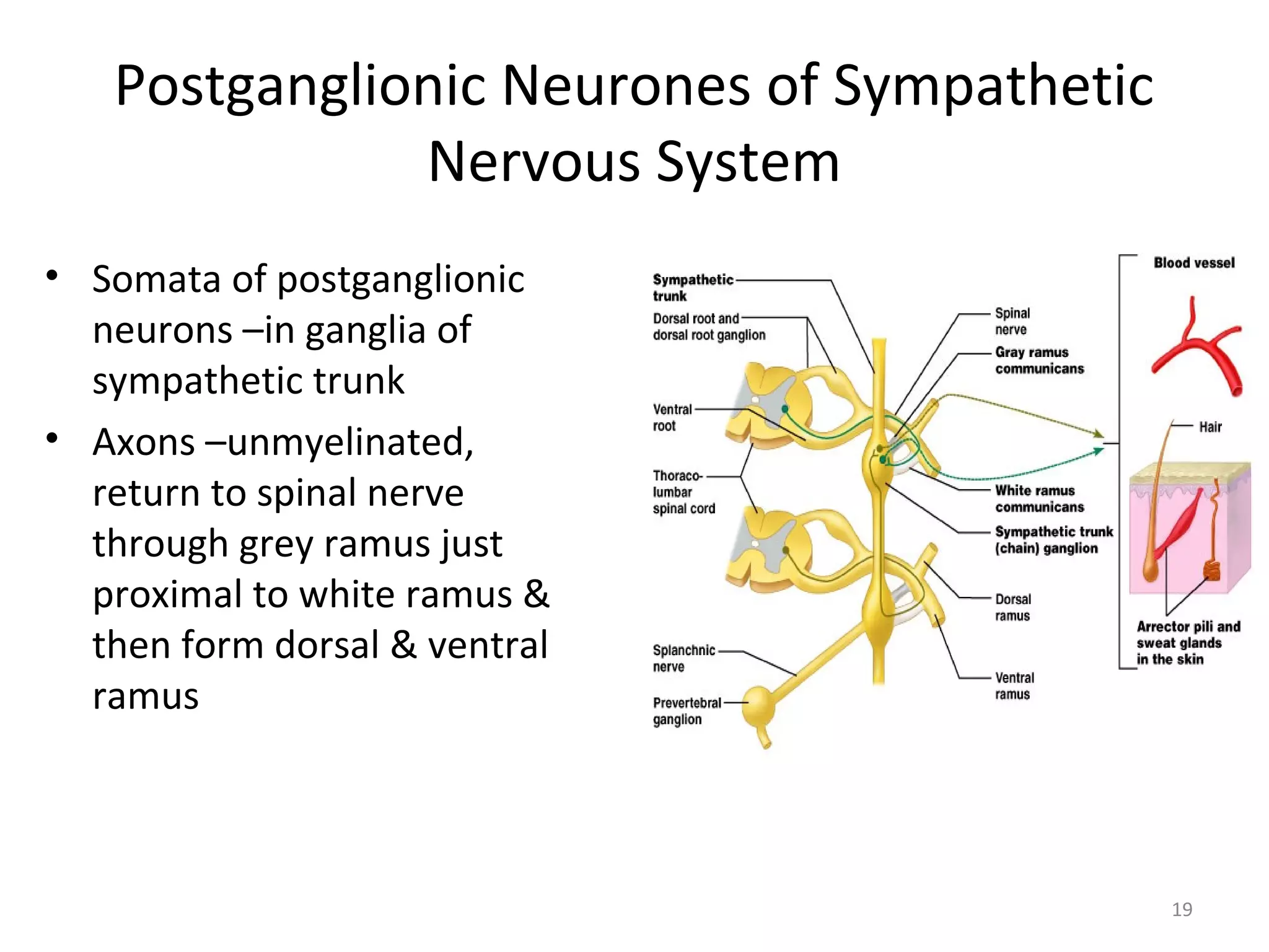
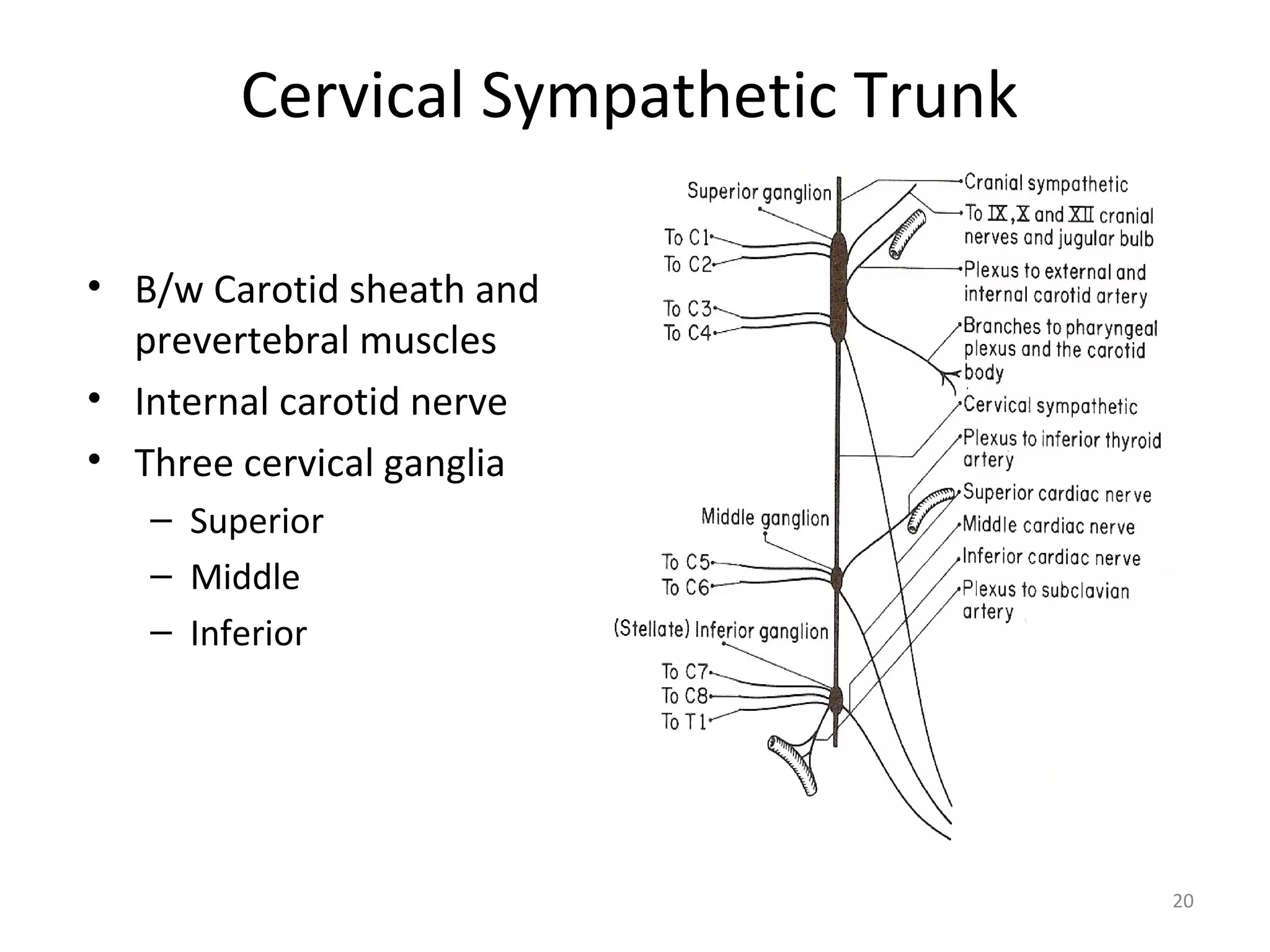
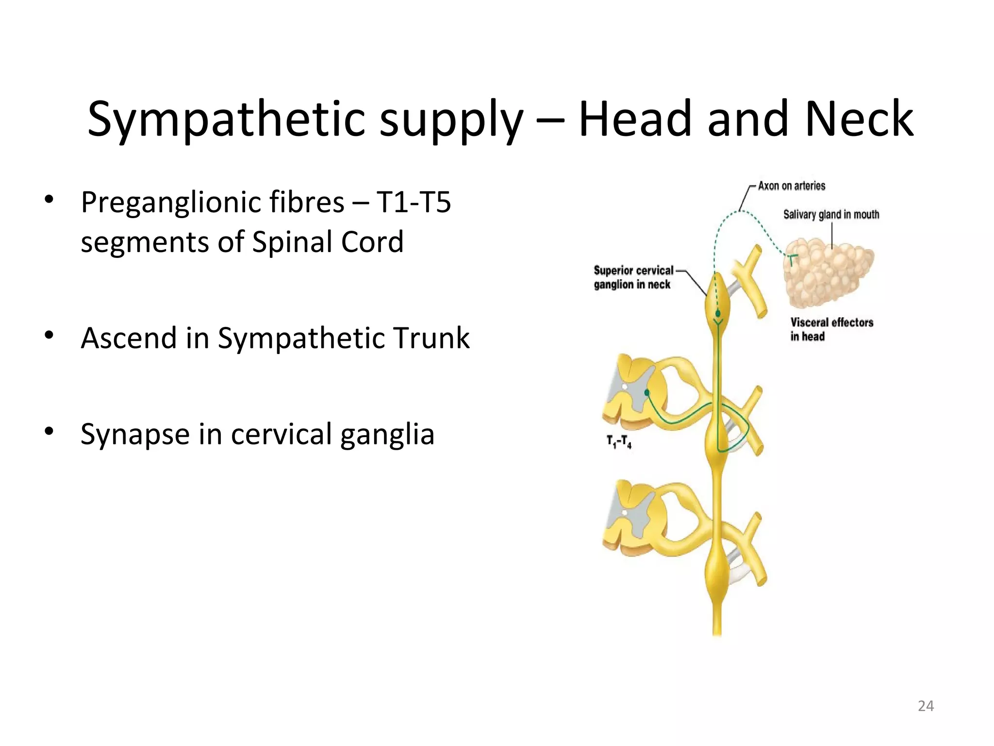
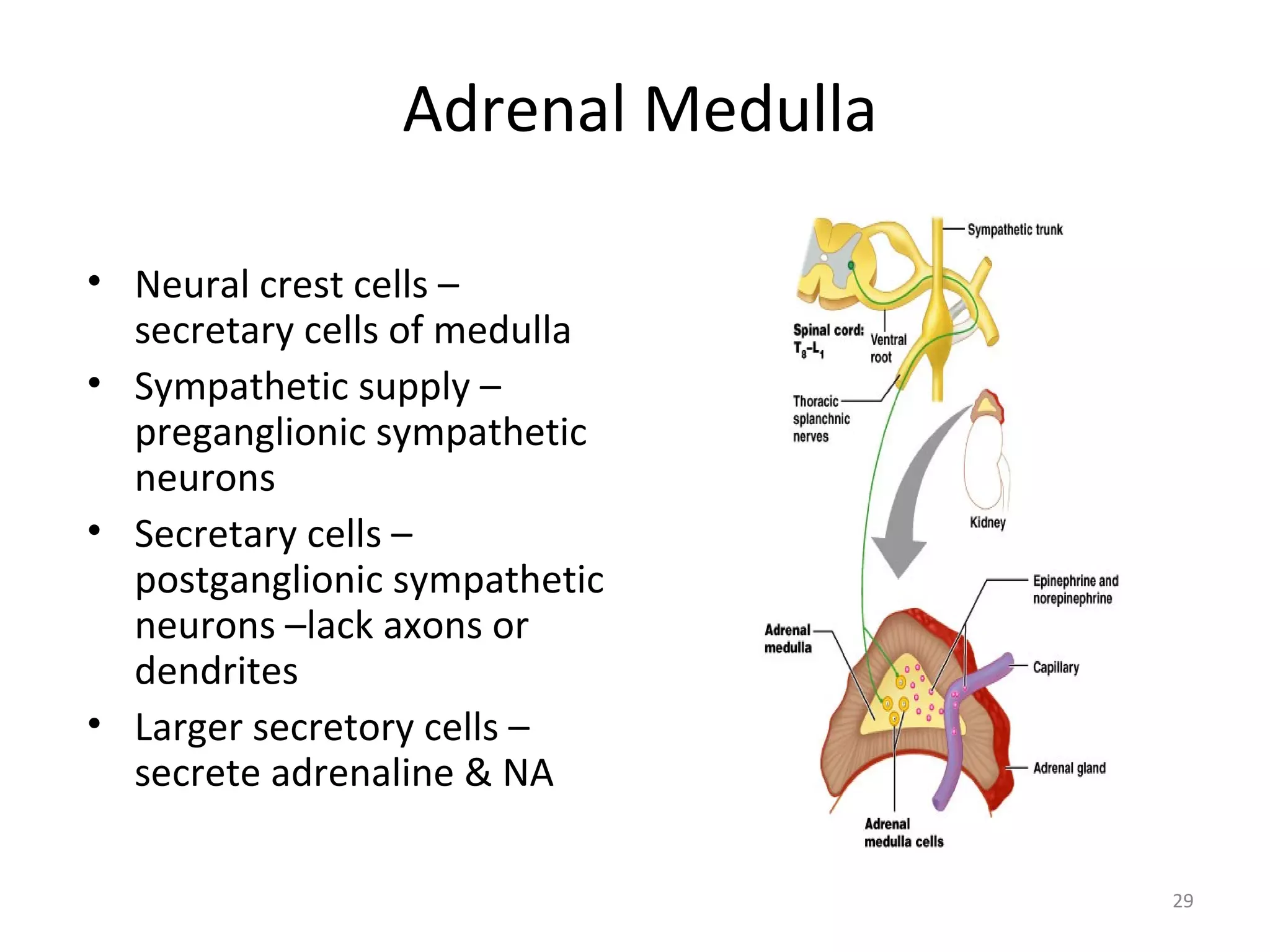
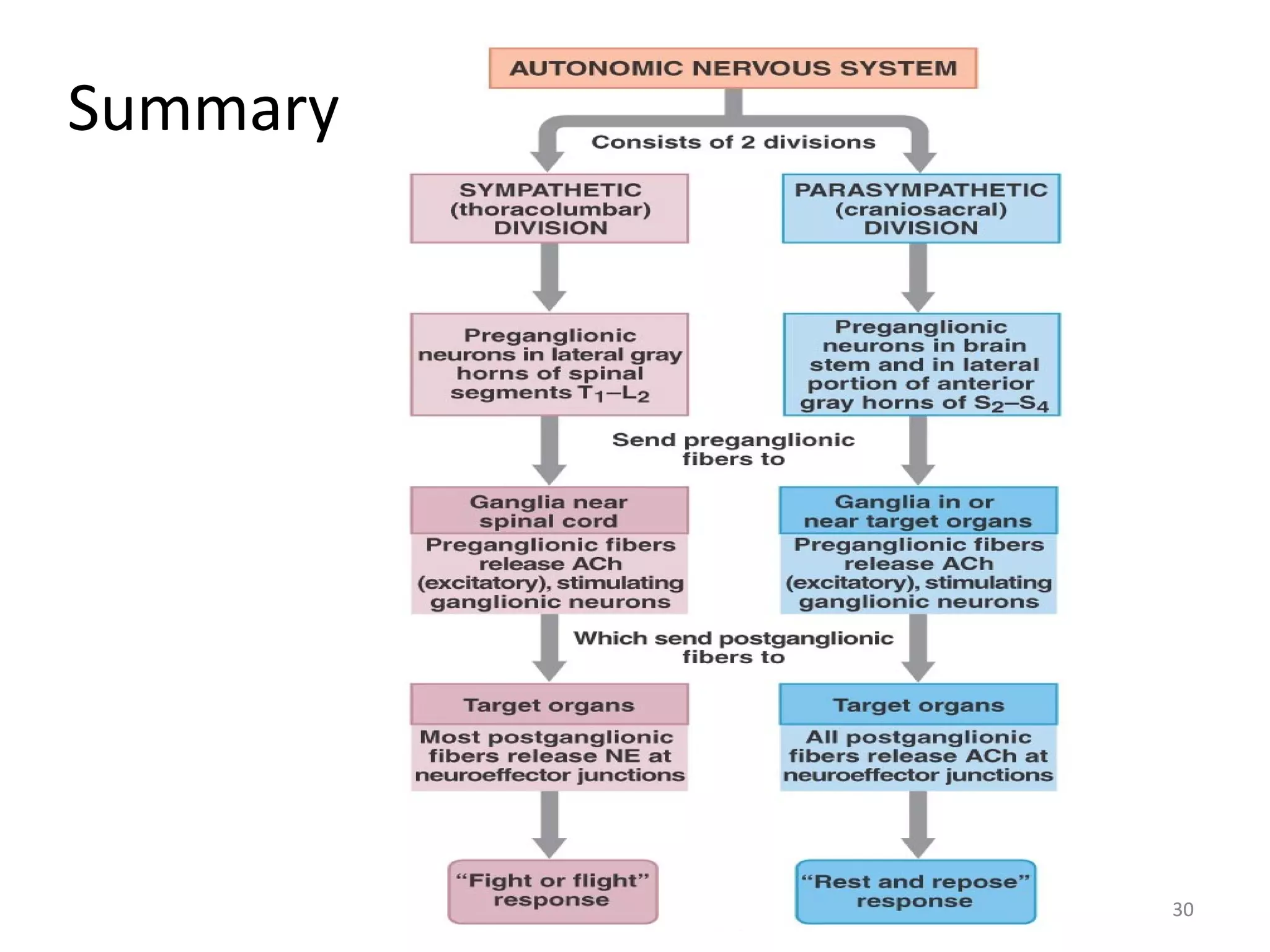
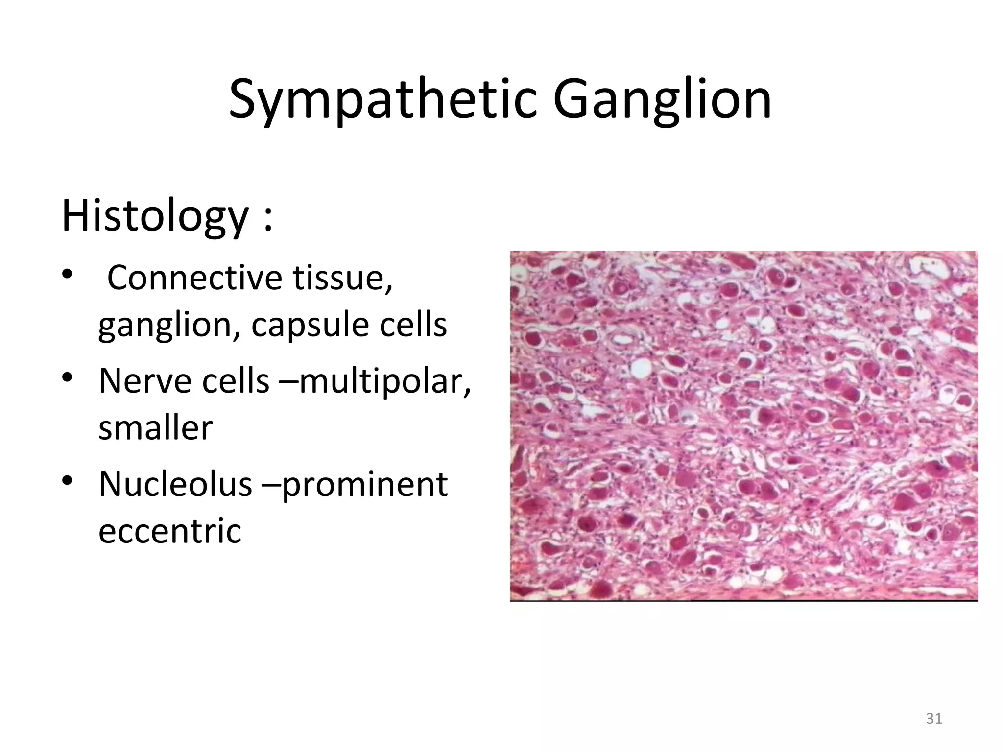
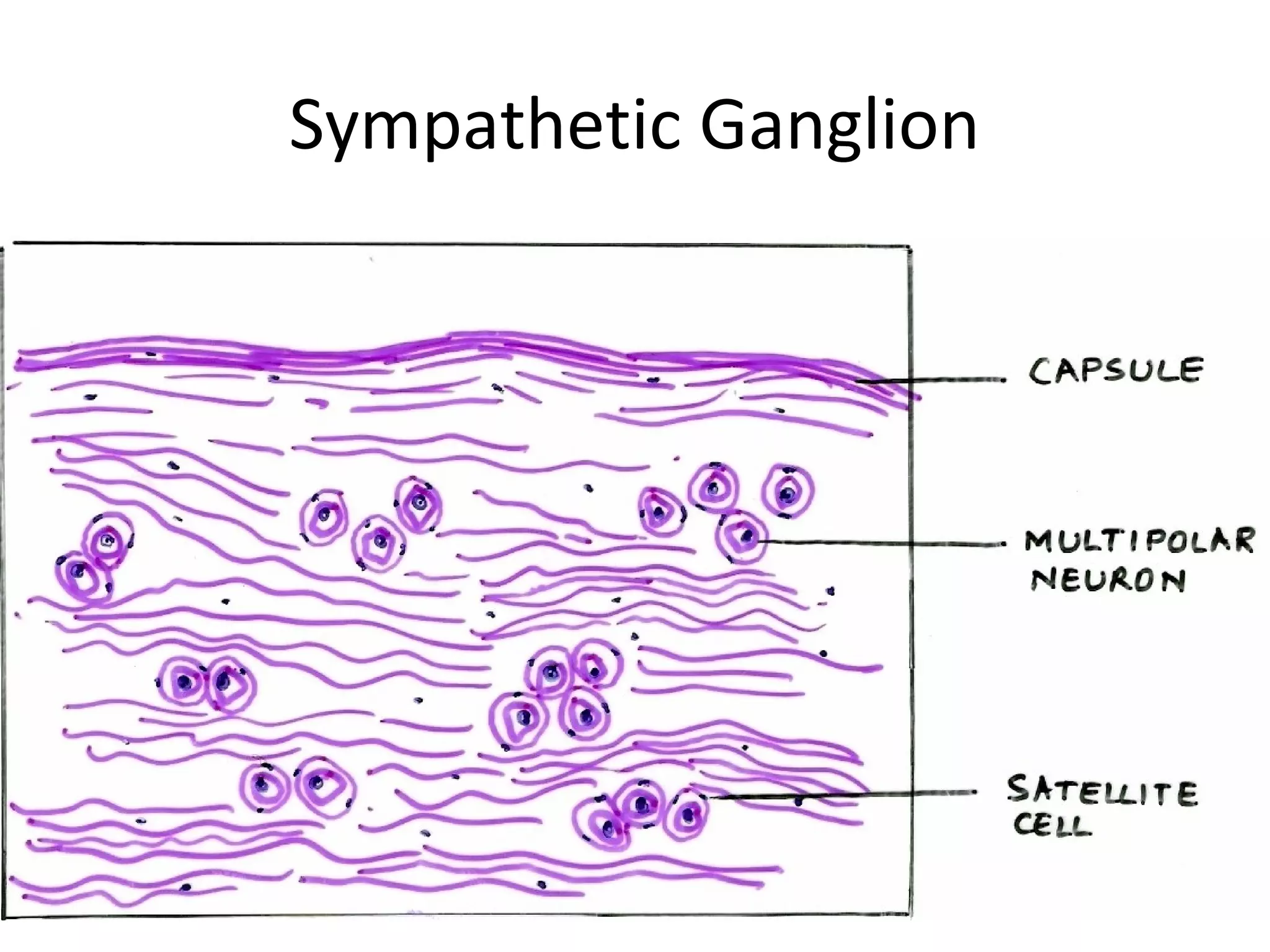
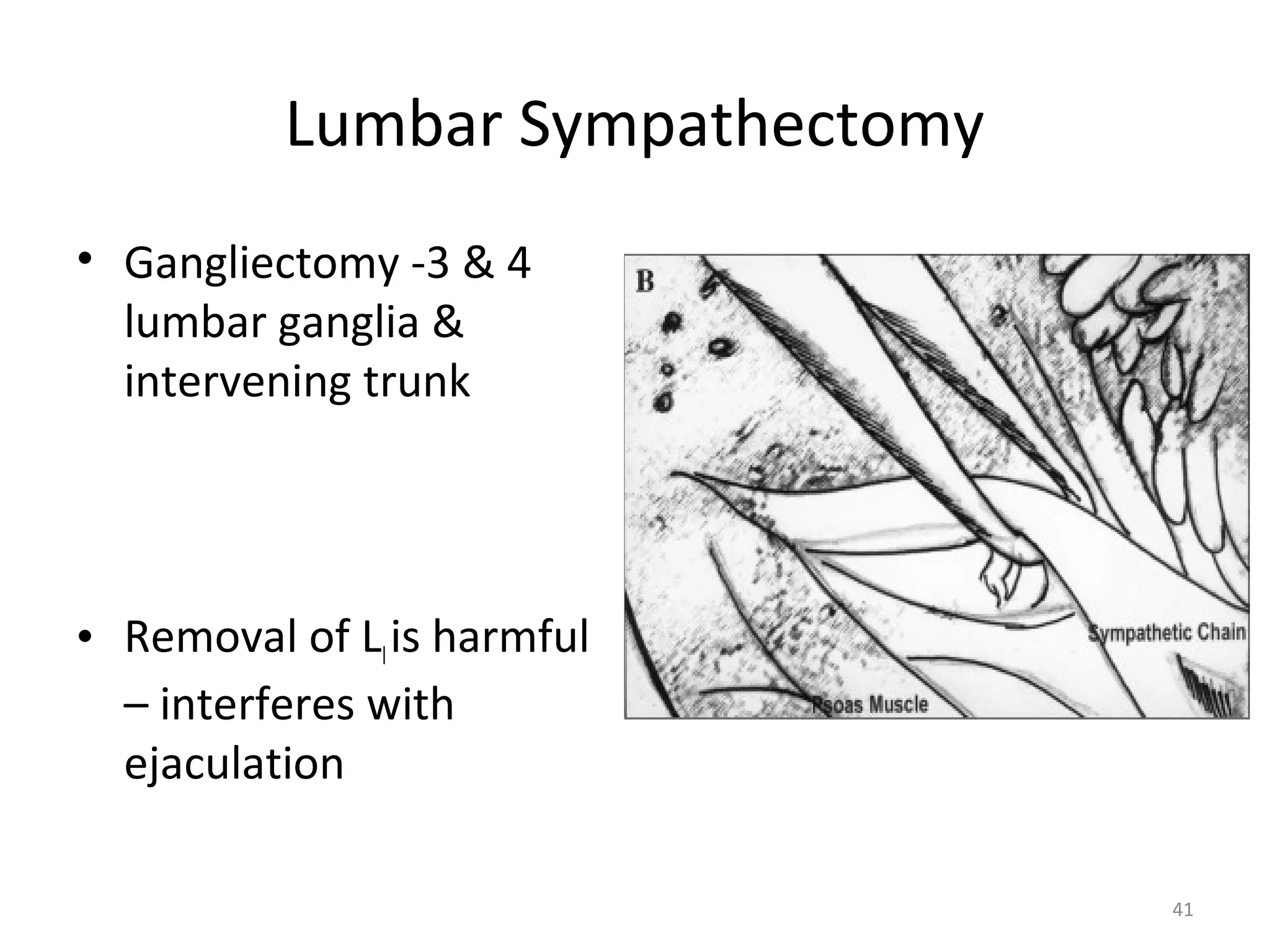
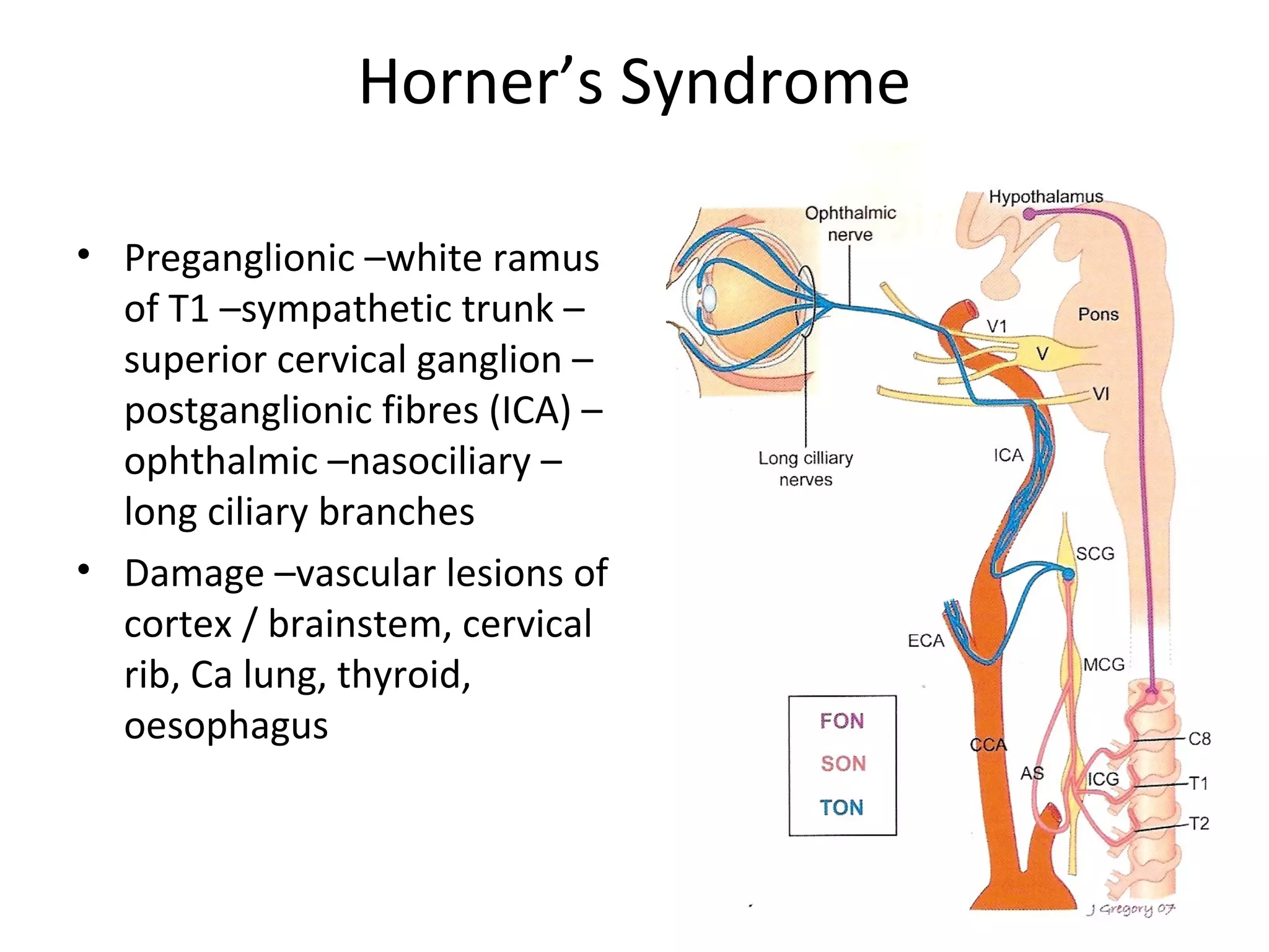
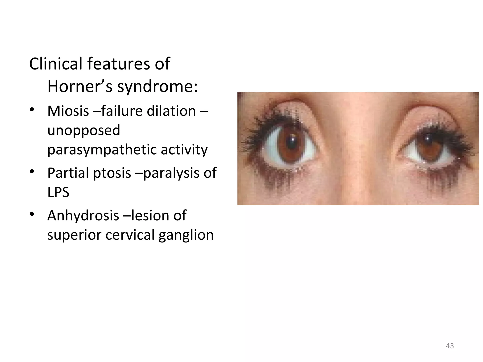
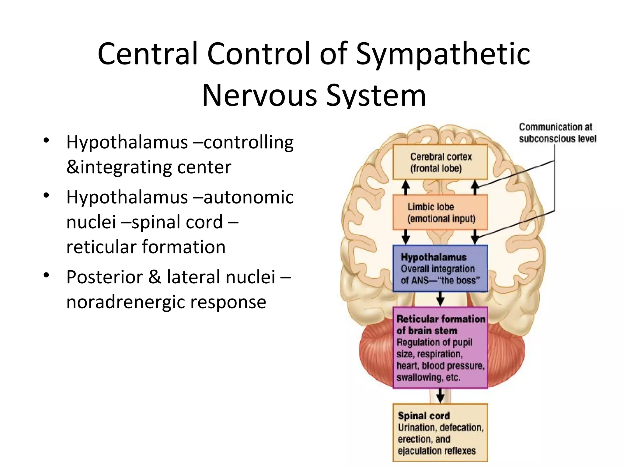
1
Autonomic nervous system
Sympathetic nervous system
Dr
Swati Patil
2
• Autonomic nervous system
-History
-Introduction
-Types :- Sympathetic –Development
-Introduction
-Course
-Types
-Applied
3
Autonomic Nervous System
• Self regulating
• History:
– 1898 - J.N.Langley assigned the term Autonomic
Nervous System
– 1921 – subdivided ANS into
• Sympathetic
• Parasympathetic
• Enteric
4
5
Autonomic Nervous System
• Introduction
– Visceral component of
nervous system ,
function closely related
to somatic nervous
system
– Visceral afferent
pathways resemble
somatic afferent
– Peripheral processes –
auotonomic ganglia –
somatic nerves
6
Visceral Afferents
-Cell bodies –unipolar –
present in cranial sensory or
dorsal root ganglia
-Central processes –with
somatic afferents into CNS
–establish connections
7
Visceral Efferent
• Visceral efferent
pathways in ANS differ
from their somatic
equivalents
• pre-ganglionic neurons:
– Somata are located in
• visceral efferent nuclei &
• in lateral grey columns
– axons are
• myelinated ,
• pass to peripheral ganglia
• synapse with
postganglionic neurons
8
Visceral Efferents
Pre-ganglionic
ganglion
Post-ganglionic
9
• Post-ganglionic neurons –axons are unmyelinated ,
more numerous
10
Subdivisions of ANS
• Sympathetic
• Parasympathetic
• Enteric
11
• Sympathetic –mass
response
- Constriction of cutaneous
arteries
- Cardiac acceleration
- Rise in blood pressure
- Contraction of sphincters
- Depression of peristalsis
• Sympathetic:
mobilization & increased
metabolism
“fight, flight or fright” or
“fight, flight or freeze”
12
Neurotransmitters:
• pre-ganglionic neurons of
both are cholinergic
• post-ganglionic
– parasympathetic –cholinergic
– sympathetic –nor-adrenergic
• principal co-transmitters
– ATP , Neuropeptide Y
13
Sympathetic Nervous System
• Development :-
-During 5th
week, neural crest
cells migrate along sides of
spinal cord ganglia -
dorsolateral to aorta
-Some neural crest cells
migrate ventral to aorta
pre-aortic ganglia – celiac
& mesenteric ganglia
14
• Other neural crest cells
migrate to heart, lungs, GIT
terminal ganglia
• Axons of sympathetic
neurones in
intermediolateral cell
column of thoracolumbar
seg of spinal cord
pass through ventral root of
spinal nerve & white ramus
communicans to reach
paravertebral ganglia
15
• Synapse with neurons –
ascend / descend in
sympathetic trunk
• Other presynaptic fibers –
pass through paravertebral
ganglia without synapsing –
splanchnic nerves to viscera
• Post synaptic fibres –grey
rami from sympathetic
ganglion into spinal nerve
• Sympathetic trunk –
ascending & descending
fibres
16
Sympathetic Trunk
• Two ganglionated nerve
cords –either side of
vertebral column
• White & grey rami
communicantes
• Location –neck ,thorax,
abdomen, pelvis
17
Preganglionic neurones:
• Cell bodies of preganglionic
sympathetic neurons –in
lateral horn
• Axons –myelinated ,diam
-1.5 - 4 microm
• Leave cord in ventral nerve
roots – pass into spinal
nerves, soon leave in white
rami communicants
18
Behaviour of Preganglionic Fibres
• Synapse with neurons in
nearest ganglion or may
ascend or descend
• Fibres terminate in single
ganglion or through
collateral branches –
synapse
• Fibres may ascend or
descend without synapsing
–emerge in branches of
sympathetic trunk –synapse
in ganglia of autonomic
plexus
19
Postganglionic Neurones of Sympathetic
Nervous System
• Somata of postganglionic
neurons –in ganglia of
sympathetic trunk
• Axons –unmyelinated,
return to spinal nerve
through grey ramus just
proximal to white ramus &
then form dorsal & ventral
ramus
20
Cervical Sympathetic Trunk
• B/w Carotid sheath and
prevertebral muscles
• Internal carotid nerve
• Three cervical ganglia
– Superior
– Middle
– Inferior
21
Superior Cervical Ganglion
• Largest ganglion
• Lies in front of transverse
processes of C2 and C3
vertebrae
• Branches
– Medial
– Lateral
22
Middle Cervical Ganglion
• Smallest of the Cervical
ganglion
• Lies on the C6
vertebra in front or behind
Inf. thyroid artery
• Branches
– grey rami communicantes
– Cardiac branch
– Vascular Branch
23
Stellate Ganglion
• Formed by the fusion of
C7,C8 andT1ganglia
• Lies b/w neck of 1st
Rib and
transverse process of C7
vertebra
• Branches
– Grey rami communicans
– Vascular branches
24
Sympathetic supply – Head and Neck
• Preganglionic fibres – T1-T5
segments of Spinal Cord
• Ascend in Sympathetic Trunk
• Synapse in cervical ganglia
25
Thoracic Sympathetic Trunk
• Comprises of 11 ganglia
• Ganglia lie against the
heads of ribs
• Branches
– Grey rami
communicans
– Pul. And cardiac
Plexus
– Splanchnic Nerves
26
Coeliac Plexus
• Situated around the origin
of coeliac artery
• Formed by greater
Splanchnic Nerves and Ist
lumbar sympathetic nerves
• Nerves from the plexus
supply abdominal viscera
via blood vessels
27
Lumbar Sympathetic Trunk
• Lies retroperitoneally on
the anterolat. surface of
lumbar vertebrae
• Rt side – overlapped by IVC
• Lt side – overlapped by
Aorta
• Branches
– Splanchnic nerves
– Grey rami communicantes
28
Hypogastric Plexuses
• Superior hypogastric plexus:-
-location
-formation
-branches
• Inferior hypogastric plexus :-
-location
-formation
-branches
29
Adrenal Medulla
• Neural crest cells –
secretary cells of medulla
• Sympathetic supply –
preganglionic sympathetic
neurons
• Secretary cells –
postganglionic sympathetic
neurons –lack axons or
dendrites
• Larger secretory cells –
secrete adrenaline & NA
30
30
Summary
31
Sympathetic Ganglion
Histology :
• Connective tissue,
ganglion, capsule cells
• Nerve cells –multipolar,
smaller
• Nucleolus –prominent
eccentric
32
Sympathetic Ganglion
33
Enteric Nervous System
• Myenteric (Auerbach’s)
plexus & submucosal
(Meissner) plexus
• Plexus –small enteric
ganglia –joined by thin
nerves –unmyelinated
• Avascular –nutrition by
diffusion
• Neurones –excitatory &
inhibitory
• Afferents to ENS -2 types –
cholinergic & NA
34
Pain Afferents
• Sensory neurones –pain
in thoracic & abdominal
organs
• Cell bodies –dorsal root
ganglia
• Peripheral processes –
white communicating
rami –sympathetic trunk
–viscera
35
• Referred pain –diffuse
localization & radiation
• Zone of reference of pain
from int organ coincides
with part of body served
by somatic sensory
neurons assoc with same
segment of spinal cord
36
Referred Pain
• Heart –middle &
inferior cervical cardiac
nerves, thoracic cardiac
branches of left
sympathetic chain
• Gall bladder –greater
splanchnic nerve ,
diaphragm –phrenic
nerve
37
• Stomach –epigastrium –
Rt & Lt greater splanchnic
nerves
• Duodenal ulcer –AAW –
T9 T10
• Appendix –lesser
splanchnic nerves –T10
• Pelvis & ureter –least
splanchnic nerves –loin &
groin
38
Surgical Sympathectomy
• Indications
– Peripheral vascular disease –sympathectomy –temporary
vasodilatation –development of collaterals
– Hyperhidrosis –sympathectomy –permanent relief
– Relief of Pain –a) visceral pain –excision / destruction –
coeliac ganglia –Ca pancreas, chronic pancreatits
– b) causlgia –intense pain
39
Upper Thoracic and Cervical
Sympathectomy
• Upper limb -2 & 3 thoracic
ganglia with rami &
intervening part
• 1st
thoracic –not removed,
preganglionic fibres not
arise above T2 (removal –
Horner’s syndrome)
40
Kuntz Nerve
• Communicating branch B/w
T1and T2 nerves
• Receives Grey rami from
Stellate and T2 ganglia
• Clinical relevance –failure
to identify during thoracic
sympathectomy
41
Lumbar Sympathectomy
• Gangliectomy -3 & 4
lumbar ganglia &
intervening trunk
• Removal of LIis harmful
– interferes with
ejaculation
42
Horner’s Syndrome
• Preganglionic –white ramus
of T1 –sympathetic trunk –
superior cervical ganglion –
postganglionic fibres (ICA) –
ophthalmic –nasociliary –
long ciliary branches
• Damage –vascular lesions of
cortex / brainstem, cervical
rib, Ca lung, thyroid,
oesophagus
43
Clinical features of
Horner’s syndrome:
• Miosis –failure dilation –
unopposed
parasympathetic activity
• Partial ptosis –paralysis of
LPS
• Anhydrosis –lesion of
superior cervical ganglion
44
Central Control of Sympathetic
Nervous System
• Hypothalamus –controlling
&integrating center
• Hypothalamus –autonomic
nuclei –spinal cord –
reticular formation
• Posterior & lateral nuclei –
noradrenergic response
45
THANK
THANK YOU…
YOU…

2-sympatheticnervoussystem-121125122947-phpapp02.pdf

  • 1.
    1 Autonomic nervous system Sympatheticnervous system Sympathetic nervous system Dr Swati Patil
  • 2.
    2 • Autonomic nervoussystem -History -Introduction -Types :- Sympathetic –Development -Introduction -Course -Types -Applied
  • 3.
    3 Autonomic Nervous System •Self regulating • History: – 1898 - J.N.Langley assigned the term Autonomic Nervous System – 1921 – subdivided ANS into • Sympathetic • Parasympathetic • Enteric
  • 4.
  • 5.
    5 Autonomic Nervous System •Introduction – Visceral component of nervous system , function closely related to somatic nervous system – Visceral afferent pathways resemble somatic afferent – Peripheral processes – auotonomic ganglia – somatic nerves
  • 6.
    6 Visceral Afferents -Cell bodies–unipolar – present in cranial sensory or dorsal root ganglia -Central processes –with somatic afferents into CNS –establish connections
  • 7.
    7 Visceral Efferent • Visceralefferent pathways in ANS differ from their somatic equivalents • pre-ganglionic neurons: – Somata are located in • visceral efferent nuclei & • in lateral grey columns – axons are • myelinated , • pass to peripheral ganglia • synapse with postganglionic neurons
  • 8.
  • 9.
    9 • Post-ganglionic neurons–axons are unmyelinated , more numerous
  • 10.
    10 Subdivisions of ANS •Sympathetic • Parasympathetic • Enteric
  • 11.
    11 • Sympathetic –mass response -Constriction of cutaneous arteries - Cardiac acceleration - Rise in blood pressure - Contraction of sphincters - Depression of peristalsis • Sympathetic: mobilization & increased metabolism “fight, flight or fright” or “fight, flight or freeze”
  • 12.
    12 Neurotransmitters: • pre-ganglionic neuronsof both are cholinergic • post-ganglionic – parasympathetic –cholinergic – sympathetic –nor-adrenergic • principal co-transmitters – ATP , Neuropeptide Y
  • 13.
    13 Sympathetic Nervous System •Development :- -During 5th week, neural crest cells migrate along sides of spinal cord ganglia - dorsolateral to aorta -Some neural crest cells migrate ventral to aorta pre-aortic ganglia – celiac & mesenteric ganglia
  • 14.
    14 • Other neuralcrest cells migrate to heart, lungs, GIT terminal ganglia • Axons of sympathetic neurones in intermediolateral cell column of thoracolumbar seg of spinal cord pass through ventral root of spinal nerve & white ramus communicans to reach paravertebral ganglia
  • 15.
    15 • Synapse withneurons – ascend / descend in sympathetic trunk • Other presynaptic fibers – pass through paravertebral ganglia without synapsing – splanchnic nerves to viscera • Post synaptic fibres –grey rami from sympathetic ganglion into spinal nerve • Sympathetic trunk – ascending & descending fibres
  • 16.
    16 Sympathetic Trunk • Twoganglionated nerve cords –either side of vertebral column • White & grey rami communicantes • Location –neck ,thorax, abdomen, pelvis
  • 17.
    17 Preganglionic neurones: • Cellbodies of preganglionic sympathetic neurons –in lateral horn • Axons –myelinated ,diam -1.5 - 4 microm • Leave cord in ventral nerve roots – pass into spinal nerves, soon leave in white rami communicants
  • 18.
    18 Behaviour of PreganglionicFibres • Synapse with neurons in nearest ganglion or may ascend or descend • Fibres terminate in single ganglion or through collateral branches – synapse • Fibres may ascend or descend without synapsing –emerge in branches of sympathetic trunk –synapse in ganglia of autonomic plexus
  • 19.
    19 Postganglionic Neurones ofSympathetic Nervous System • Somata of postganglionic neurons –in ganglia of sympathetic trunk • Axons –unmyelinated, return to spinal nerve through grey ramus just proximal to white ramus & then form dorsal & ventral ramus
  • 20.
    20 Cervical Sympathetic Trunk •B/w Carotid sheath and prevertebral muscles • Internal carotid nerve • Three cervical ganglia – Superior – Middle – Inferior
  • 21.
    21 Superior Cervical Ganglion •Largest ganglion • Lies in front of transverse processes of C2 and C3 vertebrae • Branches – Medial – Lateral
  • 22.
    22 Middle Cervical Ganglion •Smallest of the Cervical ganglion • Lies on the C6 vertebra in front or behind Inf. thyroid artery • Branches – grey rami communicantes – Cardiac branch – Vascular Branch
  • 23.
    23 Stellate Ganglion • Formedby the fusion of C7,C8 andT1ganglia • Lies b/w neck of 1st Rib and transverse process of C7 vertebra • Branches – Grey rami communicans – Vascular branches
  • 24.
    24 Sympathetic supply –Head and Neck • Preganglionic fibres – T1-T5 segments of Spinal Cord • Ascend in Sympathetic Trunk • Synapse in cervical ganglia
  • 25.
    25 Thoracic Sympathetic Trunk •Comprises of 11 ganglia • Ganglia lie against the heads of ribs • Branches – Grey rami communicans – Pul. And cardiac Plexus – Splanchnic Nerves
  • 26.
    26 Coeliac Plexus • Situatedaround the origin of coeliac artery • Formed by greater Splanchnic Nerves and Ist lumbar sympathetic nerves • Nerves from the plexus supply abdominal viscera via blood vessels
  • 27.
    27 Lumbar Sympathetic Trunk •Lies retroperitoneally on the anterolat. surface of lumbar vertebrae • Rt side – overlapped by IVC • Lt side – overlapped by Aorta • Branches – Splanchnic nerves – Grey rami communicantes
  • 28.
    28 Hypogastric Plexuses • Superiorhypogastric plexus:- -location -formation -branches • Inferior hypogastric plexus :- -location -formation -branches
  • 29.
    29 Adrenal Medulla • Neuralcrest cells – secretary cells of medulla • Sympathetic supply – preganglionic sympathetic neurons • Secretary cells – postganglionic sympathetic neurons –lack axons or dendrites • Larger secretory cells – secrete adrenaline & NA
  • 30.
  • 31.
    31 Sympathetic Ganglion Histology : •Connective tissue, ganglion, capsule cells • Nerve cells –multipolar, smaller • Nucleolus –prominent eccentric
  • 32.
  • 33.
    33 Enteric Nervous System •Myenteric (Auerbach’s) plexus & submucosal (Meissner) plexus • Plexus –small enteric ganglia –joined by thin nerves –unmyelinated • Avascular –nutrition by diffusion • Neurones –excitatory & inhibitory • Afferents to ENS -2 types – cholinergic & NA
  • 34.
    34 Pain Afferents • Sensoryneurones –pain in thoracic & abdominal organs • Cell bodies –dorsal root ganglia • Peripheral processes – white communicating rami –sympathetic trunk –viscera
  • 35.
    35 • Referred pain–diffuse localization & radiation • Zone of reference of pain from int organ coincides with part of body served by somatic sensory neurons assoc with same segment of spinal cord
  • 36.
    36 Referred Pain • Heart–middle & inferior cervical cardiac nerves, thoracic cardiac branches of left sympathetic chain • Gall bladder –greater splanchnic nerve , diaphragm –phrenic nerve
  • 37.
    37 • Stomach –epigastrium– Rt & Lt greater splanchnic nerves • Duodenal ulcer –AAW – T9 T10 • Appendix –lesser splanchnic nerves –T10 • Pelvis & ureter –least splanchnic nerves –loin & groin
  • 38.
    38 Surgical Sympathectomy • Indications –Peripheral vascular disease –sympathectomy –temporary vasodilatation –development of collaterals – Hyperhidrosis –sympathectomy –permanent relief – Relief of Pain –a) visceral pain –excision / destruction – coeliac ganglia –Ca pancreas, chronic pancreatits – b) causlgia –intense pain
  • 39.
    39 Upper Thoracic andCervical Sympathectomy • Upper limb -2 & 3 thoracic ganglia with rami & intervening part • 1st thoracic –not removed, preganglionic fibres not arise above T2 (removal – Horner’s syndrome)
  • 40.
    40 Kuntz Nerve • Communicatingbranch B/w T1and T2 nerves • Receives Grey rami from Stellate and T2 ganglia • Clinical relevance –failure to identify during thoracic sympathectomy
  • 41.
    41 Lumbar Sympathectomy • Gangliectomy-3 & 4 lumbar ganglia & intervening trunk • Removal of LIis harmful – interferes with ejaculation
  • 42.
    42 Horner’s Syndrome • Preganglionic–white ramus of T1 –sympathetic trunk – superior cervical ganglion – postganglionic fibres (ICA) – ophthalmic –nasociliary – long ciliary branches • Damage –vascular lesions of cortex / brainstem, cervical rib, Ca lung, thyroid, oesophagus
  • 43.
    43 Clinical features of Horner’ssyndrome: • Miosis –failure dilation – unopposed parasympathetic activity • Partial ptosis –paralysis of LPS • Anhydrosis –lesion of superior cervical ganglion
  • 44.
    44 Central Control ofSympathetic Nervous System • Hypothalamus –controlling &integrating center • Hypothalamus –autonomic nuclei –spinal cord – reticular formation • Posterior & lateral nuclei – noradrenergic response
  • 45.