Downloaded 678 times
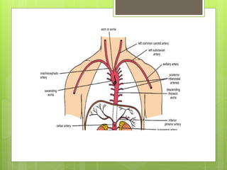
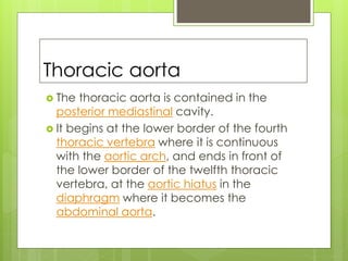
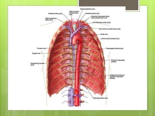

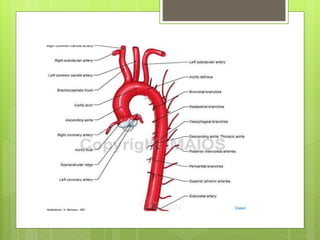
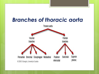


The thoracic aorta begins where the aortic arch ends at the fourth thoracic vertebrae and extends down to the diaphragm. It supplies blood to the thoracic cavity and has several important branches including the bronchial arteries which supply the lungs, esophageal arteries which supply the esophagus, and posterior intercostal arteries which supply the spaces between the ribs. The thoracic aorta also gives off mediastinal and pericardial branches before passing through the diaphragm and becoming the abdominal aorta.


