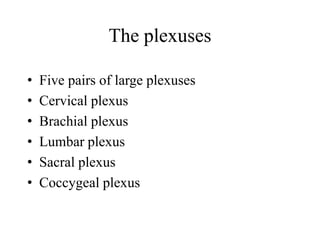
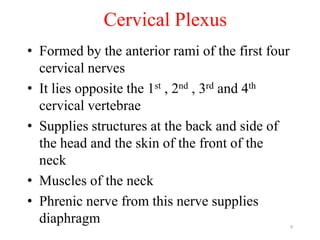
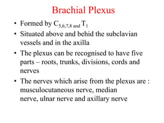
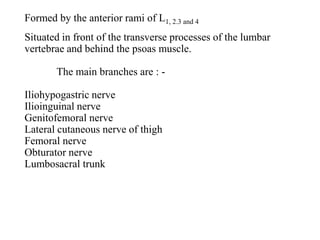
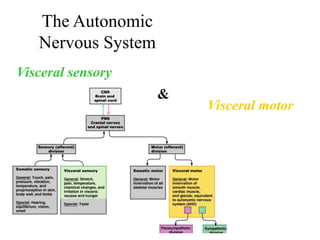
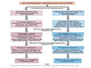
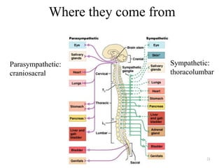
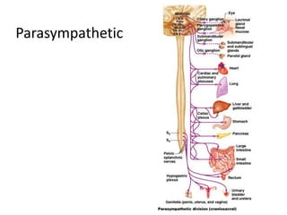
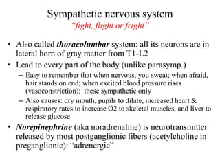
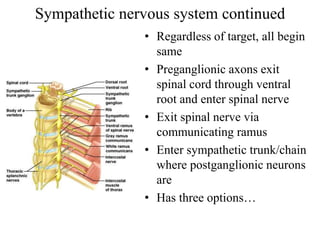
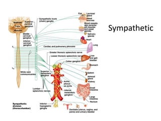
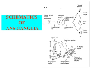
THE PERIPHERAL
NERVOUS
SYSTEM
Peripheral nervous system consists of:
• 31 pairs of spinal nerves
• 12 pairs of cranial nerves
• The autonomic nervous system
• Peripheral nervous system consists of
Sensory nerve fibres
Motor nerve fibres
Each nerve consists of
• Numerous nerve fibres collected into bundles
• Each bundle has coverings of protective
connective tissue :
1. Endoneurium - surrounds each individual fibre
2. Perineurium – surrounds each bundle
3. Epineurium – surrounds and covers a number of
bundles of nerve fibres
Nerve Roots
• Anterior nerve root consists of motor nerve
fibres from neurones in the anterior column
of grey matter
• Posterior nerve root consists of sensory
fibres
• Just outside the spinal cord there is a spinal
ganglion(posterior root ganglion) consisting
of nerve cells
• Sensory nerve fibres pass through these
ganglia before entering the spinal cord.
• The area of skin supplied by each nerve is
called a dermatome.
• For a short distance the roots have dura and
arachnoid
• No pia
• Anterior and posterior roots join to form mixed
spinal nerve
• Outside the intervertebral foramen the
spinal nerve divides into :-
1. Ramus communicans
2. a posterior ramus
3. an anterior ramus
Rami and Plexuses
• The ramus communicans is part of the autonomic nervous
system
• They join preganglionic sympthetic neurones
• The posterior ramus passes backwards to supply skin and
muscles
• Tha anterior rami supply the anterior and lateral aspects of
the neck, trunk and the upper and lower limbs.
• In the cervical, lumbar and sacral rgions the anterior rami
unite near their origins to form nerwork of nerves or
plexuses, where nerve fibres are regrouped and rearranged
before proceeding to supply skin, bones muscles, and
joints
The plexuses
• Five pairs of large plexuses
• Cervical plexus
• Brachial plexus
• Lumbar plexus
• Sacral plexus
• Coccygeal plexus
9
Cervical Plexus
• Formed by the anterior rami of the first four
cervical nerves
• It lies opposite the 1st , 2nd , 3rd and 4th
cervical vertebrae
• Supplies structures at the back and side of
the head and the skin of the front of the
neck
• Muscles of the neck
• Phrenic nerve from this nerve supplies
diaphragm
Brachial Plexus
• Formed by C5,6,7,8 and T1
• Situated above and behid the subclavian
vessels and in the axilla
• The plexus can be recognised to have five
parts – roots, trunks, divisions, cords and
nerves
• The nerves which arise from the plexus are :
musculocutaneous nerve, median
nerve, ulnar nerve and axillary nerve
Formed by the anterior rami of L1, 2.3 and 4
Situated in front of the transverse processes of the lumbar
vertebrae and behind the psoas muscle.
The main branches are : -
Iliohypogastric nerve
Ilioinguinal nerve
Genitofemoral nerve
Lateral cutaneous nerve of thigh
Femoral nerve
Obturator nerve
Lumbosacral trunk
Iliohypogastric, ilioinguinal nerves : supply muscles
and the skin in the area of the lower abdomen, upper
and medial aspects of the thigh and the inguinal region
Lateral cutaneous nerves of the thigh supplies the
lateral skin of thigh
Femoral nerve : extensor muscles of the thigh – and the
anterior; saphenous branch : medial skin of the leg,
ankle and foot
Obturator nerve : adductor muscles
Lumbosacral trunk : goes to sacral plexus
The Sacral Plexus
• Formed by by the anterior rami of the lumbosacral
trunk (L4,5) and sacral nerves (S1,2,3)
• Lies on the posterior wall of the pelvic cavity
• Branches supply the muscles and skin of the
pelvis, muscles around the hip joint
• It gives off sciatic nerve (L 4, 5 S1, 2, 3)
• Sciatic nerve : largest nerve in the body
passes through greater sciatic forameninto the
buttock then descends through the back of thigh
supplies the hamstring muscles
at the level of the middle of the femur divides into
two: -
the tibial nerve and the common peroneal nerve
14
• The tibial nerve descends through the popliteal
fossa
• Supplies the skin and muscles of the leg, sole of
the foot and toes
• Common peroneal nerve winds round the neck of
the fibula
• Divides into deep peroneal (anterior tibial) and
superficial peroneal nerves
• These two supply the skin and muscles of the leg
Pudendal nerve
Supplies the external anal sphincter, the external urethral
sphincter and adjacent skin
The Autonomic
Nervous System
Visceral sensory
Visceral motor
&
Autonomic nervous system
• The autonomic nervous system is the
subdivision of the peripheral nervous system
that regulates body activities that are generally
not under conscious control
• Visceral motor innervates non-skeletal (non-
somatic) muscles
• Visceral sensory
The autonomic nervous system, or ANS, is responsible for controlling
several body responses which are under the conscious level and which
are mostly involuntary, like breathing, digestion, sexual arousal, beating
of the heart, and many other biochemical processes.
There are two distinguishable divisions:
the parasympathetic and the sympathetic nervous systems.
ANS innervation is divided into sympathetic nervous system and
parasympathetic nervous system divisions. The sympathetic
division has thoracolumbar “outflow”, meaning that the neurons
begin at the thoracic and lumbar (T1-L2) portions of the spinal
cord. The parasympathetic division has craniosacral “outflow”,
meaning that the neurons begin at the cranial nerves (CN 3, CN7,
CN 9, CN10) and sacral (S2-S4) spinal cord.
The ANS is unique in that it requires a sequential two-neuron
efferent pathway; the preganglionic neuron must first synapse onto
a postganglionic neuron before innervating the target organ. The
preganglionic, or first, neuron will begin at the “outflow” and will
synapse at the postganglionic, or second, neuron’s cell body. The
post ganglionic neuron will then synapse at the target organ.
Divisions of the autonomic nervous system
• Parasympathetic division
• Sympathetic division
Serve most of the same organs but cause opposing or antagonistic effects
Parasysmpathetic: routine maintenance
The hypothalamus is the center of the parasympathetic nervous system. It operates
through various interconnecting systems and organs. This system is responsible in
the “thinking” phase in stressful situations and is called the “Rest and Digest”
phase because your body goes back to its normal responses.
“rest &digest”
Sympathetic: mobilization & increased metabolism
This type of the autonomic nervous system is responsible in the distribution of
essential nutrients and oxygen to the various parts of the body. The ANS is
responsible for “Flight or Fight.” The body has to respond immediately because of
the urgency of some situations. “fight, flight or fright” or “fight,
flight or freeze”
21
Where they come from
Parasympathetic:
craniosacral
Sympathetic:
thoracolumbar
Parasympathetic nervous system
“rest & digest”
• Also called the craniosacral system because
all its preganglionic neurons are in the brain
stem or sacral levels of the spinal cord
– Cranial nerves III,VII, IX and X
– In lateral horn of gray matter from S2-S4
• Only innervate internal organs (not skin)
• Acetylcholine is neurotransmitter at end organ
as well as at preganglionic synapse:
“cholinergic”
Cranial outflow
III - pupils constrict
VII - tears, nasal mucus, saliva
IX – parotid salivary gland
X (Vagus n) – visceral organs of thorax & abdomen:
Stimulates digestive glands
Increases motility of smooth muscle of digestive tract
Decreases heart rate
Causes bronchial constriction
Sacral outflow (S2-4): form pelvic splanchnic nerves
Supply 2nd half of large intestine
Supply all the pelvic (genitourinary) organs
24
Parasympathetic
Sympathetic nervous system
“fight, flight or fright”
• Also called thoracolumbar system: all its neurons are in
lateral horn of gray matter from T1-L2
• Lead to every part of the body (unlike parasymp.)
– Easy to remember that when nervous, you sweat; when afraid,
hair stands on end; when excited blood pressure rises
(vasoconstriction): these sympathetic only
– Also causes: dry mouth, pupils to dilate, increased heart &
respiratory rates to increase O2 to skeletal muscles, and liver to
release glucose
• Norepinephrine (aka noradrenaline) is neurotransmitter
released by most postganglionic fibers (acetylcholine in
preganglionic): “adrenergic”
Sympathetic nervous system continued
• Regardless of target, all begin
same
• Preganglionic axons exit
spinal cord through ventral
root and enter spinal nerve
• Exit spinal nerve via
communicating ramus
• Enter sympathetic trunk/chain
where postganglionic neurons
are
• Has three options…
Options of preganglionic axons in sympathetic trunk
1. Synapse on postganglionic neuron in chain ganglion
then return to spinal nerve and follow its branch to the
skin
2. Ascend or descend within sympathetic trunk, synapse
with a posganglionic neuron within a chain
ganglion, and return to spinal nerve at that level and
follow branches to skin
3. Enter sympathetic chain, pass through without
synapsing, form a splanchnic nerve that passes toward
thoracic or abdominal organs
– These synapse in prevertebral ganglion in front of
aorta
– Postganglionic axons follow arteries to organs
Sympathetic
Visceral sensory neurons
• Monitor temperature, pain, irritation, chemical changes and
stretch in the visceral organs
– Brain interprets as hunger, fullness, pain, nausea, well-being
• Receptors widely scattered – localization poor (e.g. which part
is giving you the gas pain?)
• Visceral sensory fibers run within autonomic nerves,
especially vagus and sympathetic nerves
– Sympathetic nerves carry most pain fibers from visceral organs of body
trunk
• Simplified pathway: sensory neurons to spinothalamic tract to
thalamus to cerebral cortex
• Visceral pain is induced by stretching, infection and cramping
of internal organs but seldom by cutting (e.g. cutting off a
colon polyp) or scraping them
Visceral sensory and autonomic neurons
participate in visceral reflex arcs
• Many are spinal reflexes such as defecation and
micturition
reflexes
• Some only
involve peripheral
neurons: spinal
cord not involved
(not shown)*
*e.g. “enteric” nervous system: 3 neuron reflex arcs entirely within the wall of the gut
Central control of the
Autonomic NS
Amygdala: main limbic
region for emotions
-Stimulates sympathetic
activity, especially previously
learned fear-related behavior
-Can be voluntary when decide to
recall frightful experience -
cerebral cortex acts through
amygdala
-Some people can regulate some
autonomic activities by gaining
extraordinary control over their
emotions
Hypothalamus: main
integration center
Reticular formation: most
direct influence over
autonomic function
SCHEMATICS
OF
ANS GANGLIA
• Ganglia are located close to the spinal
column (short pre- and long post-ganglionic
fibers)
• Ganglia are often interconnected
• Large ratio of post- to pre-ganglionic fiber
number
Sympathetic Nervous system
• The anatomy of sympathetic ans is structurally
designed to produce diffuse systemic responses by
simultaneous targetting of multiple tissues
• In addition to the routine homeostatic maintenance
of vital functions, the sympathetic nervous sytem is
designed to respond to physiological stress
PARASYMPATHETIC NERVOUS SYSTEM
• Ganglia are located close to or within the target
tissue (long pre- and short post-ganglionic
fibers)
• Ganglia are not interconnected
• Ratio of post- to pre-ganglionic fiber number =
1
PARASYMPATHETIC NERVOUS SYSTEM
• The anatomy of ans is structurally designed to
produce precise pin-point responses by targetting of
specific tissues
• In addition to the routine homeostatic maintenance of
vital functions, the parasympathetic nervous sytem is
designed to limit responses to physiological stress

Ans changed

  • 1.
  • 2.
    Peripheral nervous systemconsists of: • 31 pairs of spinal nerves • 12 pairs of cranial nerves • The autonomic nervous system • Peripheral nervous system consists of Sensory nerve fibres Motor nerve fibres
  • 3.
    Each nerve consistsof • Numerous nerve fibres collected into bundles • Each bundle has coverings of protective connective tissue : 1. Endoneurium - surrounds each individual fibre 2. Perineurium – surrounds each bundle 3. Epineurium – surrounds and covers a number of bundles of nerve fibres
  • 5.
    Nerve Roots • Anteriornerve root consists of motor nerve fibres from neurones in the anterior column of grey matter • Posterior nerve root consists of sensory fibres • Just outside the spinal cord there is a spinal ganglion(posterior root ganglion) consisting of nerve cells • Sensory nerve fibres pass through these ganglia before entering the spinal cord. • The area of skin supplied by each nerve is called a dermatome.
  • 6.
    • For ashort distance the roots have dura and arachnoid • No pia • Anterior and posterior roots join to form mixed spinal nerve • Outside the intervertebral foramen the spinal nerve divides into :- 1. Ramus communicans 2. a posterior ramus 3. an anterior ramus
  • 7.
    Rami and Plexuses •The ramus communicans is part of the autonomic nervous system • They join preganglionic sympthetic neurones • The posterior ramus passes backwards to supply skin and muscles • Tha anterior rami supply the anterior and lateral aspects of the neck, trunk and the upper and lower limbs. • In the cervical, lumbar and sacral rgions the anterior rami unite near their origins to form nerwork of nerves or plexuses, where nerve fibres are regrouped and rearranged before proceeding to supply skin, bones muscles, and joints
  • 8.
    The plexuses • Fivepairs of large plexuses • Cervical plexus • Brachial plexus • Lumbar plexus • Sacral plexus • Coccygeal plexus
  • 9.
    9 Cervical Plexus • Formedby the anterior rami of the first four cervical nerves • It lies opposite the 1st , 2nd , 3rd and 4th cervical vertebrae • Supplies structures at the back and side of the head and the skin of the front of the neck • Muscles of the neck • Phrenic nerve from this nerve supplies diaphragm
  • 10.
    Brachial Plexus • Formedby C5,6,7,8 and T1 • Situated above and behid the subclavian vessels and in the axilla • The plexus can be recognised to have five parts – roots, trunks, divisions, cords and nerves • The nerves which arise from the plexus are : musculocutaneous nerve, median nerve, ulnar nerve and axillary nerve
  • 11.
    Formed by theanterior rami of L1, 2.3 and 4 Situated in front of the transverse processes of the lumbar vertebrae and behind the psoas muscle. The main branches are : - Iliohypogastric nerve Ilioinguinal nerve Genitofemoral nerve Lateral cutaneous nerve of thigh Femoral nerve Obturator nerve Lumbosacral trunk
  • 12.
    Iliohypogastric, ilioinguinal nerves: supply muscles and the skin in the area of the lower abdomen, upper and medial aspects of the thigh and the inguinal region Lateral cutaneous nerves of the thigh supplies the lateral skin of thigh Femoral nerve : extensor muscles of the thigh – and the anterior; saphenous branch : medial skin of the leg, ankle and foot Obturator nerve : adductor muscles Lumbosacral trunk : goes to sacral plexus
  • 13.
    The Sacral Plexus •Formed by by the anterior rami of the lumbosacral trunk (L4,5) and sacral nerves (S1,2,3) • Lies on the posterior wall of the pelvic cavity • Branches supply the muscles and skin of the pelvis, muscles around the hip joint • It gives off sciatic nerve (L 4, 5 S1, 2, 3) • Sciatic nerve : largest nerve in the body passes through greater sciatic forameninto the buttock then descends through the back of thigh supplies the hamstring muscles at the level of the middle of the femur divides into two: - the tibial nerve and the common peroneal nerve
  • 14.
    14 • The tibialnerve descends through the popliteal fossa • Supplies the skin and muscles of the leg, sole of the foot and toes • Common peroneal nerve winds round the neck of the fibula • Divides into deep peroneal (anterior tibial) and superficial peroneal nerves • These two supply the skin and muscles of the leg Pudendal nerve Supplies the external anal sphincter, the external urethral sphincter and adjacent skin
  • 15.
    The Autonomic Nervous System Visceralsensory Visceral motor &
  • 16.
    Autonomic nervous system •The autonomic nervous system is the subdivision of the peripheral nervous system that regulates body activities that are generally not under conscious control • Visceral motor innervates non-skeletal (non- somatic) muscles • Visceral sensory
  • 17.
    The autonomic nervoussystem, or ANS, is responsible for controlling several body responses which are under the conscious level and which are mostly involuntary, like breathing, digestion, sexual arousal, beating of the heart, and many other biochemical processes. There are two distinguishable divisions: the parasympathetic and the sympathetic nervous systems.
  • 18.
    ANS innervation isdivided into sympathetic nervous system and parasympathetic nervous system divisions. The sympathetic division has thoracolumbar “outflow”, meaning that the neurons begin at the thoracic and lumbar (T1-L2) portions of the spinal cord. The parasympathetic division has craniosacral “outflow”, meaning that the neurons begin at the cranial nerves (CN 3, CN7, CN 9, CN10) and sacral (S2-S4) spinal cord. The ANS is unique in that it requires a sequential two-neuron efferent pathway; the preganglionic neuron must first synapse onto a postganglionic neuron before innervating the target organ. The preganglionic, or first, neuron will begin at the “outflow” and will synapse at the postganglionic, or second, neuron’s cell body. The post ganglionic neuron will then synapse at the target organ.
  • 19.
    Divisions of theautonomic nervous system • Parasympathetic division • Sympathetic division Serve most of the same organs but cause opposing or antagonistic effects Parasysmpathetic: routine maintenance The hypothalamus is the center of the parasympathetic nervous system. It operates through various interconnecting systems and organs. This system is responsible in the “thinking” phase in stressful situations and is called the “Rest and Digest” phase because your body goes back to its normal responses. “rest &digest” Sympathetic: mobilization & increased metabolism This type of the autonomic nervous system is responsible in the distribution of essential nutrients and oxygen to the various parts of the body. The ANS is responsible for “Flight or Fight.” The body has to respond immediately because of the urgency of some situations. “fight, flight or fright” or “fight, flight or freeze”
  • 21.
    21 Where they comefrom Parasympathetic: craniosacral Sympathetic: thoracolumbar
  • 22.
    Parasympathetic nervous system “rest& digest” • Also called the craniosacral system because all its preganglionic neurons are in the brain stem or sacral levels of the spinal cord – Cranial nerves III,VII, IX and X – In lateral horn of gray matter from S2-S4 • Only innervate internal organs (not skin) • Acetylcholine is neurotransmitter at end organ as well as at preganglionic synapse: “cholinergic”
  • 23.
    Cranial outflow III -pupils constrict VII - tears, nasal mucus, saliva IX – parotid salivary gland X (Vagus n) – visceral organs of thorax & abdomen: Stimulates digestive glands Increases motility of smooth muscle of digestive tract Decreases heart rate Causes bronchial constriction Sacral outflow (S2-4): form pelvic splanchnic nerves Supply 2nd half of large intestine Supply all the pelvic (genitourinary) organs
  • 24.
  • 25.
    Sympathetic nervous system “fight,flight or fright” • Also called thoracolumbar system: all its neurons are in lateral horn of gray matter from T1-L2 • Lead to every part of the body (unlike parasymp.) – Easy to remember that when nervous, you sweat; when afraid, hair stands on end; when excited blood pressure rises (vasoconstriction): these sympathetic only – Also causes: dry mouth, pupils to dilate, increased heart & respiratory rates to increase O2 to skeletal muscles, and liver to release glucose • Norepinephrine (aka noradrenaline) is neurotransmitter released by most postganglionic fibers (acetylcholine in preganglionic): “adrenergic”
  • 26.
    Sympathetic nervous systemcontinued • Regardless of target, all begin same • Preganglionic axons exit spinal cord through ventral root and enter spinal nerve • Exit spinal nerve via communicating ramus • Enter sympathetic trunk/chain where postganglionic neurons are • Has three options…
  • 27.
    Options of preganglionicaxons in sympathetic trunk 1. Synapse on postganglionic neuron in chain ganglion then return to spinal nerve and follow its branch to the skin 2. Ascend or descend within sympathetic trunk, synapse with a posganglionic neuron within a chain ganglion, and return to spinal nerve at that level and follow branches to skin 3. Enter sympathetic chain, pass through without synapsing, form a splanchnic nerve that passes toward thoracic or abdominal organs – These synapse in prevertebral ganglion in front of aorta – Postganglionic axons follow arteries to organs
  • 28.
  • 29.
    Visceral sensory neurons •Monitor temperature, pain, irritation, chemical changes and stretch in the visceral organs – Brain interprets as hunger, fullness, pain, nausea, well-being • Receptors widely scattered – localization poor (e.g. which part is giving you the gas pain?) • Visceral sensory fibers run within autonomic nerves, especially vagus and sympathetic nerves – Sympathetic nerves carry most pain fibers from visceral organs of body trunk • Simplified pathway: sensory neurons to spinothalamic tract to thalamus to cerebral cortex • Visceral pain is induced by stretching, infection and cramping of internal organs but seldom by cutting (e.g. cutting off a colon polyp) or scraping them
  • 30.
    Visceral sensory andautonomic neurons participate in visceral reflex arcs • Many are spinal reflexes such as defecation and micturition reflexes • Some only involve peripheral neurons: spinal cord not involved (not shown)* *e.g. “enteric” nervous system: 3 neuron reflex arcs entirely within the wall of the gut
  • 31.
    Central control ofthe Autonomic NS Amygdala: main limbic region for emotions -Stimulates sympathetic activity, especially previously learned fear-related behavior -Can be voluntary when decide to recall frightful experience - cerebral cortex acts through amygdala -Some people can regulate some autonomic activities by gaining extraordinary control over their emotions Hypothalamus: main integration center Reticular formation: most direct influence over autonomic function
  • 32.
  • 33.
    • Ganglia arelocated close to the spinal column (short pre- and long post-ganglionic fibers) • Ganglia are often interconnected • Large ratio of post- to pre-ganglionic fiber number Sympathetic Nervous system
  • 34.
    • The anatomyof sympathetic ans is structurally designed to produce diffuse systemic responses by simultaneous targetting of multiple tissues • In addition to the routine homeostatic maintenance of vital functions, the sympathetic nervous sytem is designed to respond to physiological stress
  • 35.
    PARASYMPATHETIC NERVOUS SYSTEM •Ganglia are located close to or within the target tissue (long pre- and short post-ganglionic fibers) • Ganglia are not interconnected • Ratio of post- to pre-ganglionic fiber number = 1
  • 36.
    PARASYMPATHETIC NERVOUS SYSTEM •The anatomy of ans is structurally designed to produce precise pin-point responses by targetting of specific tissues • In addition to the routine homeostatic maintenance of vital functions, the parasympathetic nervous sytem is designed to limit responses to physiological stress