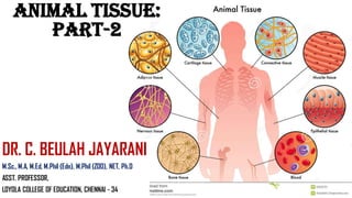
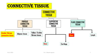
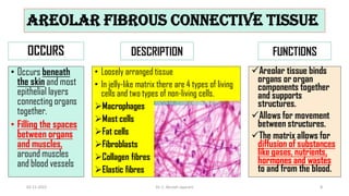
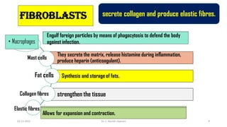
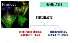
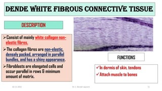
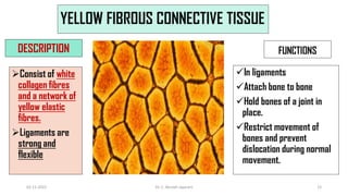
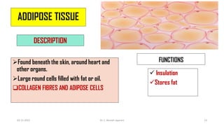
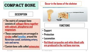
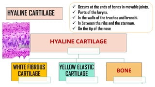
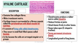
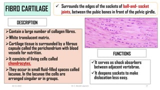
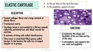
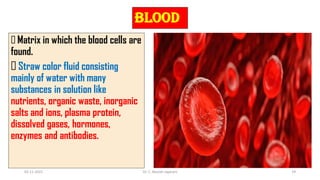
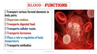
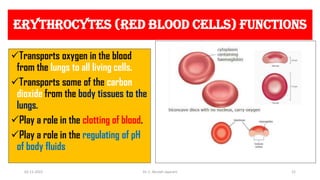
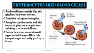
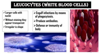
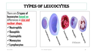
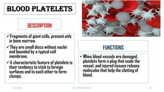
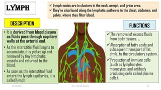
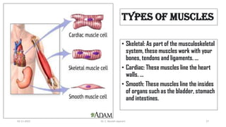
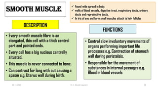
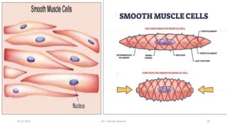
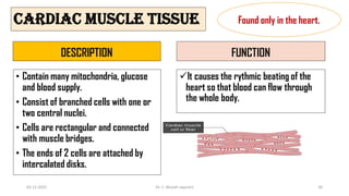
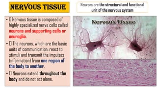
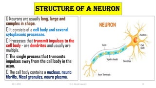
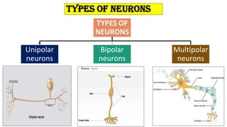
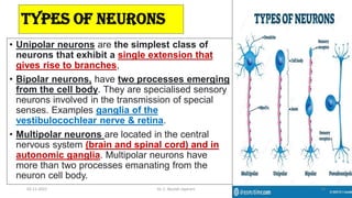
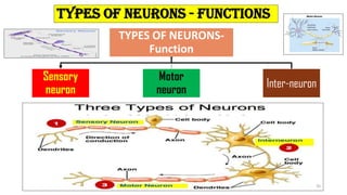
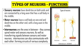
ANIMAL TISSUE:
PART-2
DR. C. BEULAH JAYARANI
M.Sc., M.A, M.Ed, M.Phil (Edn), M.Phil (ZOO), NET, Ph.D
ASST. PROFESSOR,
LOYOLA COLLEGE OF EDUCATION, CHENNAI - 34
What is Tissue?
02-11-2022 Dr. C. Beulah Jayarani 2
An assemblage of one or more
types of specialized cells held
together with extracellular material
constitute the tissue.
What is Animal tissues?
The animal cells are grouped
together to form animal tissues.
These tissues vary in their
structure, function, and origin.
02-11-2022 Dr. C. Beulah Jayarani 3
ANIMAL TISSUE
SIMPLE TISSUE COMPOUND TISSUE
TYPES OF ANIMAL TISSUE
A group of cells that are similar
in origin, form, structure and
work together to perform a
specific function is called a
simple tissue
A group of cells different in
their structure and function
but co-ordinate to perform a
specific function is called a
compound tissue.
02-11-2022 Dr. C. Beulah Jayarani 4
Animal tissues (structure and functions)
02-11-2022 Dr. C. Beulah Jayarani 5
ANIMAL TISSUES
(structure and
functions)
a. Epithelial
tissue
b. Connective
tissue
c. Muscular
tissue
d. Nervous
tissue
All these tissues originate in the embryonic stages from the three
primary germ layers namely ectoderm, mesoderm and endoderm.
6
CONNECTIVE TISSUE
CONNECTIVE
TISSUE
FIBROUS
CONNECTIVE
TISSUE
SUPPORTIVE
CONNECTIVE
TISSUE
FLUID CONNECTIVE
TISSUE
Areolar fibrous
connective tissue
Adipose tissue
Yellow & white
fibrous tissue
Cartilage
Bone
Lymph
Blood
02-11-2022 Dr. C. Beulah Jayarani 7
AREOLAR FIBROUS CONNECTIVE TISSUE
OCCURS
• Occurs beneath
the skin and most
epithelial layers
connecting organs
together.
• Filling the spaces
between organs
and muscles,
around muscles
and blood vessels
DESCRIPTION
• Loosely arranged tissue
• In jelly-like matrix there are 4 types of living
cells and two types of non-living cells.
Macrophages
Mast cells
Fat cells
Fibroblasts
Collagen fibres
Elastic fibres
FUNCTIONS
Areolar tissue binds
organs or organ
components together
and supports
structures.
Allows for movement
between structures.
The matrix allows for
diffusion of substances
like gases, nutrients,
hormones and wastes
to and from the blood.
02-11-2022 Dr. C. Beulah Jayarani 8
Fibroblasts
They secrete the matrix, release histamine during inflammation,
produce heparin (anticoagulant).
Synthesis and storage of fats.
strengthen the tissue
secrete collagen and produce elastic fibres.
Engulf foreign particles by means of phagocytosis to defend the body
against infection.
Mast cells
Fat cells
Collagen fibres
Elastic fibres
Allows for expansion and contraction.
02-11-2022 Dr. C. Beulah Jayarani 9
FIBROBLASTS
FIBROBLASTS
DENDE WHITE FIBROUS
CONNECTIVE TISSUE
YELLOW FIBROUS
CONNECTIVE TISSUE
02-11-2022 Dr. C. Beulah Jayarani 10
DENDE WHITE FIBROUS CONNECTIVE TISSUE
DESCRIPTION
Consist of mainly white collagen non-
elastic fibres.
The collagen fibres are non-elastic,
densely packed, arranged in parallel
bundles, and has a shiny appearance.
Fibroblasts are elongated cells and
occur parallel in rows & minimum
amount of matrix.
FUNCTIONS
In dermis of skin, tendons
Attach muscle to bones
02-11-2022 Dr. C. Beulah Jayarani 11
YELLOW FIBROUS CONNECTIVE TISSUE
DESCRIPTION
Consist of white
collagen fibres
and a network of
yellow elastic
fibres.
Ligaments are
strong and
flexible
FUNCTIONS
In ligaments
Attach bone to bone
Hold bones of a joint in
place.
Restrict movement of
bones and prevent
dislocation during normal
movement.
02-11-2022 Dr. C. Beulah Jayarani 12
ADDIPOSE TISSUE
DESCRIPTION
Found beneath the skin, around heart and
other organs.
Large round cells filled with fat or oil.
COLLAGEN FIBRES AND ADIPOSE CELLS
FUNCTIONS
 Insulation
Stores fat
02-11-2022 Dr. C. Beulah Jayarani 13
COMPACT BONE
DESCRIPTION
The matrix of compact bone
consists of collagen fibres together
with calcium, phosphorus and
magnesium.
These components are arranged in
circles called lamellae, around the
Haversian canal, containing artery,
vein and nerve.
Contain bone cells called osteocytes
FUNCTIONS
• Support
• Protection
• Locomotion
• Red blood corpuscles and white blood cells
are produced in the red bone marrow.
Occur in the bones of the skeleton
02-11-2022 Dr. C. Beulah Jayarani 14
HYALINE CARTILAGE
 Occurs at the ends of bones in movable joints.
 Parts of the larynx.
 In the walls of the trachea and bronchi.
 In between the ribs and the sternum.
 On the tip of the nose
HYALINE CARTILAGE
WHITE FIBROUS
CARTILAGE
YELLOW ELASTIC
CARTILAGE
BONE
15
HYALINE CARTILAGE
DESCRIPTION
Contains fine collagen fibres.
White translucent matrix.
Cartilage tissue is surrounded by a fibrous capsule
called the perichondrium with blood vessels for
nutrition.
It consists of living cells called chondrocytes.
They occur in small fluid-filled spaces called
lacunae.
In the lacunae the cells are arranged singular or in
groups.
FUNCTIONS
Chondrocytes produce a rubber
matrix called chondrin.
Reduce friction at joints
Attach bones firmly to other bones.
Keeps tubes open (C-shaped in
trachea).
Forms permanent structures
(nose).
Longitudinal growth of long bones.
02-11-2022 Dr. C. Beulah Jayarani 16
FIBRO CARTILAGE
DESCRIPTION
Contain a large number of collagen fibres.
White translucent matrix.
Cartilage tissue is surrounded by a fibrous
capsule called the perichondrium with blood
vessels for nutrition.
It consists of living cells called
chondrocytes.
They occur in small fluid-filled spaces called
lacunae. In the lacunae the cells are
arranged singular or in groups.
FUNCTIONS
It serves as shock absorbers
between adjacent vertebrae.
It deepens sockets to make
dislocation less easy.
 Surrounds the edges of the sockets of ball-and- socket
joints, between the pubic bones in front of the pelvic girdle.
02-11-2022 Dr. C. Beulah Jayarani 17
ELASTIC CARTILAGE
DESCRIPTION
Contain collagen fibres and a large network of
elastic fibres.
Translucent matrix.
Cartilage tissue is surrounded by a fibrous capsule
called the perichondrium with blood vessels for
nutrition.
It consists of living cells called chondrocytes.
They occur in small fluid-filled spaces called
lacunae. In the lacunae the cells are arranged
singular or in groups.
FUNCTIONS
• It maintains the shape and
flexibility of the ear lobe and tip
of the nose.
• It strengthens and supports the
ear, nose and epiglottis.
 In the ear lobe, at the tip of the nose
 In the epiglottis, septum of nose
02-11-2022 Dr. C. Beulah Jayarani 18
BLOOD
Matrix in which the blood cells are
found.
Straw color fluid consisting
mainly of water with many
substances in solution like
nutrients, organic waste, inorganic
salts and ions, plasma protein,
dissolved gases, hormones,
enzymes and antibodies.
02-11-2022 Dr. C. Beulah Jayarani 19
BLOOD - FUNCTIONS
Transport various formed elements to
body parts.
Dispersion medium.
Transports digested food.
Transports cellular waste.
Transports hormones.
Plays a role in regulation of body
temperature.
Transports antibodies
02-11-2022 Dr. C. Beulah Jayarani 20
ERYTHROCYTES (RED BLOOD CELLS) FUNCTIONS
Transports oxygen in the blood
from the lungs to all living cells.
Transports some of the carbon
dioxide from the body tissues to the
lungs.
Play a role in the clotting of blood.
Play a role in the regulating of pH
of body fluids
02-11-2022 Dr. C. Beulah Jayarani 21
ERYTHROCYTES (RED BLOOD CELLS)
Small round biconcave disks filled with
cytoplasm, but without a nucleus.
Contain the red pigment hemoglobin.
Hemoglobin contains 4 units, each with
the protein globin and a complex iron-
containing, structure called heme.
The iron form a loose association with
oxygen and in this way red blood cells
transport oxygen and readily give it up to
tissues.
02-11-2022 Dr. C. Beulah Jayarani 22
LEUCOCYTES (WHITE BLOOD CELLS)
Engulf infections by means
of phagocytosis.
Produce antibodies.
Defense or immunity of
body
Larger cells with
nuclei
Without staining they
appear transparent
Irregular is shape
02-11-2022 Dr. C. Beulah Jayarani 23
types of leucocytes
There are 5 types of
leucocytes based on
differences in size and
nuclear shape.
Neutrophils
Basophils
Eosinophils
Monocytes
Lymphocytes
02-11-2022 Dr. C. Beulah Jayarani 24
BLOOD PLATELETS
DESCRIPTION
Fragments of giant cells, present only
in bone marrow.
They are small discs without nuclei
and bounded by a typical cell
membrane.
A characteristic feature of platelets is
their tendency to stick to foreign
surfaces and to each other to form
clumps.
FUNCTIONS
• When blood vessels are damaged,
platelets form a plug that seals the
vessel, and injured tissues release
molecules that help the clotting of
blood.
02-11-2022 Dr. C. Beulah Jayarani 25
LYMPH
DESCRIPTION
• It is derived from blood plasma
as fluids pass through capillary
walls at the arterial end.
• As the interstitial fluid begins to
accumulate, it is picked up and
removed by tiny lymphatic
vessels and returned to the
blood.
• As soon as the interstitial fluid
enters the lymph capillaries, it is
called lymph.
FUNCTIONS
The removal of excess fluids
from body tissues. ...
Absorption of fatty acids and
subsequent transport of fat,
chyle, to the circulatory system.
Production of immune cells
(such as lymphocytes,
monocytes, and antibody
producing cells called plasma
cells).
 Lymph nodes are in clusters in the neck, armpit, and groin area.
 They're also found along the lymphatic pathways in the chest, abdomen, and
pelvis, where they filter blood.
02-11-2022 Dr. C. Beulah Jayarani 26
types of muscles
• Skeletal: As part of the musculoskeletal
system, these muscles work with your
bones, tendons and ligaments. ...
• Cardiac: These muscles line the heart
walls. ...
• Smooth: These muscles line the insides
of organs such as the bladder, stomach
and intestines.
02-11-2022 Dr. C. Beulah Jayarani 27
SMOOTH MUSCLE
DESCRIPTION
• Every smooth muscle fibre is an
elongated, thin cell with a thick central
part and pointed ends.
• Every cell has a big nucleus centrally
situated.
• This muscle is never connected to bone.
• Can contract for long with out causing a
spasm e.g. Uterus wall during birth.
FUNCTIONS
• Control slow involuntary movements of
organs performing important life
processes e.g. Contraction of stomach
wall during peristalsis.
• Responsible for the movement of
substances in internal passages e.g.
Blood in blood vessels
 Found wide spread in body.
 walls of blood vessels, digestive tract, respiratory ducts, urinary
ducts and reproductive ducts.
 In iris of eye and form small muscles attach to hair follicles
02-11-2022 Dr. C. Beulah Jayarani 28
02-11-2022 Dr. C. Beulah Jayarani 29
CARDIAC MUSCLE TISSUE
DESCRIPTION
• Contain many mitochondria, glucose
and blood supply.
• Consist of branched cells with one or
two central nuclei.
• Cells are rectangular and connected
with muscle bridges.
• The ends of 2 cells are attached by
intercalated disks.
FUNCTION
It causes the rythmic beating of the
heart so that blood can flow through
the whole body.
Found only in the heart.
02-11-2022 Dr. C. Beulah Jayarani 30
NERVOUS TISSUE
• Nervous tissue is composed of
highly specialized nerve cells called
neurons and supporting cells or
neuroglia.
• The neurons, which are the basic
units of communication, react to
stimuli and transmit the impulses
(information) from one region of
the body to another.
• Neurons extend throughout the
body and do not act alone.
Neurons are the structural and functional
unit of the nervous system
31
STRUCTURE OF A NEURON
Neurons are usually long, large and
complex in shape.
It consists of a cell body and several
cytoplasmic processes.
Processes that transmit impulses to the
cell body - are dendrites and usually are
multiple.
The single process that transmits
impulses away from the cell body in the
axon.
The cell body contains a nucleus, neuro
fibrills, Nissl granules, neuro plasma.
02-11-2022 Dr. C. Beulah Jayarani 32
types of neurons
TYPES OF
NEURONS
Unipolar
neurons
Bipolar
neurons
Multipolar
neurons
33
• Unipolar neurons are the simplest class of
neurons that exhibit a single extension that
gives rise to branches,
• Bipolar neurons, have two processes emerging
from the cell body. They are specialised sensory
neurons involved in the transmission of special
senses. Examples ganglia of the
vestibulocochlear nerve & retina.
• Multipolar neurons are located in the central
nervous system (brain and spinal cord) and in
autonomic ganglia. Multipolar neurons have
more than two processes emanating from the
neuron cell body.
types of neurons
02-11-2022 Dr. C. Beulah Jayarani 34
types of neurons - functions
TYPES OF NEURONS-
Function
Sensory
neuron
Motor
neuron
Inter-neuron
35
• Sensory neurons have dendrites on both ends and
are connected by a long axon that has a cell body in
the middle.
• Motor neurons have a cell body on one end and
dendrites on the other end, with a long axon in the
middle.
• Interneurons are the ones in between - they connect
spinal motor and sensory neurons. As well as
transferring signals between sensory and motor
neurons, interneurons can also communicate with
each other, forming circuits of various complexity.
types of neurons - functions
36
REFERENCES
“SCIENCE READER”
Tamil Nadu text books
NCERT Books
Nabor, Murray W.,
Introduction to botany
Google Images
02-11-2022 Dr. C. Beulah Jayarani 37

ANIMAL TISSUE - 2 (1).pdf

  • 1.
    ANIMAL TISSUE: PART-2 DR. C.BEULAH JAYARANI M.Sc., M.A, M.Ed, M.Phil (Edn), M.Phil (ZOO), NET, Ph.D ASST. PROFESSOR, LOYOLA COLLEGE OF EDUCATION, CHENNAI - 34
  • 2.
    What is Tissue? 02-11-2022Dr. C. Beulah Jayarani 2 An assemblage of one or more types of specialized cells held together with extracellular material constitute the tissue.
  • 3.
    What is Animaltissues? The animal cells are grouped together to form animal tissues. These tissues vary in their structure, function, and origin. 02-11-2022 Dr. C. Beulah Jayarani 3
  • 4.
    ANIMAL TISSUE SIMPLE TISSUECOMPOUND TISSUE TYPES OF ANIMAL TISSUE A group of cells that are similar in origin, form, structure and work together to perform a specific function is called a simple tissue A group of cells different in their structure and function but co-ordinate to perform a specific function is called a compound tissue. 02-11-2022 Dr. C. Beulah Jayarani 4
  • 5.
    Animal tissues (structureand functions) 02-11-2022 Dr. C. Beulah Jayarani 5 ANIMAL TISSUES (structure and functions) a. Epithelial tissue b. Connective tissue c. Muscular tissue d. Nervous tissue All these tissues originate in the embryonic stages from the three primary germ layers namely ectoderm, mesoderm and endoderm.
  • 6.
  • 7.
    CONNECTIVE TISSUE CONNECTIVE TISSUE FIBROUS CONNECTIVE TISSUE SUPPORTIVE CONNECTIVE TISSUE FLUID CONNECTIVE TISSUE Areolarfibrous connective tissue Adipose tissue Yellow & white fibrous tissue Cartilage Bone Lymph Blood 02-11-2022 Dr. C. Beulah Jayarani 7
  • 8.
    AREOLAR FIBROUS CONNECTIVETISSUE OCCURS • Occurs beneath the skin and most epithelial layers connecting organs together. • Filling the spaces between organs and muscles, around muscles and blood vessels DESCRIPTION • Loosely arranged tissue • In jelly-like matrix there are 4 types of living cells and two types of non-living cells. Macrophages Mast cells Fat cells Fibroblasts Collagen fibres Elastic fibres FUNCTIONS Areolar tissue binds organs or organ components together and supports structures. Allows for movement between structures. The matrix allows for diffusion of substances like gases, nutrients, hormones and wastes to and from the blood. 02-11-2022 Dr. C. Beulah Jayarani 8
  • 9.
    Fibroblasts They secrete thematrix, release histamine during inflammation, produce heparin (anticoagulant). Synthesis and storage of fats. strengthen the tissue secrete collagen and produce elastic fibres. Engulf foreign particles by means of phagocytosis to defend the body against infection. Mast cells Fat cells Collagen fibres Elastic fibres Allows for expansion and contraction. 02-11-2022 Dr. C. Beulah Jayarani 9
  • 10.
    FIBROBLASTS FIBROBLASTS DENDE WHITE FIBROUS CONNECTIVETISSUE YELLOW FIBROUS CONNECTIVE TISSUE 02-11-2022 Dr. C. Beulah Jayarani 10
  • 11.
    DENDE WHITE FIBROUSCONNECTIVE TISSUE DESCRIPTION Consist of mainly white collagen non- elastic fibres. The collagen fibres are non-elastic, densely packed, arranged in parallel bundles, and has a shiny appearance. Fibroblasts are elongated cells and occur parallel in rows & minimum amount of matrix. FUNCTIONS In dermis of skin, tendons Attach muscle to bones 02-11-2022 Dr. C. Beulah Jayarani 11
  • 12.
    YELLOW FIBROUS CONNECTIVETISSUE DESCRIPTION Consist of white collagen fibres and a network of yellow elastic fibres. Ligaments are strong and flexible FUNCTIONS In ligaments Attach bone to bone Hold bones of a joint in place. Restrict movement of bones and prevent dislocation during normal movement. 02-11-2022 Dr. C. Beulah Jayarani 12
  • 13.
    ADDIPOSE TISSUE DESCRIPTION Found beneaththe skin, around heart and other organs. Large round cells filled with fat or oil. COLLAGEN FIBRES AND ADIPOSE CELLS FUNCTIONS  Insulation Stores fat 02-11-2022 Dr. C. Beulah Jayarani 13
  • 14.
    COMPACT BONE DESCRIPTION The matrixof compact bone consists of collagen fibres together with calcium, phosphorus and magnesium. These components are arranged in circles called lamellae, around the Haversian canal, containing artery, vein and nerve. Contain bone cells called osteocytes FUNCTIONS • Support • Protection • Locomotion • Red blood corpuscles and white blood cells are produced in the red bone marrow. Occur in the bones of the skeleton 02-11-2022 Dr. C. Beulah Jayarani 14
  • 15.
    HYALINE CARTILAGE  Occursat the ends of bones in movable joints.  Parts of the larynx.  In the walls of the trachea and bronchi.  In between the ribs and the sternum.  On the tip of the nose HYALINE CARTILAGE WHITE FIBROUS CARTILAGE YELLOW ELASTIC CARTILAGE BONE 15
  • 16.
    HYALINE CARTILAGE DESCRIPTION Contains finecollagen fibres. White translucent matrix. Cartilage tissue is surrounded by a fibrous capsule called the perichondrium with blood vessels for nutrition. It consists of living cells called chondrocytes. They occur in small fluid-filled spaces called lacunae. In the lacunae the cells are arranged singular or in groups. FUNCTIONS Chondrocytes produce a rubber matrix called chondrin. Reduce friction at joints Attach bones firmly to other bones. Keeps tubes open (C-shaped in trachea). Forms permanent structures (nose). Longitudinal growth of long bones. 02-11-2022 Dr. C. Beulah Jayarani 16
  • 17.
    FIBRO CARTILAGE DESCRIPTION Contain alarge number of collagen fibres. White translucent matrix. Cartilage tissue is surrounded by a fibrous capsule called the perichondrium with blood vessels for nutrition. It consists of living cells called chondrocytes. They occur in small fluid-filled spaces called lacunae. In the lacunae the cells are arranged singular or in groups. FUNCTIONS It serves as shock absorbers between adjacent vertebrae. It deepens sockets to make dislocation less easy.  Surrounds the edges of the sockets of ball-and- socket joints, between the pubic bones in front of the pelvic girdle. 02-11-2022 Dr. C. Beulah Jayarani 17
  • 18.
    ELASTIC CARTILAGE DESCRIPTION Contain collagenfibres and a large network of elastic fibres. Translucent matrix. Cartilage tissue is surrounded by a fibrous capsule called the perichondrium with blood vessels for nutrition. It consists of living cells called chondrocytes. They occur in small fluid-filled spaces called lacunae. In the lacunae the cells are arranged singular or in groups. FUNCTIONS • It maintains the shape and flexibility of the ear lobe and tip of the nose. • It strengthens and supports the ear, nose and epiglottis.  In the ear lobe, at the tip of the nose  In the epiglottis, septum of nose 02-11-2022 Dr. C. Beulah Jayarani 18
  • 19.
    BLOOD Matrix in whichthe blood cells are found. Straw color fluid consisting mainly of water with many substances in solution like nutrients, organic waste, inorganic salts and ions, plasma protein, dissolved gases, hormones, enzymes and antibodies. 02-11-2022 Dr. C. Beulah Jayarani 19
  • 20.
    BLOOD - FUNCTIONS Transportvarious formed elements to body parts. Dispersion medium. Transports digested food. Transports cellular waste. Transports hormones. Plays a role in regulation of body temperature. Transports antibodies 02-11-2022 Dr. C. Beulah Jayarani 20
  • 21.
    ERYTHROCYTES (RED BLOODCELLS) FUNCTIONS Transports oxygen in the blood from the lungs to all living cells. Transports some of the carbon dioxide from the body tissues to the lungs. Play a role in the clotting of blood. Play a role in the regulating of pH of body fluids 02-11-2022 Dr. C. Beulah Jayarani 21
  • 22.
    ERYTHROCYTES (RED BLOODCELLS) Small round biconcave disks filled with cytoplasm, but without a nucleus. Contain the red pigment hemoglobin. Hemoglobin contains 4 units, each with the protein globin and a complex iron- containing, structure called heme. The iron form a loose association with oxygen and in this way red blood cells transport oxygen and readily give it up to tissues. 02-11-2022 Dr. C. Beulah Jayarani 22
  • 23.
    LEUCOCYTES (WHITE BLOODCELLS) Engulf infections by means of phagocytosis. Produce antibodies. Defense or immunity of body Larger cells with nuclei Without staining they appear transparent Irregular is shape 02-11-2022 Dr. C. Beulah Jayarani 23
  • 24.
    types of leucocytes Thereare 5 types of leucocytes based on differences in size and nuclear shape. Neutrophils Basophils Eosinophils Monocytes Lymphocytes 02-11-2022 Dr. C. Beulah Jayarani 24
  • 25.
    BLOOD PLATELETS DESCRIPTION Fragments ofgiant cells, present only in bone marrow. They are small discs without nuclei and bounded by a typical cell membrane. A characteristic feature of platelets is their tendency to stick to foreign surfaces and to each other to form clumps. FUNCTIONS • When blood vessels are damaged, platelets form a plug that seals the vessel, and injured tissues release molecules that help the clotting of blood. 02-11-2022 Dr. C. Beulah Jayarani 25
  • 26.
    LYMPH DESCRIPTION • It isderived from blood plasma as fluids pass through capillary walls at the arterial end. • As the interstitial fluid begins to accumulate, it is picked up and removed by tiny lymphatic vessels and returned to the blood. • As soon as the interstitial fluid enters the lymph capillaries, it is called lymph. FUNCTIONS The removal of excess fluids from body tissues. ... Absorption of fatty acids and subsequent transport of fat, chyle, to the circulatory system. Production of immune cells (such as lymphocytes, monocytes, and antibody producing cells called plasma cells).  Lymph nodes are in clusters in the neck, armpit, and groin area.  They're also found along the lymphatic pathways in the chest, abdomen, and pelvis, where they filter blood. 02-11-2022 Dr. C. Beulah Jayarani 26
  • 27.
    types of muscles •Skeletal: As part of the musculoskeletal system, these muscles work with your bones, tendons and ligaments. ... • Cardiac: These muscles line the heart walls. ... • Smooth: These muscles line the insides of organs such as the bladder, stomach and intestines. 02-11-2022 Dr. C. Beulah Jayarani 27
  • 28.
    SMOOTH MUSCLE DESCRIPTION • Everysmooth muscle fibre is an elongated, thin cell with a thick central part and pointed ends. • Every cell has a big nucleus centrally situated. • This muscle is never connected to bone. • Can contract for long with out causing a spasm e.g. Uterus wall during birth. FUNCTIONS • Control slow involuntary movements of organs performing important life processes e.g. Contraction of stomach wall during peristalsis. • Responsible for the movement of substances in internal passages e.g. Blood in blood vessels  Found wide spread in body.  walls of blood vessels, digestive tract, respiratory ducts, urinary ducts and reproductive ducts.  In iris of eye and form small muscles attach to hair follicles 02-11-2022 Dr. C. Beulah Jayarani 28
  • 29.
    02-11-2022 Dr. C.Beulah Jayarani 29
  • 30.
    CARDIAC MUSCLE TISSUE DESCRIPTION •Contain many mitochondria, glucose and blood supply. • Consist of branched cells with one or two central nuclei. • Cells are rectangular and connected with muscle bridges. • The ends of 2 cells are attached by intercalated disks. FUNCTION It causes the rythmic beating of the heart so that blood can flow through the whole body. Found only in the heart. 02-11-2022 Dr. C. Beulah Jayarani 30
  • 31.
    NERVOUS TISSUE • Nervoustissue is composed of highly specialized nerve cells called neurons and supporting cells or neuroglia. • The neurons, which are the basic units of communication, react to stimuli and transmit the impulses (information) from one region of the body to another. • Neurons extend throughout the body and do not act alone. Neurons are the structural and functional unit of the nervous system 31
  • 32.
    STRUCTURE OF ANEURON Neurons are usually long, large and complex in shape. It consists of a cell body and several cytoplasmic processes. Processes that transmit impulses to the cell body - are dendrites and usually are multiple. The single process that transmits impulses away from the cell body in the axon. The cell body contains a nucleus, neuro fibrills, Nissl granules, neuro plasma. 02-11-2022 Dr. C. Beulah Jayarani 32
  • 33.
    types of neurons TYPESOF NEURONS Unipolar neurons Bipolar neurons Multipolar neurons 33
  • 34.
    • Unipolar neuronsare the simplest class of neurons that exhibit a single extension that gives rise to branches, • Bipolar neurons, have two processes emerging from the cell body. They are specialised sensory neurons involved in the transmission of special senses. Examples ganglia of the vestibulocochlear nerve & retina. • Multipolar neurons are located in the central nervous system (brain and spinal cord) and in autonomic ganglia. Multipolar neurons have more than two processes emanating from the neuron cell body. types of neurons 02-11-2022 Dr. C. Beulah Jayarani 34
  • 35.
    types of neurons- functions TYPES OF NEURONS- Function Sensory neuron Motor neuron Inter-neuron 35
  • 36.
    • Sensory neuronshave dendrites on both ends and are connected by a long axon that has a cell body in the middle. • Motor neurons have a cell body on one end and dendrites on the other end, with a long axon in the middle. • Interneurons are the ones in between - they connect spinal motor and sensory neurons. As well as transferring signals between sensory and motor neurons, interneurons can also communicate with each other, forming circuits of various complexity. types of neurons - functions 36
  • 37.
    REFERENCES “SCIENCE READER” Tamil Nadutext books NCERT Books Nabor, Murray W., Introduction to botany Google Images 02-11-2022 Dr. C. Beulah Jayarani 37