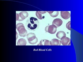
The document summarizes the four major types of tissues in the body: epithelial, connective, muscular, and nervous tissue. It provides detailed information on the structure and function of each type of tissue. Epithelial tissue forms protective layers and linings. Connective tissue includes bone, cartilage, blood, and adipose tissue that support and bind other tissues. Muscular tissue, including cardiac, smooth and skeletal muscle, allows movement. Nervous tissue is specialized for conducting electrical signals and is found in the brain, spinal cord and nerves.
Every organism is composed of several different types of human body tissue. The human body tissue is another way of describing how our cells are grouped together in a highly organized manner according to specific structure and function. These groupings of cells form tissues, which then make up organs and various parts of the body.
This power point helpful for diploma students. this presentation include classification of tissue- epithelial tissue, muscular tissue,skeletal muscle, cardiac muscle, nervous tissue ,difference between smooth muscle and skeletal muscle
Tissues definition and classification, function & location of of epithelium tissues, connective tissues, muscular tissues, nervous tissue etc.. it is usefull for the diploma in pharmacy students , bachelor of pharmacy students and doctor of pharmacy students
Tissue Definition
Tissues are groups of cells that have a similar structure and act together to perform a specific function. The word tissue comes from a form of an old French verb meaning “to weave”. There are four different types of tissues in animals: connective, muscle, nervous, and epithelial. In plants, tissues are divided into three types: vascular, ground, and epidermal. Groups of tissues make up organs in the body such as the brain and heart.
Types of Animal Tissues
Connective
Connective tissue connects or separates groups of other tissues. It is found in between all the other tissues and organs in the body. Connective tissue is made up of cells and ground substance, which is a gel that surrounds cells. Most connective tissue, except for lymph and blood, also contains fibers, which are long, narrow proteins. Fibers can be collagenous, which bind bones to tissues; elastic, which allow organs like the lungs to move; or reticular, which provide physical support to cells. Connective tissue also allows oxygen to diffuse from blood vessels into cells.
About 1 in 10 people are have a disorder involving connective tissue. Some connective tissue disorders include sarcomas, Marfan syndrome, lupus, and scurvy, which is a Vitamin C deficiency that leads to fragile connective tissue.
Muscle
Muscle tissue comprises all the muscles in the body, and the specialized nature of the tissue is what allows muscles to contract. There are three types of muscle tissue: skeletal muscle, cardiac muscle, and smooth muscle. Skeletal muscle anchors tendons to bones and allows the body to move. Cardiac muscle is found in the heart and contracts to pump blood. Smooth muscle is found in the intestines, where it helps move food through the digestive tract, and it is also found in other organs like blood vessels, the uterus, and the bladder. Skeletal and cardiac muscles are striated; this means that they contain sarcomeres (a unit of muscle tissue) that are arranged in a uniform pattern. Smooth muscle does not have sarcomeres.
Duchenne muscular dystrophy is an example of a muscle tissue disorder. It is an inherited disorder that causes muscles to atrophy over time. The muscles shorten as they atrophy, which can cause scoliosis and immobile joints. Individuals with the disorder are usually male because the gene responsible for it is found on the X chromosome (of which males have only one).
Nervous
Nervous tissue is found in the brain, spinal cord, and peripheral nerves, which are all parts of the nervous system. It is made up of neurons, which are nerve cells, and neuroglia, which are cells that help nerve impulses travel. Nervous tissue is grouped into four types: gray matter and white matter in the brain, and nerves and ganglia in the peripheral nervous system. The main difference between gray and white matter is that axons of the neurons in gray matter are unmyelinated, while white matter is myelinated. Myelin is a white, fatty substance that insulates neurons and
Title: Sense of Smell
Presenter: Dr. Faiza, Assistant Professor of Physiology
Qualifications:
MBBS (Best Graduate, AIMC Lahore)
FCPS Physiology
ICMT, CHPE, DHPE (STMU)
MPH (GC University, Faisalabad)
MBA (Virtual University of Pakistan)
Learning Objectives:
Describe the primary categories of smells and the concept of odor blindness.
Explain the structure and location of the olfactory membrane and mucosa, including the types and roles of cells involved in olfaction.
Describe the pathway and mechanisms of olfactory signal transmission from the olfactory receptors to the brain.
Illustrate the biochemical cascade triggered by odorant binding to olfactory receptors, including the role of G-proteins and second messengers in generating an action potential.
Identify different types of olfactory disorders such as anosmia, hyposmia, hyperosmia, and dysosmia, including their potential causes.
Key Topics:
Olfactory Genes:
3% of the human genome accounts for olfactory genes.
400 genes for odorant receptors.
Olfactory Membrane:
Located in the superior part of the nasal cavity.
Medially: Folds downward along the superior septum.
Laterally: Folds over the superior turbinate and upper surface of the middle turbinate.
Total surface area: 5-10 square centimeters.
Olfactory Mucosa:
Olfactory Cells: Bipolar nerve cells derived from the CNS (100 million), with 4-25 olfactory cilia per cell.
Sustentacular Cells: Produce mucus and maintain ionic and molecular environment.
Basal Cells: Replace worn-out olfactory cells with an average lifespan of 1-2 months.
Bowman’s Gland: Secretes mucus.
Stimulation of Olfactory Cells:
Odorant dissolves in mucus and attaches to receptors on olfactory cilia.
Involves a cascade effect through G-proteins and second messengers, leading to depolarization and action potential generation in the olfactory nerve.
Quality of a Good Odorant:
Small (3-20 Carbon atoms), volatile, water-soluble, and lipid-soluble.
Facilitated by odorant-binding proteins in mucus.
Membrane Potential and Action Potential:
Resting membrane potential: -55mV.
Action potential frequency in the olfactory nerve increases with odorant strength.
Adaptation Towards the Sense of Smell:
Rapid adaptation within the first second, with further slow adaptation.
Psychological adaptation greater than receptor adaptation, involving feedback inhibition from the central nervous system.
Primary Sensations of Smell:
Camphoraceous, Musky, Floral, Pepperminty, Ethereal, Pungent, Putrid.
Odor Detection Threshold:
Examples: Hydrogen sulfide (0.0005 ppm), Methyl-mercaptan (0.002 ppm).
Some toxic substances are odorless at lethal concentrations.
Characteristics of Smell:
Odor blindness for single substances due to lack of appropriate receptor protein.
Behavioral and emotional influences of smell.
Transmission of Olfactory Signals:
From olfactory cells to glomeruli in the olfactory bulb, involving lateral inhibition.
Primitive, less old, and new olfactory systems with different path
micro teaching on communication m.sc nursing.pdfAnurag Sharma
Microteaching is a unique model of practice teaching. It is a viable instrument for the. desired change in the teaching behavior or the behavior potential which, in specified types of real. classroom situations, tends to facilitate the achievement of specified types of objectives.
Title: Sense of Taste
Presenter: Dr. Faiza, Assistant Professor of Physiology
Qualifications:
MBBS (Best Graduate, AIMC Lahore)
FCPS Physiology
ICMT, CHPE, DHPE (STMU)
MPH (GC University, Faisalabad)
MBA (Virtual University of Pakistan)
Learning Objectives:
Describe the structure and function of taste buds.
Describe the relationship between the taste threshold and taste index of common substances.
Explain the chemical basis and signal transduction of taste perception for each type of primary taste sensation.
Recognize different abnormalities of taste perception and their causes.
Key Topics:
Significance of Taste Sensation:
Differentiation between pleasant and harmful food
Influence on behavior
Selection of food based on metabolic needs
Receptors of Taste:
Taste buds on the tongue
Influence of sense of smell, texture of food, and pain stimulation (e.g., by pepper)
Primary and Secondary Taste Sensations:
Primary taste sensations: Sweet, Sour, Salty, Bitter, Umami
Chemical basis and signal transduction mechanisms for each taste
Taste Threshold and Index:
Taste threshold values for Sweet (sucrose), Salty (NaCl), Sour (HCl), and Bitter (Quinine)
Taste index relationship: Inversely proportional to taste threshold
Taste Blindness:
Inability to taste certain substances, particularly thiourea compounds
Example: Phenylthiocarbamide
Structure and Function of Taste Buds:
Composition: Epithelial cells, Sustentacular/Supporting cells, Taste cells, Basal cells
Features: Taste pores, Taste hairs/microvilli, and Taste nerve fibers
Location of Taste Buds:
Found in papillae of the tongue (Fungiform, Circumvallate, Foliate)
Also present on the palate, tonsillar pillars, epiglottis, and proximal esophagus
Mechanism of Taste Stimulation:
Interaction of taste substances with receptors on microvilli
Signal transduction pathways for Umami, Sweet, Bitter, Sour, and Salty tastes
Taste Sensitivity and Adaptation:
Decrease in sensitivity with age
Rapid adaptation of taste sensation
Role of Saliva in Taste:
Dissolution of tastants to reach receptors
Washing away the stimulus
Taste Preferences and Aversions:
Mechanisms behind taste preference and aversion
Influence of receptors and neural pathways
Impact of Sensory Nerve Damage:
Degeneration of taste buds if the sensory nerve fiber is cut
Abnormalities of Taste Detection:
Conditions: Ageusia, Hypogeusia, Dysgeusia (parageusia)
Causes: Nerve damage, neurological disorders, infections, poor oral hygiene, adverse drug effects, deficiencies, aging, tobacco use, altered neurotransmitter levels
Neurotransmitters and Taste Threshold:
Effects of serotonin (5-HT) and norepinephrine (NE) on taste sensitivity
Supertasters:
25% of the population with heightened sensitivity to taste, especially bitterness
Increased number of fungiform papillae
NVBDCP.pptx Nation vector borne disease control programSapna Thakur
NVBDCP was launched in 2003-2004 . Vector-Borne Disease: Disease that results from an infection transmitted to humans and other animals by blood-feeding arthropods, such as mosquitoes, ticks, and fleas. Examples of vector-borne diseases include Dengue fever, West Nile Virus, Lyme disease, and malaria.
Basavarajeeyam is an important text for ayurvedic physician belonging to andhra pradehs. It is a popular compendium in various parts of our country as well as in andhra pradesh. The content of the text was presented in sanskrit and telugu language (Bilingual). One of the most famous book in ayurvedic pharmaceutics and therapeutics. This book contains 25 chapters called as prakaranas. Many rasaoushadis were explained, pioneer of dhatu druti, nadi pareeksha, mutra pareeksha etc. Belongs to the period of 15-16 century. New diseases like upadamsha, phiranga rogas are explained.
Report Back from SGO 2024: What’s the Latest in Cervical Cancer?bkling
Are you curious about what’s new in cervical cancer research or unsure what the findings mean? Join Dr. Emily Ko, a gynecologic oncologist at Penn Medicine, to learn about the latest updates from the Society of Gynecologic Oncology (SGO) 2024 Annual Meeting on Women’s Cancer. Dr. Ko will discuss what the research presented at the conference means for you and answer your questions about the new developments.
Ozempic: Preoperative Management of Patients on GLP-1 Receptor Agonists Saeid Safari
Preoperative Management of Patients on GLP-1 Receptor Agonists like Ozempic and Semiglutide
ASA GUIDELINE
NYSORA Guideline
2 Case Reports of Gastric Ultrasound
2. Tissue
- is a group of similar cells specialized
for the performance of a common
function.
- The study of tissues is called Histology
- the cells in multicellular animal maybe
divided into 2. Somatic cells or body
cells and Germ cells, having to do only
with reproduction and continuance of species.
3.
4. There are 4 major groups of somatic
tissues, and these are:
1. Epithelial Tissues
2. Connective Tissues
3. Muscular Tissues
4. Nervous Tissues
5. Epithelial Tissues
- form the covering or lining of all free body
surfaces, both external and internal.
- consists of renewable sheets of cells
- supported by basement membrane which
separates epithelial tissues from underlying,
adjacent tissues.
6. Functions of Epithelial Tissue:
1. Absorb (the lining of the small intestine)
2. Transport ( kidney tubules)
3. Excrete ( sweat glands)
4. Protect ( skin )
5. Contain nerve cells for sensory reception
(taste buds in the tongue)
7. Structures of Epithelial Tissues:
Layers:
1. Simple – consisting of only one layer of cells.
2. Stratified – consisting multiple slacked layers
3. Pseudostratified ciliated columnar epithilium
- possess cilia and appears layered but not.
- appears layered because of their nuclei are at two
or more levels within the cell of the tissue.
12. Connective Tissue
- binds together and supports other
structures
- divided throughout an extracellular matrix
- derived from the Mesenchyme, a
generalized embryonic tissue that can
differentiate also into vascular and smooth
muscle. (also considered the most primitive
connective tissue)
13. Two General Types of Connecticve
Tissues
1. Loose Connective Tissue
- strong, flexible fibers of protein collagen are interwoven
with fine elastic, and reticular fibers giving it its elastic
consistency and making it an exellent binding tissue.
2. Fibrous Connective Tissue
- the collagen fibers are densly packed and may lie parallel to
one another, creating very strong cords, such as tendons and
ligaments.
14. Kinds of Connective Tissues
1. Adipose Tissue
- is a type of loose connective that consists of large cells that
store lipids.
- consists scattered, rounded or branched in form, with
intercellular spaces occupied by delicate fibers.
- also known as fat tissues
- cells accumulate in large numbers to form what is commonly
known as fat.
- Two minor cell types are present; fibroblasts, which produce
fibers, and macrophages, which are phagocytes.
16. 2. Cartilage
-is a firm yet elastic matrix (chondrin) secreted by small
groups of rounded cartilage cells or Chondrocytes
embedded within it and covered by a thin, fibrous
Perichondrium.
- Is a hard yet flexible tissue that supports such structures
as the outer ear and forms the entire skeleton of such
animals as sharks and rays.
- Cells called Chondrocytes lie within spaces called
Lacunae that are sorrounded by a rubbery matrix that
chomdroblasts secrete.
- the matrix gives the cartilage its strength and elasticity.
18. 3. Bone or Osseous Tissue
- Like other connective tissues, bone consists of cells,
fibers and ground substance but differs because the extracellular
matrix is calcified.
- the rigid extracellular matrix has several functions
* Provides an internal support for the body and
attachment for muscles and tendons.
* Protects vital organs of the cranial and thoracic
cavity, and encloses the blood forming elements of the bone
marrow.
* Provides a reservvoir of ionic calcium essential
for many cellular processes of the body.
19. Macroscopic Structure of the Bone
A. Diaphysis: shaft of the bone
B. Epiphysis: end of a long bone
C. Metaphysis: area between the diaphysis and the epiphysis
D. Epiphyseal plate (growth plate)
E. Medullary Cavity: central cavity of bone occupied by bone
marrow
F. Trabeculae: irregular lattice of thin columns of bone
sorrounded by bone marrow
G. Articular Cartilage: Hyaline cartilage covering joint surfaces
H. Periosteum: external covering of bone
I. Endosteum: internal covering of bone
20. Bone Cells
A. Osteoprogenitor Cell
1. A mesenchymeal sterm cell that can undergo mitotic
division and differentiate into an osteoblast.
2. Osteoprogenitor calls are located on the immer cellular
layer of the periosteum, the endosteum and lining osteonic
canals.
3. These calls are most active during bone growth, but large
numbers are reactivated in adult life in repair of fractures.
4. They also differentiate into osteoblasts during the
continous process of bone remodeling
21. B. Osteoblasts
1. Cells that are derived from osteoprogenitor cellas and
are responsible for the synthesis of the organic components of
bonw matrix, which is called osteoid.
2. They are located on the surface of bone tissue and
resemble epithelium. When the cells are active they have a
cuboidal appearance; and when their activity declines, they
flatten
3. They have the structure expected of cells that are
actively engaged on protein synthesis, such as extensive rough
ER, well developed Golgi complex and numerous secretory
vesicles.
4. The cells have cytoplasmic processes that bring them in
contact with neighboring cells.
22. D. Osteocytes
1. When osteoblast has completely sorrounded itself and
its cytoplasmic processes with matrix, the cell is now termed an
osteocyte.
2. The space on which the cell resides os termed lacuna.
The thin cylindrical spaces that house cytoplasmic processes are
called canaliculi. Canaliculi also contain extracellular fluid
carrying nutrients to nourish osteocytes.
3. Processes of adjacent cells make contact via gap
junctions, which allow ions and small molecules to travel from
cell to cell.
23. D. Osteoclasts
1. Large, motile, multinucleated, bone-resorbing cells
derived from blood monocytes that occupy depressions in the
bone matrix undergoing active resorption.
2. Adhere tightly to established bone matrix and acidify
the surface by the use of a proton pump that actively transports H
ionsonto the surface of the bone. Lysosomal enzymes are
released by exocytosis and degrade the organic components of
bone.
3. The degraded minerals on organic components are
endocytosed by the osteoclast and delivered to nearby capillaries
to enter the circulation.
24. Bone Coverings
A. Periosteum:
1. The external covering of bone, except in areas where
tendons and ligaments insert into bone and on the surfaces
covered by articular cartilage.
2. Consists of 2 layers
a. Outer fibrous layer – collagenous connective tissue that
contains many blood vessels.
b. Inner Cellular Layer – layer that contains
osteoprogenitor cells that have osteogenic potential
3. Sharpey’s Fibers: Bundles of periostal collgen fibers that
penetrate the bone matrix and strongly adheres the periosteum to
bone.
25. B. Endosteum
-Thin layer of osteoprogenitor cells, osteoblasts and a
small amount of connective tissue that lines all internal surfaces
of cavities within bone including the osteonic canals and marrow
spaces.
27. 4. Blood
- is a connectie tissue in which a fluid called plasma
suspends specialized re and white blood cells plus platelets.
- a viscous fluid pumped all throughout the body
- mesoderm in origin
- transports various substances throughout the bodies of
animals.
- Erythrocytes (Red Blood Cells)
- composed of cells, fibers and amorphous ground
substance.
28. Functions:
1. Carry oxygen and nutrients to the cell and carry waste
materials – CO2 away from the cells to kidneys and lungs.
2. Immune System
3. Homeostasis in the body.
Ground Substance/Matrix:
- fluid and protein in the plasma
Albumin = are to maintain the osmotic
pressure of the blood.
29. Cells:
1. Erythrocytes (RBC)
2. Leukocytes (WBC)
3. Thrombocytes/ blood platelets
Fibers:
- these are the potential fibers of fibrinogen which are
converted to fibrils fiber during blood clotting.
- Clotted solid. Which rapidly stops blood flow from the
wound.
Note - blood clots occur when platelets interact with and
respond to collagen fibers that they encounter when they leave
the circulaing system in response to a wound.
30. Human Red Blood Cells
Platelets and T-lymphocyte
(Erythocytes = red; platelets = yellow; T-lymphocyte = light green)
32. Muscle Tissue
- is the driving force, the power behind movement in most
vertebrates and invertebrates.
- specialized for contractility
- most common tissue in the body of most animals.
- made of elongated cells made for contraction.
- allows movement.
3 Important Properties of Muscle Tissue
1. Excitability (or irritability)- the capacity to receive and
respond stimulus.
2. Extensibility –the ability to be stretched
3. Elasticity –the ability to return to its original shape
after being stretche or contracted.
34. Smooth Muscle
- is formed of spindle –shaped cells, each containing a
single nucleus
- cells are arranged closely to form sheets
- not striated
- mostly found in the walls of hollow organs
- are organized into sheets of muscle circling the wall of
the alimentary canal, blood vessels, respiratiry passages,
and urinary and genital ducts.
- moves substances or objects( foodstuffs, urine, baby)
along internal passageways
- also called involuntary muscle because higher brain
centers do not control its contractions.
36. Skeletal Muscle
-is composed of striated muscle fibers (cells) containing
many peripheral nuclei.
- are those attached to the skeleton and are typically
organized into sturdy, compact bundles or bands.
- extremely long, cylindrical, multinucleate cells that may
reach from one end of the muscle to the other.
- the characteristic light and dark bands(striations)
represents the fine structure of the myofibrils that make up the
fibers.
- a.k.a. voluntary muscle because nercous system
consciously controls its contractions.
- for voluntary movement or locomotion
38. Cardiac Muscle
- is the muscle of the vertebrate heart
- consists of closely opposed, but separate, uninucleate cell
fibers that appear branching and interconnected.
- contains specialized cell junctions called Interlaced
Disks that allow ions to (action potential) to move quikly
from cell to cell.
- can be found in the walls of the heart
- as the walls of the heart contract, cardiac muscle tissue
propels blood onto the circulation; involuntary control.
41. Nervous Tissue
- is composed of several different types of cells:
- is highly specialized for the property of irritability and
conductivity.
- can be found in the brain, spinal cord, and nerves.
- transmits electrical signals from sensory receptors to the
spinal cord or brain, and from the spinal cord or brain to
effectors(muscles and glands)
Nerve – group of fibers or process bound together by a
connective tissue.
42. Neurons
- Impulse conducting cells
- Structural and functional unit of the nervous system.
- arranged in chains
Synapse – point of contact between neurons
Dendrite – the process that transmits stimuli to the cell
body
Axon – carries impulses away from the cell body
Bipolar cells – one dendrite and one axon
Multipolar Cells – multiple dendrites and single axon
Ganglion – a group of nerve cell bodies, with their
conspicuous nuclei when outside the central nervous system.
43. Neuroglia
- cells involved with protection and, support, and
nourishment
- serve as delicate packing to hold neurons apart and may
also aid in nutrition of neurons.
*Nerve fibers are sheathed by special cells called Schwann Cells.
*Nodes of Ranvier- marks the end of the Schwann call and the
beginning of another.
Peripheral Glial Cells
- cells that form sheaths and help protect, nourish,
and maintain the peripheral nervous system.