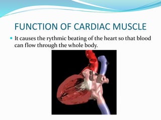
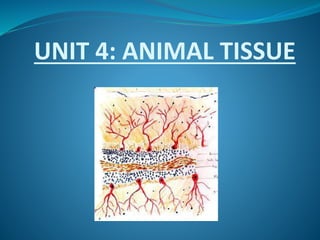
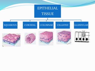
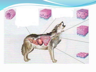
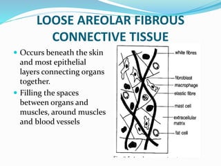
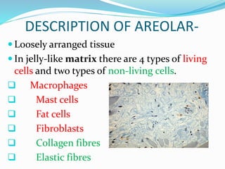
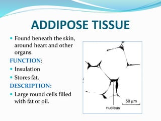
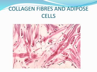
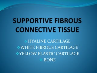
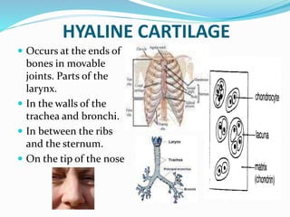
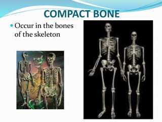
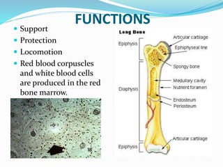
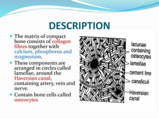
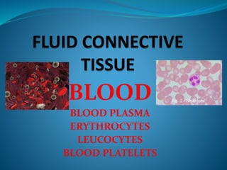
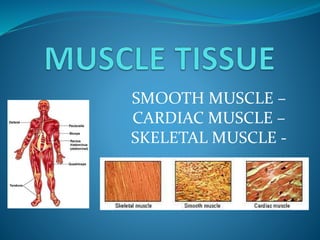
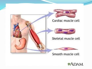
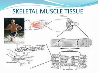
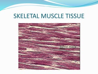
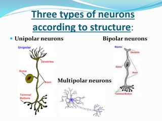
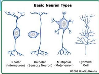
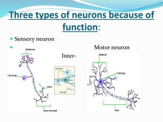
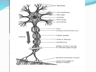
SlideShare a Scribd company logo
UNIT 4: ANIMAL TISSUE
WHERE DO TISSUES FIT IN
THE LIVING ORGANISM?
 Life begins as a single cell called a zygote
after fertilization
 This cell differentiates into a variety of
tissues. (Group of cells with the same
function)
 A group of tissues with the same function
forms an organ.
 Group of organs – Organ system
 Group of organ systems - Organism
WHAT IS TISSUE?
SPECIALIZED CELL of the
same type that perform a
common function in the
body.
MAJOR TYPES OF ANIMAL
TISSUE
 EPITHELIAL TISSUE: Covers or lines
body cavity and forms glands.
 CONNECTIVE TISSUE: Binds different
organs together and support the body.
 MUSCLE TISSUE: Movement
 NERVOUS TISSUE: Senses and transmits
impulses
EPITHELIAL
TISSUE
SQUAMOUS CUBOIDAL COLUMNAR CILLIATED GLANDULAR
SQUAMOUS EPITHELIAL TISSUE
STRUCTURE
 Single layer thin, large, flattened cells
 Cells have irregular shape and are closely packed in
mosaic form.
 Cells are attached to a basement membrane.
FUNCTIONS
 Diffusion of gasses can occur easily over surface.
 Supply friction free surface for blood flow.
 Protects and supports underlying tissue
SQUAMOUS EPITELIAL TISSUE
WHERE?
•Lining of lungs
and blood vessels
•In mouth and
esophagus.
CUBOIDAL EPITHELIAL TISSUE
STRUCTUTE
 Square cells, closely packed in a single layer.
 Cells attached to a basement membrane.
FUNCTION
 Absorbs molecules
CUBOIDAL EPITHELIAL CELLS
WHERE?
•Lining of
kidney tubules
•Lining
various
glands.
COLUMNAR EPITHELIAL TISSUE
STRUCTURE
 Tall elongated, column-shaped cells.
 Nucleus near the base of the cell.
 Cells are attached to a basement membrane.
FUNCTIONS
 Plays a role in absorption of nutrients.
 Protects and supports underlying tissues.
COLUMNAR EPITHELIAL TISSUE
WHERE?
•Lining of
small
intestine,
stomach
and
oviducts
CILLIATED CULUMNAR EPITHELIAL
TISSUE
STRUCTURE
 Comprises of columnar cells with cilia at their free
edges
FUNCTIONS
 Sweeps impurities towards throat
 Improves movement of substance through the ducts.
CILLIATED COLUMNAR EPITHELIAL
TISSUE
WHERE?
•Lining of
trachea and
nostrils,
bronchi
and sperm
ducts)
GLANDULAR COLUMNAR
EPITHELIAL TISSUE
STRUCTURE
 Consist of cuboidal and columnar epithelial cells.
 Unicellular glandular cells with goblet cells scattered
among non-glandular cells.
FUNCTIONS
 Produce mucus which serves to lubricate the intestine
and make movement of food easier.
 Keep respiratory passage moist.
 Secrete enzymes, hormones, sweat, wax and saliva.
GLANDULAR COLUMNAR
EPITHELIAL TISSUE
WHERE?
• In small
intestine and
respirative
passages.
• Occur in
salivary glands,
thyroid gland
and sebaceous
glands in the
skin.
• FIBROUS CONNECTIVE TISSUE
• SUPPORTIVE CONNECTIVE
TISSUE
• FLUID CONNECTIVE TISSUE
 LOOSE AREOLAR FIBROUS
CONNECTIVE TISSUE
DENSE WHITE FIBROUS CONNECTIVE
TISSUE
DENSE YELLOW FIBROUS
CONNECTIVE TISSUE
ADIPOSE TISSUE
LOOSE AREOLAR FIBROUS
CONNECTIVE TISSUE
 Occurs beneath the skin
and most epithelial
layers connecting organs
together.
 Filling the spaces
between organs and
muscles, around muscles
and blood vessels
DESCRIPTION OF AREOLAR-
 Loosely arranged tissue
 In jelly-like matrix there are 4 types of living
cells and two types of non-living cells.
 Macrophages
 Mast cells
 Fat cells
 Fibroblasts
 Collagen fibres
 Elastic fibres
FUNCTIONS OF AREOLAR-
Areolar tissue binds organs or organ
components together and supports
structures.
Allows for movement between
structures.
The matrix allows for diffusion of
substances like gases, nutrients,
hormones and wastes to and from the
blood.
 Fibroblasts – secrete collagen and produce
elastic fibres.
 Macrophages – Engulf foreign particles by
means of phagocytosis to defend the body
against infection.
 Mast cells – They secrete the matrix, release
histamine during inflammation, produce
heparin (anticoagulant).
 Fat cells – Synthesis and storage of fats.
 Collagen fibres strengthen the tissue.
 Elastic fibres – Allows for expansion and
contraction.
MACROPHAGES, PLASMA CELLS
MAST CELLS AND ELASTIC FIBRES
FIBROBLASTS
DENSE WHITE FIBROUS
CONNECTIVE TISSUE
 In dermis of skin,
tendons
FUNCTIONS:
Attach muscle to bones
DESCRIPTION OF DENSE WHITE-
 Consist of mainly white collagen non-elastic
fibres.
 The collagen fibres are non-elastic,
densely packed, arranged in parallel
bundles, and has a shiny appearance.
 Fibroblasts are elongated cells and occur
parallel in rows.
 Minimum amount of matrix.
YELLOW FIBROUS CONNECTIVE
TISSUE
 In ligaments
FUNCTIONS:
 Attach bone to bone
 Hold bones of a joint in
place.
 Restrict movement of
bones and prevent
dislocation during
normal movement.
DESCRIPTION OF DENSE YELLOW-
 Consist of white collagen fibres and a
network of yellow elastic fibres.
 Ligaments are strong and flexible.
ADDIPOSE TISSUE
 Found beneath the skin,
around heart and other
organs.
FUNCTION:
 Insulation
 Stores fat.
DESCRIPTION:
 Large round cells filled
with fat or oil.
COLLAGEN FIBRES AND ADIPOSE
CELLS
 HYALINE CARTILAGE
WHITE FIBROUS CARTILAGE
YELLOW ELASTIC CARTILAGE
 BONE
HYALINE CARTILAGE
 Occurs at the ends of
bones in movable
joints. Parts of the
larynx.
 In the walls of the
trachea and bronchi.
 In between the ribs
and the sternum.
 On the tip of the nose
DESCRIPTION
 Contains fine collagen fibres.
 White translucent matrix.
 Cartilage tissue is surrounded by a fibrous
capsule called the perichondrium with
blood vessels for nutrition.
 It consists of living cells called
chondrocytes.
 They occur in small fluid-filled spaces called
lacunae. In the lacunae the cells are
arranged singular or in groups.
FUNCTIONS
 Chondrocytes produce a
rubber matrix called
chondrin.
 Reduce friction at joints
 Attach bones firmly to other
bones.
 Keeps tubes open (C-shaped in
trachea).
 Forms permanent structures
(nose).
 Longitudinal growth of long
bones.
White FIBROCARTILAGE
 Occurs as disks between
the vertebrae.
 Surrounds the edges of
the sockets of ball-and-
socket joints.
 Between the pubic bones
in front of the pelvic
girdle.
FUNCTIONS
 It serves as shock
absorbers between
adjacent vertebrae.
 It deepens sockets to
make dislocation less
easy.
DESCRIPTION
 Contain a large number of collagen
fibres.
 White translucent matrix.
 Cartilage tissue is surrounded by a
fibrous capsule called the
perichondrium with blood vessels
for nutrition.
 It consists of living cells called
chondrocytes.
 They occur in small fluid-filled
spaces called lacunae. In the
lacunae the cells are arranged
singular or in groups.
Yellow ELASTIC CARTILAGE
 In the ear lobe
 At the tip of the nose
 In the epiglottis.
 Septum of nose
FUNCTIONS
 It maintains the shape
and flexibility of the ear
lobe and tip of the nose.
 It strengthens and
supports the ear, nose
and epiglottis.
DESCRIPTION
 Contain collagen fibres and a large network of elastic
fibres.
 Translucent matrix.
 Cartilage tissue is surrounded by a fibrous capsule
called the perichondrium with blood vessels for
nutrition.
 It consists of living cells called chondrocytes.
 They occur in small fluid-filled spaces called lacunae.
In the lacunae the cells are arranged singular or in
groups.
COMPACT BONE
 Occur in the bones
of the skeleton
FUNCTIONS Support
 Protection
 Locomotion
 Red blood corpuscles
and white blood cells
are produced in the red
bone marrow.
DESCRIPTION
 The matrix of compact
bone consists of collagen
fibres together with
calcium, phosphorus and
magnesium.
 These components are
arranged in circles called
lamellae, around the
Haversian canal,
containing artery, vein and
nerve.
 Contain bone cells called
osteocytes
BLOOD
BLOOD PLASMA
ERYTHROCYTES
LEUCOCYTES
BLOOD PLATELETS
BLOODPLASMA
WHERE?
 Matrix in which the blood
cells are found.
FUNCTIONS:
 Transport various formed
elements to body parts.
 Dispersion medium.
 Transports digested food.
 Transports cellular waste.
 Transports hormones.
 Plays a role in regulation of
body temperature.
 Transports antibodies
DESCRIPTION
 Straw color fluid
consisting mainly of
water with many
substances in solution
like nutrients, organic
waste, inorganic salts
and ions, plasma
protein, dissolved
gases, hormones,
enzymes and
antibodies.
ERYTHROCYTES (RED BLOOD
CELLS)FUNCTIONS:
 Transports oxygen in the
blood from the lungs to
all living cells.
 Transports some of the
carbon dioxide from the
body tissues the lungs.
 Play a role in the clotting
of blood.
 Play a role in the
regulating of pH of body
fluids
DESCRIPTION
 Small round biconcave disks filled with cytoplasm,
but without a nucleus.
 Contain the red pigment hemoglobin.
 Hemoglobin contains 4 units, each with the
protein globin and a complex iron-containing,
structure called heme. The iron form a loose
association with oxygen and in this way red blood
cells transport oxygen and readily give it up to
tissues.
LEUCOCYTES
(WHITE BLOOD CELLS)
FUNCTIONS:
 Engulf infections by
means of
phagocytosis.
 Produce antibodies.
 Defense or
immunity of body
DESCRIPTION
 Larger cells with nuclei.
 Without staining they
appear transparent.
 Irregular is shape. – there are
5 types of leucocytes based
on differences in size and
nuclear shape:
 Neutrophils
 Eosinophils
 Basophils
 Monocytes.
 Lymphocytes.
BLOOD PLATELETS
DESCRIPTION:
 Fragments of giant cells,
present only in bone
marrow.
 They are small discs
without nuclei and
bounded by a typical cell
membrane.
 A characteristic feature of
platelets is their tendency
to stick to foreign surfaces
and to each other to form
clumps.
FUNCTIONS;
 When blood vessels are
damaged, platelets
form a plug that seals
the vessel, and injured
tissues release
molecules that help
the clotting of blood.
SMOOTH MUSCLE –
CARDIAC MUSCLE –
SKELETAL MUSCLE -
SMOOTH MUSCLE
 Found wide spread in
body.
 Found in the walls of
bloodvessels, digestive
tract, respiratory ducts,
urinary ducts and
reproductive ducts.
 In iris of eye and form
small muscles attach to
hair follicles
FUNCTIONS OF SMOOTH -
 Control slow involuntary
movements of organs
performing important life
processes e.g. Contraction
of stomach wall during
peristalsis.
 Responsible for the
movement of substances
in internal passages e.g.
Blood in blood vessels.
DESCRIPTION OF SMOOTH -
 Every smooth muscle fibre is an elongated, thin cell
with a thick central part and pointed ends.
 Every cell has a big nucleus centrally situated.
 This muscle is never connected to bone.
 Can contract for long with out causing a spasm e.g.
Uterus wall during birth.
CARDIAC MUSCLE TISSUE
 Found only in the heart.
DESCRIPTION OF CARDIAC -
 Contain many mitochondria, glucose and blood
supply.
 Consist of branched cells with one or two central
nuclei.
 Cells are rectangular and connected with muscle
bridges.
 The ends of 2 cells are attached by intercalated disks.
FUNCTION OF CARDIAC MUSCLE
 It causes the rythmic beating of the heart so that blood
can flow through the whole body.
SKELETAL MUSCLE TISSUE
 Skeletal muscle is voluntarily controlled.
 skeletal muscles are attached to bones by bundles of
collagen fibers known as tendons.
 Skeletal muscle is made up of individual components
known as myocytes, or "muscle cells“ = "muscle fibers"
 These long, cylindrical, multinucleated cells are also
called myofibers.
 The term muscle refers to multiple bundles of muscle
fibers held together by connective tissue.
 The plasma membrane is called the sarcolemma with
the cytoplasm known as the sarcoplasm.
 In the sarcoplasm are the myofibrils.
 Pressed against the inside of the sarcolemma are the
unusual flattened nuclei.
 Between the myofibrils are the mitochondria.
NERVOUS TISSUE
 Nervous tissue is composed of highly specialized nerve
cells called neurons and supporting cells or
neuroglia.
 The neurons, which are the basic units of
communication, react to stimuli and transmit the
impulses (information) form one region of the body to
another.
 Neurons extend throughout the body and do not act
alone.
STRUCTURE OF A NEURON
 Neurons are usually long, large and complex in shape.
 It consists of a cell body and several cytoplasmic
processes.
 Processes that transmit impulses to the cell body are
dendrites and usually are multiple.
 The single process that transmits impulses away forms
the cell body in the axon.
 The cell body contains a nucleus, neurofibrills, Nissl
granules, neuroplasma.
Three types of neurons
according to structure:
 Unipolar neurons Bipolar neurons
Multipolar neurons
Three types of neurons because of
function:
 Sensory neuron
 Motor neuron
 Inter-
Animal tissue

More Related Content

What's hot

HUMAN TISSUE ANATOMY & PHYSIOLOGY
HUMAN TISSUE ANATOMY & PHYSIOLOGYHUMAN TISSUE ANATOMY & PHYSIOLOGY
HUMAN TISSUE ANATOMY & PHYSIOLOGY
Kameshwaran Sugavanam
 
4 tissue types and epithelium
4 tissue types and epithelium4 tissue types and epithelium
4 tissue types and epithelium
James H. Workman
 
Epithelium
EpitheliumEpithelium
Epithelium
K BHATTACHARJEE
 
Animal Tissues
Animal  TissuesAnimal  Tissues
Animal Tissues
Nick Mintalar
 
Epithelial tissue
Epithelial tissueEpithelial tissue
Epithelial tissue
anisha Deshmukh
 
Oral-Epithelium by DR. C.P. ARYA ( B.Sc. ;B.D.S. ;M.D.S. ;P.M.S. ;R.N.T.C.P.)
Oral-Epithelium by DR. C.P. ARYA ( B.Sc. ;B.D.S. ;M.D.S. ;P.M.S. ;R.N.T.C.P.)Oral-Epithelium by DR. C.P. ARYA ( B.Sc. ;B.D.S. ;M.D.S. ;P.M.S. ;R.N.T.C.P.)
Oral-Epithelium by DR. C.P. ARYA ( B.Sc. ;B.D.S. ;M.D.S. ;P.M.S. ;R.N.T.C.P.)
DR. C. P. ARYA
 
Epithelial tissue 1
Epithelial tissue 1Epithelial tissue 1
Epithelial tissue 1
derick lumaban
 
00introhistologytext
00introhistologytext00introhistologytext
00introhistologytext
Eddy Mwachenje
 
Epithelial tissue
Epithelial tissueEpithelial tissue
Epithelial tissue
clarence witty mendoza
 
Eithelial tissue,connective tissue by Dr Jyoti Prakash Pani Associate Profess...
Eithelial tissue,connective tissue by Dr Jyoti Prakash Pani Associate Profess...Eithelial tissue,connective tissue by Dr Jyoti Prakash Pani Associate Profess...
Eithelial tissue,connective tissue by Dr Jyoti Prakash Pani Associate Profess...
phupu
 
Tissues
TissuesTissues
Tissue Level of Organization (Anatomy)
Tissue Level of Organization (Anatomy)Tissue Level of Organization (Anatomy)
Tissue Level of Organization (Anatomy)
Yogananta Ramadhan, MD
 
Tissues
TissuesTissues
Chapter 4,5,17 notes
Chapter 4,5,17  notesChapter 4,5,17  notes
Chapter 4,5,17 notes
Tia Hohler
 
Tissues
TissuesTissues
Tissues
Abay Alem
 
Animal Tissue
Animal TissueAnimal Tissue
Animal Tissue
DrDeepaliNayar
 
Tissue System/Collection of Cells
Tissue System/Collection of CellsTissue System/Collection of Cells
Tissue System/Collection of Cells
Pharmacy Universe
 
Tissues3
Tissues3Tissues3
Tissues3
guestb4650d
 
Anatomy and physiology Tissue Chapter
Anatomy and physiology Tissue ChapterAnatomy and physiology Tissue Chapter
Anatomy and physiology Tissue Chapter
mrhunterspage
 

What's hot (19)

HUMAN TISSUE ANATOMY & PHYSIOLOGY
HUMAN TISSUE ANATOMY & PHYSIOLOGYHUMAN TISSUE ANATOMY & PHYSIOLOGY
HUMAN TISSUE ANATOMY & PHYSIOLOGY
 
4 tissue types and epithelium
4 tissue types and epithelium4 tissue types and epithelium
4 tissue types and epithelium
 
Epithelium
EpitheliumEpithelium
Epithelium
 
Animal Tissues
Animal  TissuesAnimal  Tissues
Animal Tissues
 
Epithelial tissue
Epithelial tissueEpithelial tissue
Epithelial tissue
 
Oral-Epithelium by DR. C.P. ARYA ( B.Sc. ;B.D.S. ;M.D.S. ;P.M.S. ;R.N.T.C.P.)
Oral-Epithelium by DR. C.P. ARYA ( B.Sc. ;B.D.S. ;M.D.S. ;P.M.S. ;R.N.T.C.P.)Oral-Epithelium by DR. C.P. ARYA ( B.Sc. ;B.D.S. ;M.D.S. ;P.M.S. ;R.N.T.C.P.)
Oral-Epithelium by DR. C.P. ARYA ( B.Sc. ;B.D.S. ;M.D.S. ;P.M.S. ;R.N.T.C.P.)
 
Epithelial tissue 1
Epithelial tissue 1Epithelial tissue 1
Epithelial tissue 1
 
00introhistologytext
00introhistologytext00introhistologytext
00introhistologytext
 
Epithelial tissue
Epithelial tissueEpithelial tissue
Epithelial tissue
 
Eithelial tissue,connective tissue by Dr Jyoti Prakash Pani Associate Profess...
Eithelial tissue,connective tissue by Dr Jyoti Prakash Pani Associate Profess...Eithelial tissue,connective tissue by Dr Jyoti Prakash Pani Associate Profess...
Eithelial tissue,connective tissue by Dr Jyoti Prakash Pani Associate Profess...
 
Tissues
TissuesTissues
Tissues
 
Tissue Level of Organization (Anatomy)
Tissue Level of Organization (Anatomy)Tissue Level of Organization (Anatomy)
Tissue Level of Organization (Anatomy)
 
Tissues
TissuesTissues
Tissues
 
Chapter 4,5,17 notes
Chapter 4,5,17  notesChapter 4,5,17  notes
Chapter 4,5,17 notes
 
Tissues
TissuesTissues
Tissues
 
Animal Tissue
Animal TissueAnimal Tissue
Animal Tissue
 
Tissue System/Collection of Cells
Tissue System/Collection of CellsTissue System/Collection of Cells
Tissue System/Collection of Cells
 
Tissues3
Tissues3Tissues3
Tissues3
 
Anatomy and physiology Tissue Chapter
Anatomy and physiology Tissue ChapterAnatomy and physiology Tissue Chapter
Anatomy and physiology Tissue Chapter
 

Similar to Animal tissue

animal tissue
animal tissueanimal tissue
animal tissue
ZAMIKHAYA PRINCE
 
Tissues
TissuesTissues
Tissues
gkeswani
 
Unit-I, Chapter-3 Tissue level of organization.
Unit-I, Chapter-3 Tissue level of organization.Unit-I, Chapter-3 Tissue level of organization.
Unit-I, Chapter-3 Tissue level of organization.
Audumbar Mali
 
Transitional epithelium.pptx
Transitional epithelium.pptxTransitional epithelium.pptx
Transitional epithelium.pptx
ChinjuJoseSajith
 
Human Tissue & its types
Human Tissue & its typesHuman Tissue & its types
Human Tissue & its types
Yatendra Singh
 
STRUCTURAL-ORGANIZATION-IN-ANIMALS-7.pptx
STRUCTURAL-ORGANIZATION-IN-ANIMALS-7.pptxSTRUCTURAL-ORGANIZATION-IN-ANIMALS-7.pptx
STRUCTURAL-ORGANIZATION-IN-ANIMALS-7.pptx
musiclovers17
 
tissue and it's types
tissue and it's typestissue and it's types
tissue and it's types
abhilasha chaudhary
 
Anatomy Project
Anatomy  ProjectAnatomy  Project
Anatomy Project
guestc78ec4
 
Levels of Organization of the Human Body 2.pptx
Levels of Organization of the Human Body 2.pptxLevels of Organization of the Human Body 2.pptx
Levels of Organization of the Human Body 2.pptx
JOHNMICHAELSERAA
 
Animal Tissue - NAS102
Animal Tissue - NAS102 Animal Tissue - NAS102
Animal Tissue - NAS102
emalyn25
 
human tissue.pdf
human tissue.pdfhuman tissue.pdf
human tissue.pdf
Imtiyaz60
 
TISSUE
TISSUETISSUE
anatomy and physiology of tissue and the functional character
anatomy and physiology of tissue and the functional characteranatomy and physiology of tissue and the functional character
anatomy and physiology of tissue and the functional character
Kalinga institute of nursing sciences
 
Tissues Cole Wilson
Tissues Cole WilsonTissues Cole Wilson
Tissues Cole Wilson
guest5fedde
 
Tissues Cole Wilson
Tissues Cole WilsonTissues Cole Wilson
Tissues Cole Wilson
guest5fedde
 
types of animal tissues class 9
types of animal tissues class 9types of animal tissues class 9
types of animal tissues class 9
KanupriyaGarg1
 
3.tissues of body
3.tissues of body3.tissues of body
3.tissues of body
M Humayun Jamil
 
Tissues
TissuesTissues
Tissues
ZENITH PARMAR
 
Tissues B L A K E
Tissues B L A K ETissues B L A K E
Tissues B L A K E
guestcd741c
 
ANIMAL TISSUE.docx
ANIMAL TISSUE.docxANIMAL TISSUE.docx
ANIMAL TISSUE.docx
MOUMITASENGUPTA18
 

Similar to Animal tissue (20)

animal tissue
animal tissueanimal tissue
animal tissue
 
Tissues
TissuesTissues
Tissues
 
Unit-I, Chapter-3 Tissue level of organization.
Unit-I, Chapter-3 Tissue level of organization.Unit-I, Chapter-3 Tissue level of organization.
Unit-I, Chapter-3 Tissue level of organization.
 
Transitional epithelium.pptx
Transitional epithelium.pptxTransitional epithelium.pptx
Transitional epithelium.pptx
 
Human Tissue & its types
Human Tissue & its typesHuman Tissue & its types
Human Tissue & its types
 
STRUCTURAL-ORGANIZATION-IN-ANIMALS-7.pptx
STRUCTURAL-ORGANIZATION-IN-ANIMALS-7.pptxSTRUCTURAL-ORGANIZATION-IN-ANIMALS-7.pptx
STRUCTURAL-ORGANIZATION-IN-ANIMALS-7.pptx
 
tissue and it's types
tissue and it's typestissue and it's types
tissue and it's types
 
Anatomy Project
Anatomy  ProjectAnatomy  Project
Anatomy Project
 
Levels of Organization of the Human Body 2.pptx
Levels of Organization of the Human Body 2.pptxLevels of Organization of the Human Body 2.pptx
Levels of Organization of the Human Body 2.pptx
 
Animal Tissue - NAS102
Animal Tissue - NAS102 Animal Tissue - NAS102
Animal Tissue - NAS102
 
human tissue.pdf
human tissue.pdfhuman tissue.pdf
human tissue.pdf
 
TISSUE
TISSUETISSUE
TISSUE
 
anatomy and physiology of tissue and the functional character
anatomy and physiology of tissue and the functional characteranatomy and physiology of tissue and the functional character
anatomy and physiology of tissue and the functional character
 
Tissues Cole Wilson
Tissues Cole WilsonTissues Cole Wilson
Tissues Cole Wilson
 
Tissues Cole Wilson
Tissues Cole WilsonTissues Cole Wilson
Tissues Cole Wilson
 
types of animal tissues class 9
types of animal tissues class 9types of animal tissues class 9
types of animal tissues class 9
 
3.tissues of body
3.tissues of body3.tissues of body
3.tissues of body
 
Tissues
TissuesTissues
Tissues
 
Tissues B L A K E
Tissues B L A K ETissues B L A K E
Tissues B L A K E
 
ANIMAL TISSUE.docx
ANIMAL TISSUE.docxANIMAL TISSUE.docx
ANIMAL TISSUE.docx
 

Recently uploaded

Exploiting Artificial Intelligence for Empowering Researchers and Faculty, In...
Exploiting Artificial Intelligence for Empowering Researchers and Faculty, In...Exploiting Artificial Intelligence for Empowering Researchers and Faculty, In...
Exploiting Artificial Intelligence for Empowering Researchers and Faculty, In...
Dr. Vinod Kumar Kanvaria
 
clinical examination of hip joint (1).pdf
clinical examination of hip joint (1).pdfclinical examination of hip joint (1).pdf
clinical examination of hip joint (1).pdf
Priyankaranawat4
 
Community pharmacy- Social and preventive pharmacy UNIT 5
Community pharmacy- Social and preventive pharmacy UNIT 5Community pharmacy- Social and preventive pharmacy UNIT 5
Community pharmacy- Social and preventive pharmacy UNIT 5
sayalidalavi006
 
The simplified electron and muon model, Oscillating Spacetime: The Foundation...
The simplified electron and muon model, Oscillating Spacetime: The Foundation...The simplified electron and muon model, Oscillating Spacetime: The Foundation...
The simplified electron and muon model, Oscillating Spacetime: The Foundation...
RitikBhardwaj56
 
writing about opinions about Australia the movie
writing about opinions about Australia the moviewriting about opinions about Australia the movie
writing about opinions about Australia the movie
Nicholas Montgomery
 
ISO/IEC 27001, ISO/IEC 42001, and GDPR: Best Practices for Implementation and...
ISO/IEC 27001, ISO/IEC 42001, and GDPR: Best Practices for Implementation and...ISO/IEC 27001, ISO/IEC 42001, and GDPR: Best Practices for Implementation and...
ISO/IEC 27001, ISO/IEC 42001, and GDPR: Best Practices for Implementation and...
PECB
 
C1 Rubenstein AP HuG xxxxxxxxxxxxxx.pptx
C1 Rubenstein AP HuG xxxxxxxxxxxxxx.pptxC1 Rubenstein AP HuG xxxxxxxxxxxxxx.pptx
C1 Rubenstein AP HuG xxxxxxxxxxxxxx.pptx
mulvey2
 
What is Digital Literacy? A guest blog from Andy McLaughlin, University of Ab...
What is Digital Literacy? A guest blog from Andy McLaughlin, University of Ab...What is Digital Literacy? A guest blog from Andy McLaughlin, University of Ab...
What is Digital Literacy? A guest blog from Andy McLaughlin, University of Ab...
GeorgeMilliken2
 
World environment day ppt For 5 June 2024
World environment day ppt For 5 June 2024World environment day ppt For 5 June 2024
World environment day ppt For 5 June 2024
ak6969907
 
How to Make a Field Mandatory in Odoo 17
How to Make a Field Mandatory in Odoo 17How to Make a Field Mandatory in Odoo 17
How to Make a Field Mandatory in Odoo 17
Celine George
 
DRUGS AND ITS classification slide share
DRUGS AND ITS classification slide shareDRUGS AND ITS classification slide share
DRUGS AND ITS classification slide share
taiba qazi
 
Digital Artifact 1 - 10VCD Environments Unit
Digital Artifact 1 - 10VCD Environments UnitDigital Artifact 1 - 10VCD Environments Unit
Digital Artifact 1 - 10VCD Environments Unit
chanes7
 
The Diamonds of 2023-2024 in the IGRA collection
The Diamonds of 2023-2024 in the IGRA collectionThe Diamonds of 2023-2024 in the IGRA collection
The Diamonds of 2023-2024 in the IGRA collection
Israel Genealogy Research Association
 
Main Java[All of the Base Concepts}.docx
Main Java[All of the Base Concepts}.docxMain Java[All of the Base Concepts}.docx
Main Java[All of the Base Concepts}.docx
adhitya5119
 
Chapter 4 - Islamic Financial Institutions in Malaysia.pptx
Chapter 4 - Islamic Financial Institutions in Malaysia.pptxChapter 4 - Islamic Financial Institutions in Malaysia.pptx
Chapter 4 - Islamic Financial Institutions in Malaysia.pptx
Mohd Adib Abd Muin, Senior Lecturer at Universiti Utara Malaysia
 
A Independência da América Espanhola LAPBOOK.pdf
A Independência da América Espanhola LAPBOOK.pdfA Independência da América Espanhola LAPBOOK.pdf
A Independência da América Espanhola LAPBOOK.pdf
Jean Carlos Nunes Paixão
 
How to Fix the Import Error in the Odoo 17
How to Fix the Import Error in the Odoo 17How to Fix the Import Error in the Odoo 17
How to Fix the Import Error in the Odoo 17
Celine George
 
Advanced Java[Extra Concepts, Not Difficult].docx
Advanced Java[Extra Concepts, Not Difficult].docxAdvanced Java[Extra Concepts, Not Difficult].docx
Advanced Java[Extra Concepts, Not Difficult].docx
adhitya5119
 
How to Build a Module in Odoo 17 Using the Scaffold Method
How to Build a Module in Odoo 17 Using the Scaffold MethodHow to Build a Module in Odoo 17 Using the Scaffold Method
How to Build a Module in Odoo 17 Using the Scaffold Method
Celine George
 
Executive Directors Chat Leveraging AI for Diversity, Equity, and Inclusion
Executive Directors Chat  Leveraging AI for Diversity, Equity, and InclusionExecutive Directors Chat  Leveraging AI for Diversity, Equity, and Inclusion
Executive Directors Chat Leveraging AI for Diversity, Equity, and Inclusion
TechSoup
 

Recently uploaded (20)

Exploiting Artificial Intelligence for Empowering Researchers and Faculty, In...
Exploiting Artificial Intelligence for Empowering Researchers and Faculty, In...Exploiting Artificial Intelligence for Empowering Researchers and Faculty, In...
Exploiting Artificial Intelligence for Empowering Researchers and Faculty, In...
 
clinical examination of hip joint (1).pdf
clinical examination of hip joint (1).pdfclinical examination of hip joint (1).pdf
clinical examination of hip joint (1).pdf
 
Community pharmacy- Social and preventive pharmacy UNIT 5
Community pharmacy- Social and preventive pharmacy UNIT 5Community pharmacy- Social and preventive pharmacy UNIT 5
Community pharmacy- Social and preventive pharmacy UNIT 5
 
The simplified electron and muon model, Oscillating Spacetime: The Foundation...
The simplified electron and muon model, Oscillating Spacetime: The Foundation...The simplified electron and muon model, Oscillating Spacetime: The Foundation...
The simplified electron and muon model, Oscillating Spacetime: The Foundation...
 
writing about opinions about Australia the movie
writing about opinions about Australia the moviewriting about opinions about Australia the movie
writing about opinions about Australia the movie
 
ISO/IEC 27001, ISO/IEC 42001, and GDPR: Best Practices for Implementation and...
ISO/IEC 27001, ISO/IEC 42001, and GDPR: Best Practices for Implementation and...ISO/IEC 27001, ISO/IEC 42001, and GDPR: Best Practices for Implementation and...
ISO/IEC 27001, ISO/IEC 42001, and GDPR: Best Practices for Implementation and...
 
C1 Rubenstein AP HuG xxxxxxxxxxxxxx.pptx
C1 Rubenstein AP HuG xxxxxxxxxxxxxx.pptxC1 Rubenstein AP HuG xxxxxxxxxxxxxx.pptx
C1 Rubenstein AP HuG xxxxxxxxxxxxxx.pptx
 
What is Digital Literacy? A guest blog from Andy McLaughlin, University of Ab...
What is Digital Literacy? A guest blog from Andy McLaughlin, University of Ab...What is Digital Literacy? A guest blog from Andy McLaughlin, University of Ab...
What is Digital Literacy? A guest blog from Andy McLaughlin, University of Ab...
 
World environment day ppt For 5 June 2024
World environment day ppt For 5 June 2024World environment day ppt For 5 June 2024
World environment day ppt For 5 June 2024
 
How to Make a Field Mandatory in Odoo 17
How to Make a Field Mandatory in Odoo 17How to Make a Field Mandatory in Odoo 17
How to Make a Field Mandatory in Odoo 17
 
DRUGS AND ITS classification slide share
DRUGS AND ITS classification slide shareDRUGS AND ITS classification slide share
DRUGS AND ITS classification slide share
 
Digital Artifact 1 - 10VCD Environments Unit
Digital Artifact 1 - 10VCD Environments UnitDigital Artifact 1 - 10VCD Environments Unit
Digital Artifact 1 - 10VCD Environments Unit
 
The Diamonds of 2023-2024 in the IGRA collection
The Diamonds of 2023-2024 in the IGRA collectionThe Diamonds of 2023-2024 in the IGRA collection
The Diamonds of 2023-2024 in the IGRA collection
 
Main Java[All of the Base Concepts}.docx
Main Java[All of the Base Concepts}.docxMain Java[All of the Base Concepts}.docx
Main Java[All of the Base Concepts}.docx
 
Chapter 4 - Islamic Financial Institutions in Malaysia.pptx
Chapter 4 - Islamic Financial Institutions in Malaysia.pptxChapter 4 - Islamic Financial Institutions in Malaysia.pptx
Chapter 4 - Islamic Financial Institutions in Malaysia.pptx
 
A Independência da América Espanhola LAPBOOK.pdf
A Independência da América Espanhola LAPBOOK.pdfA Independência da América Espanhola LAPBOOK.pdf
A Independência da América Espanhola LAPBOOK.pdf
 
How to Fix the Import Error in the Odoo 17
How to Fix the Import Error in the Odoo 17How to Fix the Import Error in the Odoo 17
How to Fix the Import Error in the Odoo 17
 
Advanced Java[Extra Concepts, Not Difficult].docx
Advanced Java[Extra Concepts, Not Difficult].docxAdvanced Java[Extra Concepts, Not Difficult].docx
Advanced Java[Extra Concepts, Not Difficult].docx
 
How to Build a Module in Odoo 17 Using the Scaffold Method
How to Build a Module in Odoo 17 Using the Scaffold MethodHow to Build a Module in Odoo 17 Using the Scaffold Method
How to Build a Module in Odoo 17 Using the Scaffold Method
 
Executive Directors Chat Leveraging AI for Diversity, Equity, and Inclusion
Executive Directors Chat  Leveraging AI for Diversity, Equity, and InclusionExecutive Directors Chat  Leveraging AI for Diversity, Equity, and Inclusion
Executive Directors Chat Leveraging AI for Diversity, Equity, and Inclusion
 

Animal tissue

  • 1. UNIT 4: ANIMAL TISSUE
  • 2. WHERE DO TISSUES FIT IN THE LIVING ORGANISM?  Life begins as a single cell called a zygote after fertilization  This cell differentiates into a variety of tissues. (Group of cells with the same function)  A group of tissues with the same function forms an organ.  Group of organs – Organ system  Group of organ systems - Organism
  • 3. WHAT IS TISSUE? SPECIALIZED CELL of the same type that perform a common function in the body.
  • 4. MAJOR TYPES OF ANIMAL TISSUE  EPITHELIAL TISSUE: Covers or lines body cavity and forms glands.  CONNECTIVE TISSUE: Binds different organs together and support the body.  MUSCLE TISSUE: Movement  NERVOUS TISSUE: Senses and transmits impulses
  • 6.
  • 7. SQUAMOUS EPITHELIAL TISSUE STRUCTURE  Single layer thin, large, flattened cells  Cells have irregular shape and are closely packed in mosaic form.  Cells are attached to a basement membrane. FUNCTIONS  Diffusion of gasses can occur easily over surface.  Supply friction free surface for blood flow.  Protects and supports underlying tissue
  • 8. SQUAMOUS EPITELIAL TISSUE WHERE? •Lining of lungs and blood vessels •In mouth and esophagus.
  • 9. CUBOIDAL EPITHELIAL TISSUE STRUCTUTE  Square cells, closely packed in a single layer.  Cells attached to a basement membrane. FUNCTION  Absorbs molecules
  • 10. CUBOIDAL EPITHELIAL CELLS WHERE? •Lining of kidney tubules •Lining various glands.
  • 11. COLUMNAR EPITHELIAL TISSUE STRUCTURE  Tall elongated, column-shaped cells.  Nucleus near the base of the cell.  Cells are attached to a basement membrane. FUNCTIONS  Plays a role in absorption of nutrients.  Protects and supports underlying tissues.
  • 12. COLUMNAR EPITHELIAL TISSUE WHERE? •Lining of small intestine, stomach and oviducts
  • 13. CILLIATED CULUMNAR EPITHELIAL TISSUE STRUCTURE  Comprises of columnar cells with cilia at their free edges FUNCTIONS  Sweeps impurities towards throat  Improves movement of substance through the ducts.
  • 14. CILLIATED COLUMNAR EPITHELIAL TISSUE WHERE? •Lining of trachea and nostrils, bronchi and sperm ducts)
  • 15. GLANDULAR COLUMNAR EPITHELIAL TISSUE STRUCTURE  Consist of cuboidal and columnar epithelial cells.  Unicellular glandular cells with goblet cells scattered among non-glandular cells. FUNCTIONS  Produce mucus which serves to lubricate the intestine and make movement of food easier.  Keep respiratory passage moist.  Secrete enzymes, hormones, sweat, wax and saliva.
  • 16. GLANDULAR COLUMNAR EPITHELIAL TISSUE WHERE? • In small intestine and respirative passages. • Occur in salivary glands, thyroid gland and sebaceous glands in the skin.
  • 17. • FIBROUS CONNECTIVE TISSUE • SUPPORTIVE CONNECTIVE TISSUE • FLUID CONNECTIVE TISSUE
  • 18.  LOOSE AREOLAR FIBROUS CONNECTIVE TISSUE DENSE WHITE FIBROUS CONNECTIVE TISSUE DENSE YELLOW FIBROUS CONNECTIVE TISSUE ADIPOSE TISSUE
  • 19. LOOSE AREOLAR FIBROUS CONNECTIVE TISSUE  Occurs beneath the skin and most epithelial layers connecting organs together.  Filling the spaces between organs and muscles, around muscles and blood vessels
  • 20. DESCRIPTION OF AREOLAR-  Loosely arranged tissue  In jelly-like matrix there are 4 types of living cells and two types of non-living cells.  Macrophages  Mast cells  Fat cells  Fibroblasts  Collagen fibres  Elastic fibres
  • 21. FUNCTIONS OF AREOLAR- Areolar tissue binds organs or organ components together and supports structures. Allows for movement between structures. The matrix allows for diffusion of substances like gases, nutrients, hormones and wastes to and from the blood.
  • 22.  Fibroblasts – secrete collagen and produce elastic fibres.  Macrophages – Engulf foreign particles by means of phagocytosis to defend the body against infection.  Mast cells – They secrete the matrix, release histamine during inflammation, produce heparin (anticoagulant).  Fat cells – Synthesis and storage of fats.  Collagen fibres strengthen the tissue.  Elastic fibres – Allows for expansion and contraction.
  • 23. MACROPHAGES, PLASMA CELLS MAST CELLS AND ELASTIC FIBRES
  • 25. DENSE WHITE FIBROUS CONNECTIVE TISSUE  In dermis of skin, tendons FUNCTIONS: Attach muscle to bones
  • 26. DESCRIPTION OF DENSE WHITE-  Consist of mainly white collagen non-elastic fibres.  The collagen fibres are non-elastic, densely packed, arranged in parallel bundles, and has a shiny appearance.  Fibroblasts are elongated cells and occur parallel in rows.  Minimum amount of matrix.
  • 27. YELLOW FIBROUS CONNECTIVE TISSUE  In ligaments FUNCTIONS:  Attach bone to bone  Hold bones of a joint in place.  Restrict movement of bones and prevent dislocation during normal movement.
  • 28. DESCRIPTION OF DENSE YELLOW-  Consist of white collagen fibres and a network of yellow elastic fibres.  Ligaments are strong and flexible.
  • 29. ADDIPOSE TISSUE  Found beneath the skin, around heart and other organs. FUNCTION:  Insulation  Stores fat. DESCRIPTION:  Large round cells filled with fat or oil.
  • 30. COLLAGEN FIBRES AND ADIPOSE CELLS
  • 31.  HYALINE CARTILAGE WHITE FIBROUS CARTILAGE YELLOW ELASTIC CARTILAGE  BONE
  • 32. HYALINE CARTILAGE  Occurs at the ends of bones in movable joints. Parts of the larynx.  In the walls of the trachea and bronchi.  In between the ribs and the sternum.  On the tip of the nose
  • 33. DESCRIPTION  Contains fine collagen fibres.  White translucent matrix.  Cartilage tissue is surrounded by a fibrous capsule called the perichondrium with blood vessels for nutrition.  It consists of living cells called chondrocytes.  They occur in small fluid-filled spaces called lacunae. In the lacunae the cells are arranged singular or in groups.
  • 34. FUNCTIONS  Chondrocytes produce a rubber matrix called chondrin.  Reduce friction at joints  Attach bones firmly to other bones.  Keeps tubes open (C-shaped in trachea).  Forms permanent structures (nose).  Longitudinal growth of long bones.
  • 35. White FIBROCARTILAGE  Occurs as disks between the vertebrae.  Surrounds the edges of the sockets of ball-and- socket joints.  Between the pubic bones in front of the pelvic girdle.
  • 36. FUNCTIONS  It serves as shock absorbers between adjacent vertebrae.  It deepens sockets to make dislocation less easy.
  • 37. DESCRIPTION  Contain a large number of collagen fibres.  White translucent matrix.  Cartilage tissue is surrounded by a fibrous capsule called the perichondrium with blood vessels for nutrition.  It consists of living cells called chondrocytes.  They occur in small fluid-filled spaces called lacunae. In the lacunae the cells are arranged singular or in groups.
  • 38. Yellow ELASTIC CARTILAGE  In the ear lobe  At the tip of the nose  In the epiglottis.  Septum of nose
  • 39. FUNCTIONS  It maintains the shape and flexibility of the ear lobe and tip of the nose.  It strengthens and supports the ear, nose and epiglottis.
  • 40. DESCRIPTION  Contain collagen fibres and a large network of elastic fibres.  Translucent matrix.  Cartilage tissue is surrounded by a fibrous capsule called the perichondrium with blood vessels for nutrition.  It consists of living cells called chondrocytes.  They occur in small fluid-filled spaces called lacunae. In the lacunae the cells are arranged singular or in groups.
  • 41. COMPACT BONE  Occur in the bones of the skeleton
  • 42. FUNCTIONS Support  Protection  Locomotion  Red blood corpuscles and white blood cells are produced in the red bone marrow.
  • 43. DESCRIPTION  The matrix of compact bone consists of collagen fibres together with calcium, phosphorus and magnesium.  These components are arranged in circles called lamellae, around the Haversian canal, containing artery, vein and nerve.  Contain bone cells called osteocytes
  • 45. BLOODPLASMA WHERE?  Matrix in which the blood cells are found. FUNCTIONS:  Transport various formed elements to body parts.  Dispersion medium.  Transports digested food.  Transports cellular waste.  Transports hormones.  Plays a role in regulation of body temperature.  Transports antibodies DESCRIPTION  Straw color fluid consisting mainly of water with many substances in solution like nutrients, organic waste, inorganic salts and ions, plasma protein, dissolved gases, hormones, enzymes and antibodies.
  • 46. ERYTHROCYTES (RED BLOOD CELLS)FUNCTIONS:  Transports oxygen in the blood from the lungs to all living cells.  Transports some of the carbon dioxide from the body tissues the lungs.  Play a role in the clotting of blood.  Play a role in the regulating of pH of body fluids
  • 47. DESCRIPTION  Small round biconcave disks filled with cytoplasm, but without a nucleus.  Contain the red pigment hemoglobin.  Hemoglobin contains 4 units, each with the protein globin and a complex iron-containing, structure called heme. The iron form a loose association with oxygen and in this way red blood cells transport oxygen and readily give it up to tissues.
  • 48. LEUCOCYTES (WHITE BLOOD CELLS) FUNCTIONS:  Engulf infections by means of phagocytosis.  Produce antibodies.  Defense or immunity of body
  • 49.
  • 50. DESCRIPTION  Larger cells with nuclei.  Without staining they appear transparent.  Irregular is shape. – there are 5 types of leucocytes based on differences in size and nuclear shape:  Neutrophils  Eosinophils  Basophils  Monocytes.  Lymphocytes.
  • 51. BLOOD PLATELETS DESCRIPTION:  Fragments of giant cells, present only in bone marrow.  They are small discs without nuclei and bounded by a typical cell membrane.  A characteristic feature of platelets is their tendency to stick to foreign surfaces and to each other to form clumps. FUNCTIONS;  When blood vessels are damaged, platelets form a plug that seals the vessel, and injured tissues release molecules that help the clotting of blood.
  • 52.
  • 53. SMOOTH MUSCLE – CARDIAC MUSCLE – SKELETAL MUSCLE -
  • 54.
  • 55. SMOOTH MUSCLE  Found wide spread in body.  Found in the walls of bloodvessels, digestive tract, respiratory ducts, urinary ducts and reproductive ducts.  In iris of eye and form small muscles attach to hair follicles
  • 56. FUNCTIONS OF SMOOTH -  Control slow involuntary movements of organs performing important life processes e.g. Contraction of stomach wall during peristalsis.  Responsible for the movement of substances in internal passages e.g. Blood in blood vessels.
  • 57. DESCRIPTION OF SMOOTH -  Every smooth muscle fibre is an elongated, thin cell with a thick central part and pointed ends.  Every cell has a big nucleus centrally situated.  This muscle is never connected to bone.  Can contract for long with out causing a spasm e.g. Uterus wall during birth.
  • 58. CARDIAC MUSCLE TISSUE  Found only in the heart.
  • 59. DESCRIPTION OF CARDIAC -  Contain many mitochondria, glucose and blood supply.  Consist of branched cells with one or two central nuclei.  Cells are rectangular and connected with muscle bridges.  The ends of 2 cells are attached by intercalated disks.
  • 60.
  • 61. FUNCTION OF CARDIAC MUSCLE  It causes the rythmic beating of the heart so that blood can flow through the whole body.
  • 64. SKELETAL MUSCLE TISSUE  Skeletal muscle is voluntarily controlled.  skeletal muscles are attached to bones by bundles of collagen fibers known as tendons.  Skeletal muscle is made up of individual components known as myocytes, or "muscle cells“ = "muscle fibers"  These long, cylindrical, multinucleated cells are also called myofibers.  The term muscle refers to multiple bundles of muscle fibers held together by connective tissue.
  • 65.  The plasma membrane is called the sarcolemma with the cytoplasm known as the sarcoplasm.  In the sarcoplasm are the myofibrils.  Pressed against the inside of the sarcolemma are the unusual flattened nuclei.  Between the myofibrils are the mitochondria.
  • 66.
  • 67. NERVOUS TISSUE  Nervous tissue is composed of highly specialized nerve cells called neurons and supporting cells or neuroglia.  The neurons, which are the basic units of communication, react to stimuli and transmit the impulses (information) form one region of the body to another.  Neurons extend throughout the body and do not act alone.
  • 68. STRUCTURE OF A NEURON  Neurons are usually long, large and complex in shape.  It consists of a cell body and several cytoplasmic processes.  Processes that transmit impulses to the cell body are dendrites and usually are multiple.  The single process that transmits impulses away forms the cell body in the axon.  The cell body contains a nucleus, neurofibrills, Nissl granules, neuroplasma.
  • 69. Three types of neurons according to structure:  Unipolar neurons Bipolar neurons Multipolar neurons
  • 70.
  • 71. Three types of neurons because of function:  Sensory neuron  Motor neuron  Inter-