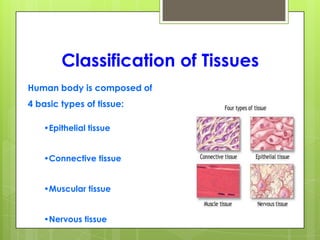
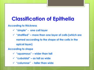
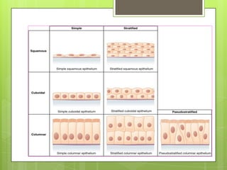
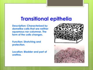
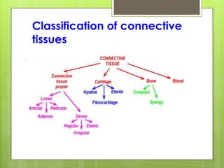
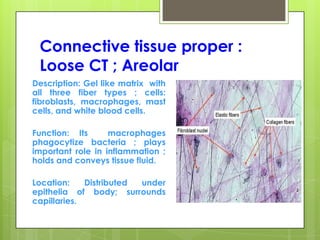
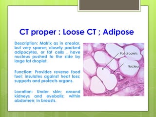
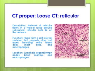
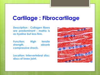
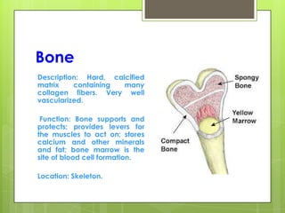
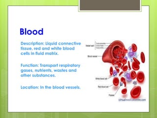
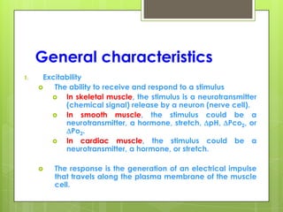
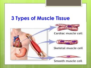
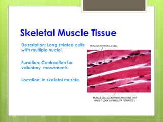
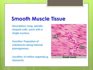
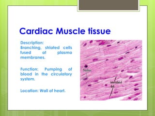
This document provides an overview of the four basic types of tissues in the human body: epithelial, connective, muscular, and nervous tissue. It describes their general characteristics, classifications, and locations. Epithelial tissue covers and lines body surfaces and organs. It is classified based on cell layers and shapes. Connective tissue connects and supports other tissues. Its classifications include connective tissue proper, cartilage, bone, and blood. Muscular tissue is excitable and contractile, with three types: skeletal, smooth, and cardiac. Nervous tissue is the most complex and forms the brain, spinal cord, and nerves to regulate body functions.
. Definition of Tissues Biological tissue is a collection of interconnected cells that perform a similar function within an organism. In other words, it is a group of cells working together mainly inside an organ.
3. Classification of Tissues Human body is composed of 4 basic types of tissue: •Epithelial tissue •Connective tissue •Muscular tissue •Nervous tissue
4. Origin of Tissue A fertilized egg divides to produce 3 primary germ cell layers. These layers differentiate to form the tissues of the body.
5. Epithelial Tissue Epithelial cells cover or line all body surfaces, cavities and tubes. So, These are called covering epithelia. Epithelial cells form the functional units of secretory glands. So, These are called glandular epithelia.
The muscular system is composed of specialized cells called muscle fibers. Their predominant function is contractibility. Muscles, attached to bones or internal organs and blood vessels, are responsible for movement. Nearly all movement in the body is the result of muscle contraction.
This power point helpful for diploma students. this presentation include classification of tissue- epithelial tissue, muscular tissue,skeletal muscle, cardiac muscle, nervous tissue ,difference between smooth muscle and skeletal muscle
Tissues, types and functions(Anatomy)- Easy explanationSwatilekha Das
Easy explanation on Tissues , types of tissues and functions of tissues with pictures.......
Easy anatomy topic for 1 st yera GNM and B.Sc nursing students.....
Skeletal system. anatomy and physiology of skeletal system. appendicular skel...mamtabisht10
SKELETAL SYSTEM
bones, cartilage and ligaments are tightly joined to form a strong, flexible framework called skeletal system
anatomy and physiology of axial and appendicular skeletal system
Axial Skeleton: The axial skeleton includes the skull, spine, ribs and sternum.
Appendicular Skeleton:
The appendicular skeleton includes the appendages of the body, which are the shoulders, arms, hips, and legs.
Cells and its components(Anatomy) Easy explanationSwatilekha Das
Cells and its components,discussion on cell membrane, cytoplasm, nucleus with pictures....
easy explanation of anatomy topic for 1 st year GNM & B.Sc nursing students...
Comment to get explanation on your required topics.....
please like and share and follow.....
detail notes on connective tissue..
Connective tissue (CT) is one of the four basic types of animal tissue, along with epithelial tissue, muscle tissue, and nervous tissue. It develops from the mesoderm. Connective tissue is found in between other tissues everywhere in the body, including the nervous system. In the central nervous system, the three outer membranes (the meninges) that envelop the brain and spinal cord are composed of connective tissue.
All connective tissue consists of three main components: fibers (elastic and collagenous fibers), ground substance and cells. Not all authorities include blood or lymph as connective tissue because they lack the fiber component. All are immersed in the body water.
a quick visual understanding of what actually nervous tissue is made up of at cellular level its functions nerve cell types chemical synapse detailed structure of neuron
HUMAN CYTOLOGY ( CELL AND TISSUE )
PRESENTED BY
BY SONU SHARMA. ROLL NO. 504/16
3RD YEAR B.SC RADIOGRAPHY & IMAGING TECHNOLOGY DEPARTMENT OF RADIO DIAGNOSIS
PANDIT BHAGWAT DAYAL SHARMA
UNIVERSITY OF MEDICAL SCIENCE AND RESEARCH P.G.I.M.S. ROHTAK,HARYANA.
Contact : - +91-7015728336
The cell is the basic structural, functional, and biological unit of all known organisms. A cell is the smallest unit of life. Cells are often called the "building blocks of life". The study of cells is called cell biology, cellular biology, or cytology.
Disassembling Of the old working pc
In this Instruction, I will give a full documentation of how to disassemble a desktop PC, for parts. The specific computer that I will demonstrate with is an HP Media Center PC m7640n with a dead motherboard. Every computer is different, but this is a general guide.
If you don't fully grasp what I mean when I am describing something, please refer to the pictures!
Step 1.
Selection the required tools and equipment:
1. Screw Driver.
2. Multimeter.
3. Soldering iron.
4. Contract cleaner.
5. Brush.
Step 2.
To turn off the power:
If the PC being running, must shut down the pc before disassembling.
There are many ways to shut down your PC—you can turn the PC off completely.
Turn your PC off completely
To shut down:
For Windows 10 Select the Start button and then select Power > Shut down.
For Windows 8.1 / Windows RT 8.1
Move your mouse to the lower left-hand corner of the screen and right-click the Start button or press Windows logo key + X on your keyboard. Tap or click Shut down or sign out and choose Shut down.
For Windows 7 Click the Start button and then click the Shut down button. Click the Start button and then click the Shut down button.
Step 3: Unplugging
The first thing you do, is unplug every cable that's plugged in to your computer. That includes the following cables:
________________________________________
• Power
• USB
• Firewire
• Mouse
• Keyboard
• Internet
• Ethernet
• Modem
• AM\FM Antenna
• Cable TV
• etc...
So pretty much just unplug every cable from your computer.
Step 2: Outer Shell/Casing
Now that your computer is fully unplugged, move your PC to a clean work space, preferably a carpet. The carpet is better than tile, because screws and other small parts will roll around.
NOTICE:
If you are working on a carpet, about every five minutes touch something that is grounded (Sink faucet\pipe, wire coming from the ground part of a wall outlet). This is so you don't shock your motherboard or other parts.
First off, unscrew the four screws on the back of the computer. On most computer cases, there will be large knobs that you can unscrew by hand or by screw driver on the back-right side of the computer. The left side has small screws because on that side you can't access much on the inside.
Once the screws are removed, you can remove the side panels. On most computers, they just slide off. Start with the left side panel (the side that once had the knobs), slide it towards the back of the computer. Now you can remove the left panel. Just like the other one, slide it towards the back of the computer.
Please refer to the pictures if you are confused!
Step 3: Outer Shell/Casing (cont.)
In the last step I removed both side panels. In this step, I will be removing the front and top panels.
________________________________________
Wait For Next Part...
Biological tissue is a collection of interconnected
cells that perform a similar function within an
organism.
In other words, it is a group of cells working together mainly inside an organ.Epithelial cells cover or line all body surfaces, cavities and tubes. So, These are called covering epithelia.Epithelial cells form the functional units of secretory glands. So, These are called glandular epithelia.
Tissues definition and classification, function & location of of epithelium tissues, connective tissues, muscular tissues, nervous tissue etc.. it is usefull for the diploma in pharmacy students , bachelor of pharmacy students and doctor of pharmacy students
Flu Vaccine Alert in Bangalore Karnatakaaddon Scans
As flu season approaches, health officials in Bangalore, Karnataka, are urging residents to get their flu vaccinations. The seasonal flu, while common, can lead to severe health complications, particularly for vulnerable populations such as young children, the elderly, and those with underlying health conditions.
Dr. Vidisha Kumari, a leading epidemiologist in Bangalore, emphasizes the importance of getting vaccinated. "The flu vaccine is our best defense against the influenza virus. It not only protects individuals but also helps prevent the spread of the virus in our communities," he says.
This year, the flu season is expected to coincide with a potential increase in other respiratory illnesses. The Karnataka Health Department has launched an awareness campaign highlighting the significance of flu vaccinations. They have set up multiple vaccination centers across Bangalore, making it convenient for residents to receive their shots.
To encourage widespread vaccination, the government is also collaborating with local schools, workplaces, and community centers to facilitate vaccination drives. Special attention is being given to ensuring that the vaccine is accessible to all, including marginalized communities who may have limited access to healthcare.
Residents are reminded that the flu vaccine is safe and effective. Common side effects are mild and may include soreness at the injection site, mild fever, or muscle aches. These side effects are generally short-lived and far less severe than the flu itself.
Healthcare providers are also stressing the importance of continuing COVID-19 precautions. Wearing masks, practicing good hand hygiene, and maintaining social distancing are still crucial, especially in crowded places.
Protect yourself and your loved ones by getting vaccinated. Together, we can help keep Bangalore healthy and safe this flu season. For more information on vaccination centers and schedules, residents can visit the Karnataka Health Department’s official website or follow their social media pages.
Stay informed, stay safe, and get your flu shot today!
Factory Supply Best Quality Pmk Oil CAS 28578–16–7 PMK Powder in Stockrebeccabio
Factory Supply Best Quality Pmk Oil CAS 28578–16–7 PMK Powder in Stock
Telegram: bmksupplier
signal: +85264872720
threema: TUD4A6YC
You can contact me on Telegram or Threema
Communicate promptly and reply
Free of customs clearance, Double Clearance 100% pass delivery to USA, Canada, Spain, Germany, Netherland, Poland, Italy, Sweden, UK, Czech Republic, Australia, Mexico, Russia, Ukraine, Kazakhstan.Door to door service
Hot Selling Organic intermediates
The prostate is an exocrine gland of the male mammalian reproductive system
It is a walnut-sized gland that forms part of the male reproductive system and is located in front of the rectum and just below the urinary bladder
Function is to store and secrete a clear, slightly alkaline fluid that constitutes 10-30% of the volume of the seminal fluid that along with the spermatozoa, constitutes semen
A healthy human prostate measures (4cm-vertical, by 3cm-horizontal, 2cm ant-post ).
It surrounds the urethra just below the urinary bladder. It has anterior, median, posterior and two lateral lobes
It’s work is regulated by androgens which are responsible for male sex characteristics
Generalised disease of the prostate due to hormonal derangement which leads to non malignant enlargement of the gland (increase in the number of epithelial cells and stromal tissue)to cause compression of the urethra leading to symptoms (LUTS
micro teaching on communication m.sc nursing.pdfAnurag Sharma
Microteaching is a unique model of practice teaching. It is a viable instrument for the. desired change in the teaching behavior or the behavior potential which, in specified types of real. classroom situations, tends to facilitate the achievement of specified types of objectives.
MANAGEMENT OF ATRIOVENTRICULAR CONDUCTION BLOCK.pdfJim Jacob Roy
Cardiac conduction defects can occur due to various causes.
Atrioventricular conduction blocks ( AV blocks ) are classified into 3 types.
This document describes the acute management of AV block.
Anti ulcer drugs and their Advance pharmacology ||
Anti-ulcer drugs are medications used to prevent and treat ulcers in the stomach and upper part of the small intestine (duodenal ulcers). These ulcers are often caused by an imbalance between stomach acid and the mucosal lining, which protects the stomach lining.
||Scope: Overview of various classes of anti-ulcer drugs, their mechanisms of action, indications, side effects, and clinical considerations.
- Video recording of this lecture in English language: https://youtu.be/lK81BzxMqdo
- Video recording of this lecture in Arabic language: https://youtu.be/Ve4P0COk9OI
- Link to download the book free: https://nephrotube.blogspot.com/p/nephrotube-nephrology-books.html
- Link to NephroTube website: www.NephroTube.com
- Link to NephroTube social media accounts: https://nephrotube.blogspot.com/p/join-nephrotube-on-social-media.html
Explore natural remedies for syphilis treatment in Singapore. Discover alternative therapies, herbal remedies, and lifestyle changes that may complement conventional treatments. Learn about holistic approaches to managing syphilis symptoms and supporting overall health.
These simplified slides by Dr. Sidra Arshad present an overview of the non-respiratory functions of the respiratory tract.
Learning objectives:
1. Enlist the non-respiratory functions of the respiratory tract
2. Briefly explain how these functions are carried out
3. Discuss the significance of dead space
4. Differentiate between minute ventilation and alveolar ventilation
5. Describe the cough and sneeze reflexes
Study Resources:
1. Chapter 39, Guyton and Hall Textbook of Medical Physiology, 14th edition
2. Chapter 34, Ganong’s Review of Medical Physiology, 26th edition
3. Chapter 17, Human Physiology by Lauralee Sherwood, 9th edition
4. Non-respiratory functions of the lungs https://academic.oup.com/bjaed/article/13/3/98/278874
Title: Sense of Smell
Presenter: Dr. Faiza, Assistant Professor of Physiology
Qualifications:
MBBS (Best Graduate, AIMC Lahore)
FCPS Physiology
ICMT, CHPE, DHPE (STMU)
MPH (GC University, Faisalabad)
MBA (Virtual University of Pakistan)
Learning Objectives:
Describe the primary categories of smells and the concept of odor blindness.
Explain the structure and location of the olfactory membrane and mucosa, including the types and roles of cells involved in olfaction.
Describe the pathway and mechanisms of olfactory signal transmission from the olfactory receptors to the brain.
Illustrate the biochemical cascade triggered by odorant binding to olfactory receptors, including the role of G-proteins and second messengers in generating an action potential.
Identify different types of olfactory disorders such as anosmia, hyposmia, hyperosmia, and dysosmia, including their potential causes.
Key Topics:
Olfactory Genes:
3% of the human genome accounts for olfactory genes.
400 genes for odorant receptors.
Olfactory Membrane:
Located in the superior part of the nasal cavity.
Medially: Folds downward along the superior septum.
Laterally: Folds over the superior turbinate and upper surface of the middle turbinate.
Total surface area: 5-10 square centimeters.
Olfactory Mucosa:
Olfactory Cells: Bipolar nerve cells derived from the CNS (100 million), with 4-25 olfactory cilia per cell.
Sustentacular Cells: Produce mucus and maintain ionic and molecular environment.
Basal Cells: Replace worn-out olfactory cells with an average lifespan of 1-2 months.
Bowman’s Gland: Secretes mucus.
Stimulation of Olfactory Cells:
Odorant dissolves in mucus and attaches to receptors on olfactory cilia.
Involves a cascade effect through G-proteins and second messengers, leading to depolarization and action potential generation in the olfactory nerve.
Quality of a Good Odorant:
Small (3-20 Carbon atoms), volatile, water-soluble, and lipid-soluble.
Facilitated by odorant-binding proteins in mucus.
Membrane Potential and Action Potential:
Resting membrane potential: -55mV.
Action potential frequency in the olfactory nerve increases with odorant strength.
Adaptation Towards the Sense of Smell:
Rapid adaptation within the first second, with further slow adaptation.
Psychological adaptation greater than receptor adaptation, involving feedback inhibition from the central nervous system.
Primary Sensations of Smell:
Camphoraceous, Musky, Floral, Pepperminty, Ethereal, Pungent, Putrid.
Odor Detection Threshold:
Examples: Hydrogen sulfide (0.0005 ppm), Methyl-mercaptan (0.002 ppm).
Some toxic substances are odorless at lethal concentrations.
Characteristics of Smell:
Odor blindness for single substances due to lack of appropriate receptor protein.
Behavioral and emotional influences of smell.
Transmission of Olfactory Signals:
From olfactory cells to glomeruli in the olfactory bulb, involving lateral inhibition.
Primitive, less old, and new olfactory systems with different path
Prix Galien International 2024 Forum ProgramLevi Shapiro
June 20, 2024, Prix Galien International and Jerusalem Ethics Forum in ROME. Detailed agenda including panels:
- ADVANCES IN CARDIOLOGY: A NEW PARADIGM IS COMING
- WOMEN’S HEALTH: FERTILITY PRESERVATION
- WHAT’S NEW IN THE TREATMENT OF INFECTIOUS,
ONCOLOGICAL AND INFLAMMATORY SKIN DISEASES?
- ARTIFICIAL INTELLIGENCE AND ETHICS
- GENE THERAPY
- BEYOND BORDERS: GLOBAL INITIATIVES FOR DEMOCRATIZING LIFE SCIENCE TECHNOLOGIES AND PROMOTING ACCESS TO HEALTHCARE
- ETHICAL CHALLENGES IN LIFE SCIENCES
- Prix Galien International Awards Ceremony
2. Definition of Tissues
Biological tissue is a collection of interconnected
cells that perform a similar function within an
organism.
In other words, it is a group of cells working
together mainly inside an organ.
3. Classification of Tissues
Human body is composed of
4 basic types of tissue:
•Epithelial tissue
•Connective tissue
•Muscular tissue
•Nervous tissue
4. Origin of Tissue
A fertilized egg divides to produce 3 primary germ cell
layers. These layers differentiate to form the tissues of the
body.
5. Epithelial Tissue
Epithelial cells cover or line all body surfaces, cavities and
tubes. So, These are called covering epithelia.
Epithelial cells form the functional units of secretory glands.
So, These are called glandular epithelia.
6. General Characteristic
Closely attached to each other forming a protective barrier.
Always has one free (apical) surface open to outside the body or
inside (cavity) an internal organ.
Always has one fixed (basal) section attached to underlying
connective tissue.
Has no blood vessels but can soak up nutrients from blood vessels
in connective tissue underneath.
Can have lots of nerves in it (innervated).
Very good at regenerating (fixing itself). i.e. sunburn, skinned
knee.
7. Functions
To protect the tissues that lie beneath it
from radiation, desiccation, toxins, invasion by pathogens, and
physical trauma.
The regulation and exchange of chemicals between the underlying
tissues and a body cavity.
The secretion of hormones into the blood vascular system, and/or
the secretion of sweat, mucus, enzymes, and other products that are
delivered by ducts glandular epithelium.
To provide sensation.
Absorbs stomach and intestinal lining (gut).
Filters the kidney.
8. Classification of Epithelia
According to thickness
“simple” - one cell layer
“stratified” – more than one layer of cells (which are
named according to the shape of the cells in the
apical layer)
According to shape
“squamous” – wider than tall
“cuboidal” – as tall as wide
“columnar” - taller than wide
9.
10. Simple squamous epithelium
Description: Single layer of
flattened cells with disc-shaped
central nuclei and sparse
cytoplasm; the simplest of the
epithelia.
Function: Passive transport of gases
and fluids.
Location:Alveoli of lungs, lining
body cavities (mesothelium), lining
blood vessels (endothelium)
11. Simple cuboidal epithelia
Description : Single layer of
cubelike cells with large,
spherical central nuclei.
Function : Secretion and
absorption.
Location: Kidney tubules;
ducts and secretory portions
of small glands; ovary
surface.
12. Simple columnar epithelia
Description: Single layer of tall
cells with round to oval
nuclei.
Types: (i)Ciliated columnar
epithelia.
(ii)Non-ciliated
columnar epithelia.
Function: Absorption;
secretion of mucus,
Enzymes and other
substances.
Location: Digestive tract, gall
bladder etc.
14. Stratified cuboidal epithelia
Description: Generally two
layers of cube-like cells.
Function: Protection.
Location: Large ducts of sweat
glands, mammary glands,
and salivary glands.
16. Pseudo stratified columnar
epithelia
Description: Single cell layered,
all cell attach to the basement
membrane but not all reach the
free surface. Nuclei at varying
depth.
Function: Secretion of mucus,
propulsion of mucus by ciliary
action.
Location: Lines of trachea.
17. Transitional epithelia
Description: Characterized by
domelike cells that are neither
squamous nor columnar. The
form of the cells changes.
Function: Stretching and
protection.
Location: Bladder and part of
urethra.
19. General characteristic
The intercellular material is maximum where as the
cellular component is minimum.
Unlike the other tissues, (e.g. epithelium, muscle and
nerve) which are formed mainly by cells, the major
constituent of connective tissue is ECM (Extra-cellular
matrix).
Possess cells, fibers and ground substances.
20. Basic Functions
Support and binding of other tissues
Holding body fluids
Defending the body against infection
macrophages, plasma cells, mast cells, WBCs
Storing nutrients as fat
22. Connective tissue proper :
Loose CT ; Areolar
Description: Gel like matrix with
all three fiber types ; cells:
fibroblasts, macrophages, mast
cells, and white blood cells.
Function: Its macrophages
phagocytize bacteria ; plays
important role in inflammation ;
holds and conveys tissue fluid.
Location: Distributed under
epithelia of body; surrounds
capillaries.
23. CT proper : Loose CT ; Adipose
Description: Matrix as in areolar,
but very sparse; closely packed
adipocytes, or fat cells , have
nucleus pushed to the side by
large fat droplet.
Function: Provides reverse food
fuel; insulates against heat loss;
supports and protects organs.
Location: Under skin; around
kidneys and eyeballs; within
abdomen; in breasts.
Fat droplets
Nucleus
24. CT proper: Loose CT; reticular
Description: Network of reticular
fibers in a typical loose ground
substance; reticular cells lie on
the network.
Function: Fibers form a soft internal
skeleton that supports other cell
types including white blood
cells, mast cells, and
macrophages.
Location: Lymphoid organs(lymph
nodes, bone marrow, and
macrophages.
25. CT proper: dense CT; Irregular
Description: Primarily irregularly
arranged collagen fibers; some
elastic fibers; major cell type is
the fibroblast.
Function: Elasticity and structural
support.
Location: Dermis of the skin;
submucosa of digestive tract and
joints.
Collagen
fibers
26. CT proper: dense CT; Regular
Description: Primarily parallel
collagen fibers; a few elastin
fibers; major cell type is the
fibroblast.
Function: Attaches muscles to
bones and bones to bones.
Location: Tendons and in most
ligaments.
27. Cartilage: Hyaline
Description: Amorphous but
firm matrix; chondroblasts
produce the matrix and when
mature lie in lacunae.
Function: Supports and
reinforces; resists compressive
stress.
Location: Forms most of the
embryonic skeleton; ends of
long bones; cartilages of nose.
28. Cartilage: Elastic
Description: Similar to hyaline
cartilage, but more elastic
fibers in matrix.
Function: Maintains the shape
and allows flexibility.
Location: Supports the external
ear(pinna).
29. Cartilage : Fibrocartilage
Description : Collagen fibers
are predominant ; matrix is
as hyaline but less firm.
Function: High tensile
strength, absorb
compressive shock.
Location: Intervertebral disc;
discs of knee joint.
30. Bone
Description: Hard, calcified
matrix containing many
collagen fibers. Very well
vascularized.
Function: Bone supports and
protects; provides levers for
the muscles to act on; stores
calcium and other minerals
and fat; bone marrow is the
site of blood cell formation.
Location: Skeleton.
31. Blood
Description: Liquid connective
tissue, red and white blood
cells in fluid matrix.
Function: Transport respiratory
gases, nutrients, wastes and
other substances.
Location: In the blood vessels.
32. Muscle Tissue
Muscle is one of our 4 tissue types and muscle tissue
combined with nerves, blood vessels, and various
connective tissues.
Muscles are quite complex and as we’ll find out, they
are a marvel of both biology and physics.
33. General characteristics
1. Excitability
The ability to receive and respond to a stimulus
In skeletal muscle, the stimulus is a neurotransmitter
(chemical signal) release by a neuron (nerve cell).
In smooth muscle, the stimulus could be a
neurotransmitter, a hormone, stretch, pH, Pco2, or
Po2.
In cardiac muscle, the stimulus could be a
neurotransmitter, a hormone, or stretch.
The response is the generation of an electrical impulse
that travels along the plasma membrane of the muscle
cell.
34. 2. Contractility
The ability to shorten forcibly when adequately
stimulated.
This is the defining property of muscle tissue.
2. Extensibility
The ability to be stretched (Extended)
4. Elasticity
The ability to recoil and resume original length
after being stretched.
37. Skeletal Muscle Tissue
Description: Long striated cells
with multiple nuclei.
Function: Contraction for
voluntary movements.
Location: In skeletal muscle.
38. Smooth Muscle Tissue
Description: Long, spindle-
shaped cells, each with a
single nucleus.
Function: Propulsion of
substances along internal
passageways.
Location: In hollow organs(e.g.
stomach)
40. Nerve Tissue
By far the most complex tissue in the human body is
nerve tissue.
Formed by a network of more than 100 million nerve
cells, assisted by many more glial cells.
Each neuron has, on an average , at least a
thousand interconnection with other neurons forming
a very complex nervous system.
42. Composition of Nerve Tissue
The nerve tissue is composed of two elements:
1. The nerve cell or neuron
2. The neuroglia
43. Neuron
Description: Neurons are
branching cells; cell processes
that may be quite long extend
from the nucleus-containing cell
body.
Function: Transmit electrical
signals from sensory receptors
and to effectors(muscles and
glands) that control their
activity.
Location: Brain, spinal cord and
nerves.
44. Glial cells
Glia carry nutrients, speed
repair, provide myelin for
axons, support the blood-
brain barrier, and may form
their own communication
network. They are also
involved in neurogenesis.