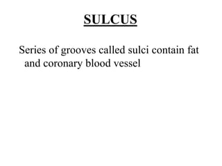
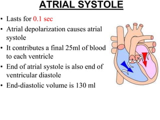
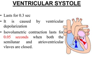
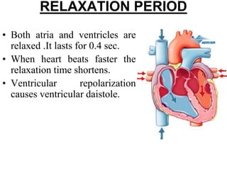
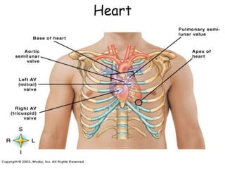
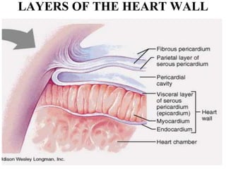
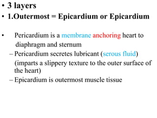
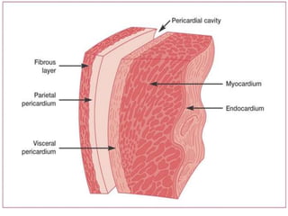
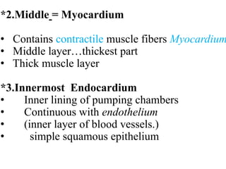
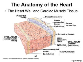
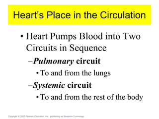
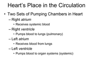
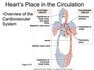
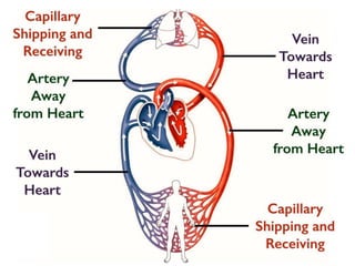
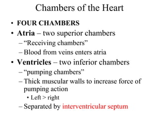
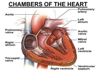
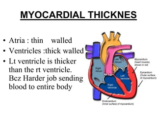
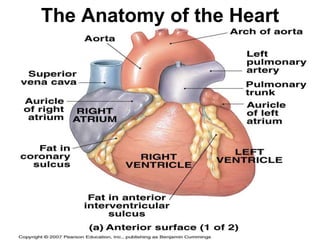
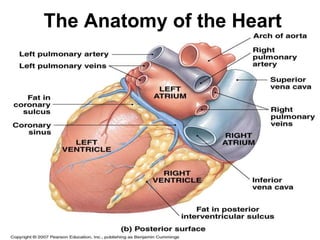
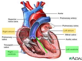
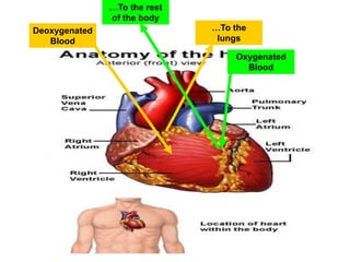
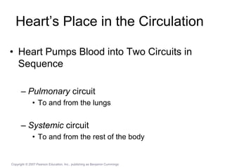
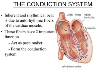
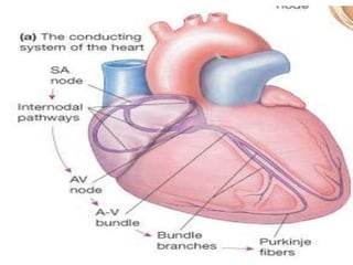
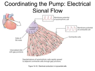
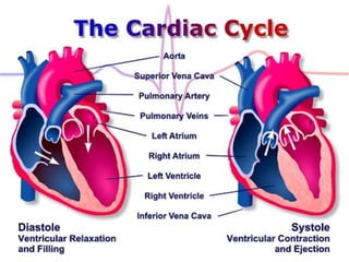
Heart Anatomy &
Basic Function
Presented By
Vijay Salvekar
Dept. of Pharmacology
GRY Institute of Pharmacy
Heart Location
• 4 chamber muscular organ
• Comparable to the size of a closed fist
• Located in the mediastinum
• (MIDLINE OF THE THORACIC CAVITY)
– Behind sternum
– Between 2nd and 6th ribs
– Between T5-T8
• Apex – base of heart
– Located at the 5th intercostal space
-300 grams (size of a fist)
Heart
Cardiovascular Function
• Cardiovascular = Heart, Arteries, Veins, Blood
• Function:
–Transportation
–Blood = transport vehicle
–Carries oxygen, nutrients, wastes, and
hormones
–Movement provided by pumping of heart
• PERICARDIUM – loose fitting sac surrounding the
heart.
• Function: Allows sufficient freedom of movement.
• CONSIST 2 PARTS
• 1.Fibrous pericardium 2.Serous pericardium
• 1.Fibrous pericardium
Tough, loose-fitting, inelastic thin and dense
irregular connective tissue
• Function : Helps in protection, anchors heart to
mediastinum
Coverings of the Heart
*2.Serous pericardium
Thinner, more delicate divided into Parietal
and visceral
i. Parietal layer: lines the inside of the fibrous
pericardium
ii. Visceral layer: adheres to outside of the heart
*Pericardial space: between parietal and visceral
layer
Filled with 10-15mL of pericardial fluid
Decreases friction
LAYERS OF THE HEART WALL
• 3 layers
• 1.Outermost = Epicardium or Epicardium
• Pericardium is a membrane anchoring heart to
diaphragm and sternum
– Pericardium secretes lubricant (serous fluid)
(imparts a slippery texture to the outer surface of
the heart)
– Epicardium is outermost muscle tissue
*2.Middle = Myocardium
• Contains contractile muscle fibers Myocardium
• Middle layer…thickest part
• Thick muscle layer
*3.Innermost Endocardium
• Inner lining of pumping chambers
• Continuous with endothelium
• (inner layer of blood vessels.)
• simple squamous epithelium
The Anatomy of the Heart
• The Heart Wall and Cardiac Muscle Tissue
Figure 12-4(a)
Heart’s Place in the Circulation
• Three Kinds of Blood Vessels
– Arteries
• Carry blood Away from heart and carry it to the capillaries
– Capillaries
• Microscopic vessels where exchange between cells and
blood takes place
– Veins
• Receive blood from capillaries and carry it back to the heart
Heart’s Place in the Circulation
• Heart Pumps Blood into Two
Circuits in Sequence
–Pulmonary circuit
•To and from the lungs
–Systemic circuit
•To and from the rest of the body
Copyright © 2007 Pearson Education, Inc., publishing as Benjamin Cummings
Heart’s Place in the Circulation
• Two Sets of Pumping Chambers in Heart
– Right atrium
• Receives systemic blood
– Right ventricle
• Pumps blood to lungs (pulmonary)
– Left atrium
• Receives blood from lungs
– Left ventricle
• Pumps blood to organ systems (systemic)
Heart’s Place in the Circulation
•Overview of the
Cardiovascular
System
Figure 12-1
Superior vena cava
inferior vena cava
aorta
Chambers of the Heart
• FOUR CHAMBERS
• Atria – two superior chambers
– “Receiving chambers”
– Blood from veins enters atria
• Ventricles – two inferior chambers
– “pumping chambers”
– Thick muscular walls to increase force of
pumping action
• Left > right
– Separated by interventricular septum
CHAMBERS OF THE HEART
Valves of the Heart
• Permit blood flow in one direction during
circulation
1.Atrioventricular valves (AV valves)
– Also cuspid valves
– Between atria and ventricles
2.Semilunar (SL valves)
– Between R ventricle and pulmonary arteries
and L ventricle and aorta
Atrioventricular Valves
• Tricuspid valve
– Between R atrium and ventricle
– 3 flaps of endocardium
– Connected to ventricular papillary muscle
via chordae tendinae
• Bicuspid valve
– Between L atrium and ventricle
– Also called mitral valve
– Two flaps of endocardium
Semilunar Valves
• Pulmonary semilunar valve
– Btwn R ventricle and pulmonary trunk
• Aorta semilunar valve
– Btwn L ventricle and aorta
The Anatomy of the Heart
• The Valves of the Heart
Figure 12-6(a)
• The Valves of the Heart
Figure 12-6(b)
Series of grooves called sulci contain fat
and coronary blood vessel
SULCUS
SULCUS
MYOCARDIAL THICKNES
• Atria : thin walled
• Ventricles :thick walled
• Lt ventricle is thicker
than the rt ventricle.
Bcz Harder job sending
blood to entire body
The Anatomy of the Heart
Deoxygenated
Blood
…To the
lungs
Oxygenated
Blood
…To the rest
of the body
Heart’s Place in the Circulation
• Heart Pumps Blood into Two Circuits in
Sequence
– Pulmonary circuit
• To and from the lungs
– Systemic circuit
• To and from the rest of the body
Copyright © 2007 Pearson Education, Inc., publishing as Benjamin Cummings
(4) Pulmonary Circulation
• Pulmonary = Deoxygenated Blood
• Involves Right Side of Heart
• Pathway:
1. Superior / Inferior Vena Cava
2. Right Atrium  Tricuspid Valve
3. Right Ventricle  Pulmonary Semilunar Valve
4. Left Pulmonary Artery
5. Lungs
(5) Systemic Circulation
• Systemic = Oxygenated Blood
• Involves Left Side of Heart
• Pathway:
1. Left Pulmonary Vein
2. Left Atrium  Bicuspid Valve
3. Left Ventricle  Aortic Semilunar Valve
4. Aorta
5. All Other Tissues
The Blood Supply to the Heart
. Coronary Arteries
left main coronary artery
left anterior descending artery: serves the
interventricular septum and anterior walls
of both ventricles
circumflex artery: serves the left atrium and
posterior wall of the left ventricle
Coronary Arteries
the coronary arteries arise from the base of the aorta and
actively deliver blood only when the heart is relaxed
the heart is 0.5% of body weight and receives 5% of the
body's blood supply (most to the left ventricle)
•Coronary Arteries
•Right main coronary artery
•posterior descending artery: serves the
posterior walls of both ventricles
•marginal artery: lateral wall of the right side
of the heart
•Cardiac Veins follow arteries and join at the
Coronary Sinus which empties blood into the
right atrium
•Coronary circulation meets heavy demands of myocardium for
oxygen, nutrients
•.Coronary arteries (right, left) branch from aorta base
Drainage is to right atrium
•.Great, middle cardiac veins drain capillaries into coronary
sinus
• The Coronary Circulation
Figure 12-7(a)
• The Coronary Circulation
Figure 12-7(b)
What causes a heart attack?
• Embolism = a blood clot in
an artery
• Build-up of “plaque” in
walls of an artery
• These can lead to restriction
or cessation of blood flow
in the afflicted artery.
• A portion of the mycardium
may die (myocardial
infarction).
THE CONDUCTION SYSTEM
• Inherent and rhythmical beat
is due to autorhythmic fibers
of the cardiac muscle.
• These fibers have 2 important
function
- Act as pace maker
- Form the conduction
system
• SA node would inititates action potential about
every 0.6 sec or 100 times/min
• The ANS alters the strength and timing of
heart beats.
Heart Physiology
– Electrical Events
• Intrinsic Conduction System of the Heart
– the ability of cardiac muscle to depolarize
and contract is intrinsic (no nervous
stimulation is required)
– nerve impulses can alter the basic rhythm of
heart activity set by intrinsic factors
Heart Physiology
– Electrical Events
• Action Potential Generated by
Autorhythmic Cells
– Sequence of Excitation
» Sinoatrial Node
» Atrioventricular Node
» Atrioventricular Bundle (bundle of
His)
» Bundle Branches
» Purkinje Fibers
Heart Physiology
– Electrical Events
• Extrinsic Innervation of the Heart
– fibers of autonomic nervous system
accelerate or inhibit the basic rate of
heartbeat set by the intrinsic conduction
system
Heart Physiology
– Electrical Events
• Electrocardiography
 electrical currents generated and
transmitted through the heart spread
throughout the body and can be
monitored
 the graphic recording of electrical
changes during heart activity is called an
electrocardiogram (ECG or EKG)
Heart Physiology
– Electrical Events
• Electrocardiography
 the ECG consists of series of three waves
» P Wave: atrial depolarization starting at
the SA node
» QRS Complex: ventricular
depolarization
Heart Physiology
– Electrical Events
• Electrocardiography
» P-R (P-Q) interval: time from the
beginning of atrial excitation to the
beginning of ventricular excitation and
includes the contraction of the atria
and the passage of the depolarization
wave through the rest of the
conduction system
Heart Physiology
–Electrical Events
• Electrocardiography
»T Wave: ventricular repolarization
»Q-T interval: time from the beginning of
the ventricular depolarization through
their repolarization and includes the
contraction of the ventricles
Coordinating the Pump: Electrical
Signal Flow
Figure 14-18: Electrical conduction in myocardial cells
PHYSIOLOGIC
CHARACTERISTICS OF THE
CONDUCTION CELLS
• AUTOMATICITY
• EXCITABILITY
• CONDUCTIVITY
• RHYTHMICITY
• CONTRACTILITY
• TONICITY
CARDIAC CYCLE
ATRIAL SYSTOLE
• Lasts for 0.1 sec
• Atrial depolarization causes atrial
systole
• It contributes a final 25ml of blood
to each ventricle
• End of atrial systole is also end of
ventricular diastole
• End-diastolic volume is 130 ml
VENTRICULAR SYSTOLE
• Lasts for 0.3 sec
• It is caused by ventricular
depolarization
• Isovolumetric contraction lasts for
0.05 seconds when both the
semilunar and atrioventricular
vlaves are closed.
• The sl valves open when
-the left ventricular pressures surpasses aortic
pressure(80 mm of mercury)
-The right ventricular pressure rises above
pulmonary pressure (20 mmhg)
• Sl valves open for 0.25 sec
• The left ventricle ejects about 70 ml into the
aorta
• The right ventricle ejects the same volume into
the pulmonary trunk.
• End systolic volume is 60ml in each ventricle .
RELAXATION PERIOD
• Both atria and ventricles are
relaxed .It lasts for 0.4 sec.
• When heart beats faster the
relaxation time shortens.
• Ventricular repolarization
causes ventricular daistole.
Thankyou

Human heart

  • 1.
    Heart Anatomy & BasicFunction Presented By Vijay Salvekar Dept. of Pharmacology GRY Institute of Pharmacy
  • 2.
    Heart Location • 4chamber muscular organ • Comparable to the size of a closed fist • Located in the mediastinum • (MIDLINE OF THE THORACIC CAVITY) – Behind sternum – Between 2nd and 6th ribs – Between T5-T8 • Apex – base of heart – Located at the 5th intercostal space -300 grams (size of a fist)
  • 3.
  • 4.
    Cardiovascular Function • Cardiovascular= Heart, Arteries, Veins, Blood • Function: –Transportation –Blood = transport vehicle –Carries oxygen, nutrients, wastes, and hormones –Movement provided by pumping of heart
  • 5.
    • PERICARDIUM –loose fitting sac surrounding the heart. • Function: Allows sufficient freedom of movement. • CONSIST 2 PARTS • 1.Fibrous pericardium 2.Serous pericardium • 1.Fibrous pericardium Tough, loose-fitting, inelastic thin and dense irregular connective tissue • Function : Helps in protection, anchors heart to mediastinum Coverings of the Heart
  • 6.
    *2.Serous pericardium Thinner, moredelicate divided into Parietal and visceral i. Parietal layer: lines the inside of the fibrous pericardium ii. Visceral layer: adheres to outside of the heart *Pericardial space: between parietal and visceral layer Filled with 10-15mL of pericardial fluid Decreases friction
  • 9.
    LAYERS OF THEHEART WALL
  • 10.
    • 3 layers •1.Outermost = Epicardium or Epicardium • Pericardium is a membrane anchoring heart to diaphragm and sternum – Pericardium secretes lubricant (serous fluid) (imparts a slippery texture to the outer surface of the heart) – Epicardium is outermost muscle tissue
  • 12.
    *2.Middle = Myocardium •Contains contractile muscle fibers Myocardium • Middle layer…thickest part • Thick muscle layer *3.Innermost Endocardium • Inner lining of pumping chambers • Continuous with endothelium • (inner layer of blood vessels.) • simple squamous epithelium
  • 13.
    The Anatomy ofthe Heart • The Heart Wall and Cardiac Muscle Tissue Figure 12-4(a)
  • 14.
    Heart’s Place inthe Circulation • Three Kinds of Blood Vessels – Arteries • Carry blood Away from heart and carry it to the capillaries – Capillaries • Microscopic vessels where exchange between cells and blood takes place – Veins • Receive blood from capillaries and carry it back to the heart
  • 15.
    Heart’s Place inthe Circulation • Heart Pumps Blood into Two Circuits in Sequence –Pulmonary circuit •To and from the lungs –Systemic circuit •To and from the rest of the body Copyright © 2007 Pearson Education, Inc., publishing as Benjamin Cummings
  • 16.
    Heart’s Place inthe Circulation • Two Sets of Pumping Chambers in Heart – Right atrium • Receives systemic blood – Right ventricle • Pumps blood to lungs (pulmonary) – Left atrium • Receives blood from lungs – Left ventricle • Pumps blood to organ systems (systemic)
  • 17.
    Heart’s Place inthe Circulation •Overview of the Cardiovascular System Figure 12-1 Superior vena cava inferior vena cava aorta
  • 19.
    Chambers of theHeart • FOUR CHAMBERS • Atria – two superior chambers – “Receiving chambers” – Blood from veins enters atria • Ventricles – two inferior chambers – “pumping chambers” – Thick muscular walls to increase force of pumping action • Left > right – Separated by interventricular septum
  • 20.
  • 21.
    Valves of theHeart • Permit blood flow in one direction during circulation 1.Atrioventricular valves (AV valves) – Also cuspid valves – Between atria and ventricles 2.Semilunar (SL valves) – Between R ventricle and pulmonary arteries and L ventricle and aorta
  • 22.
    Atrioventricular Valves • Tricuspidvalve – Between R atrium and ventricle – 3 flaps of endocardium – Connected to ventricular papillary muscle via chordae tendinae • Bicuspid valve – Between L atrium and ventricle – Also called mitral valve – Two flaps of endocardium
  • 23.
    Semilunar Valves • Pulmonarysemilunar valve – Btwn R ventricle and pulmonary trunk • Aorta semilunar valve – Btwn L ventricle and aorta
  • 24.
    The Anatomy ofthe Heart • The Valves of the Heart Figure 12-6(a)
  • 25.
    • The Valvesof the Heart Figure 12-6(b)
  • 26.
    Series of groovescalled sulci contain fat and coronary blood vessel SULCUS
  • 27.
  • 28.
    MYOCARDIAL THICKNES • Atria: thin walled • Ventricles :thick walled • Lt ventricle is thicker than the rt ventricle. Bcz Harder job sending blood to entire body
  • 29.
    The Anatomy ofthe Heart
  • 30.
    The Anatomy ofthe Heart
  • 32.
  • 33.
    Heart’s Place inthe Circulation • Heart Pumps Blood into Two Circuits in Sequence – Pulmonary circuit • To and from the lungs – Systemic circuit • To and from the rest of the body Copyright © 2007 Pearson Education, Inc., publishing as Benjamin Cummings
  • 34.
    (4) Pulmonary Circulation •Pulmonary = Deoxygenated Blood • Involves Right Side of Heart • Pathway: 1. Superior / Inferior Vena Cava 2. Right Atrium  Tricuspid Valve 3. Right Ventricle  Pulmonary Semilunar Valve 4. Left Pulmonary Artery 5. Lungs
  • 36.
    (5) Systemic Circulation •Systemic = Oxygenated Blood • Involves Left Side of Heart • Pathway: 1. Left Pulmonary Vein 2. Left Atrium  Bicuspid Valve 3. Left Ventricle  Aortic Semilunar Valve 4. Aorta 5. All Other Tissues
  • 38.
    The Blood Supplyto the Heart . Coronary Arteries left main coronary artery left anterior descending artery: serves the interventricular septum and anterior walls of both ventricles circumflex artery: serves the left atrium and posterior wall of the left ventricle Coronary Arteries the coronary arteries arise from the base of the aorta and actively deliver blood only when the heart is relaxed the heart is 0.5% of body weight and receives 5% of the body's blood supply (most to the left ventricle)
  • 39.
    •Coronary Arteries •Right maincoronary artery •posterior descending artery: serves the posterior walls of both ventricles •marginal artery: lateral wall of the right side of the heart •Cardiac Veins follow arteries and join at the Coronary Sinus which empties blood into the right atrium
  • 40.
    •Coronary circulation meetsheavy demands of myocardium for oxygen, nutrients •.Coronary arteries (right, left) branch from aorta base Drainage is to right atrium •.Great, middle cardiac veins drain capillaries into coronary sinus
  • 42.
    • The CoronaryCirculation Figure 12-7(a)
  • 43.
    • The CoronaryCirculation Figure 12-7(b)
  • 44.
    What causes aheart attack? • Embolism = a blood clot in an artery • Build-up of “plaque” in walls of an artery • These can lead to restriction or cessation of blood flow in the afflicted artery. • A portion of the mycardium may die (myocardial infarction).
  • 46.
    THE CONDUCTION SYSTEM •Inherent and rhythmical beat is due to autorhythmic fibers of the cardiac muscle. • These fibers have 2 important function - Act as pace maker - Form the conduction system
  • 47.
    • SA nodewould inititates action potential about every 0.6 sec or 100 times/min • The ANS alters the strength and timing of heart beats.
  • 48.
    Heart Physiology – ElectricalEvents • Intrinsic Conduction System of the Heart – the ability of cardiac muscle to depolarize and contract is intrinsic (no nervous stimulation is required) – nerve impulses can alter the basic rhythm of heart activity set by intrinsic factors
  • 49.
    Heart Physiology – ElectricalEvents • Action Potential Generated by Autorhythmic Cells – Sequence of Excitation » Sinoatrial Node » Atrioventricular Node » Atrioventricular Bundle (bundle of His) » Bundle Branches » Purkinje Fibers
  • 50.
    Heart Physiology – ElectricalEvents • Extrinsic Innervation of the Heart – fibers of autonomic nervous system accelerate or inhibit the basic rate of heartbeat set by the intrinsic conduction system
  • 51.
    Heart Physiology – ElectricalEvents • Electrocardiography  electrical currents generated and transmitted through the heart spread throughout the body and can be monitored  the graphic recording of electrical changes during heart activity is called an electrocardiogram (ECG or EKG)
  • 52.
    Heart Physiology – ElectricalEvents • Electrocardiography  the ECG consists of series of three waves » P Wave: atrial depolarization starting at the SA node » QRS Complex: ventricular depolarization
  • 53.
    Heart Physiology – ElectricalEvents • Electrocardiography » P-R (P-Q) interval: time from the beginning of atrial excitation to the beginning of ventricular excitation and includes the contraction of the atria and the passage of the depolarization wave through the rest of the conduction system
  • 54.
    Heart Physiology –Electrical Events •Electrocardiography »T Wave: ventricular repolarization »Q-T interval: time from the beginning of the ventricular depolarization through their repolarization and includes the contraction of the ventricles
  • 56.
    Coordinating the Pump:Electrical Signal Flow Figure 14-18: Electrical conduction in myocardial cells
  • 57.
    PHYSIOLOGIC CHARACTERISTICS OF THE CONDUCTIONCELLS • AUTOMATICITY • EXCITABILITY • CONDUCTIVITY • RHYTHMICITY • CONTRACTILITY • TONICITY
  • 58.
  • 59.
    ATRIAL SYSTOLE • Lastsfor 0.1 sec • Atrial depolarization causes atrial systole • It contributes a final 25ml of blood to each ventricle • End of atrial systole is also end of ventricular diastole • End-diastolic volume is 130 ml
  • 60.
    VENTRICULAR SYSTOLE • Lastsfor 0.3 sec • It is caused by ventricular depolarization • Isovolumetric contraction lasts for 0.05 seconds when both the semilunar and atrioventricular vlaves are closed.
  • 61.
    • The slvalves open when -the left ventricular pressures surpasses aortic pressure(80 mm of mercury) -The right ventricular pressure rises above pulmonary pressure (20 mmhg) • Sl valves open for 0.25 sec
  • 62.
    • The leftventricle ejects about 70 ml into the aorta • The right ventricle ejects the same volume into the pulmonary trunk. • End systolic volume is 60ml in each ventricle .
  • 63.
    RELAXATION PERIOD • Bothatria and ventricles are relaxed .It lasts for 0.4 sec. • When heart beats faster the relaxation time shortens. • Ventricular repolarization causes ventricular daistole.
  • 64.