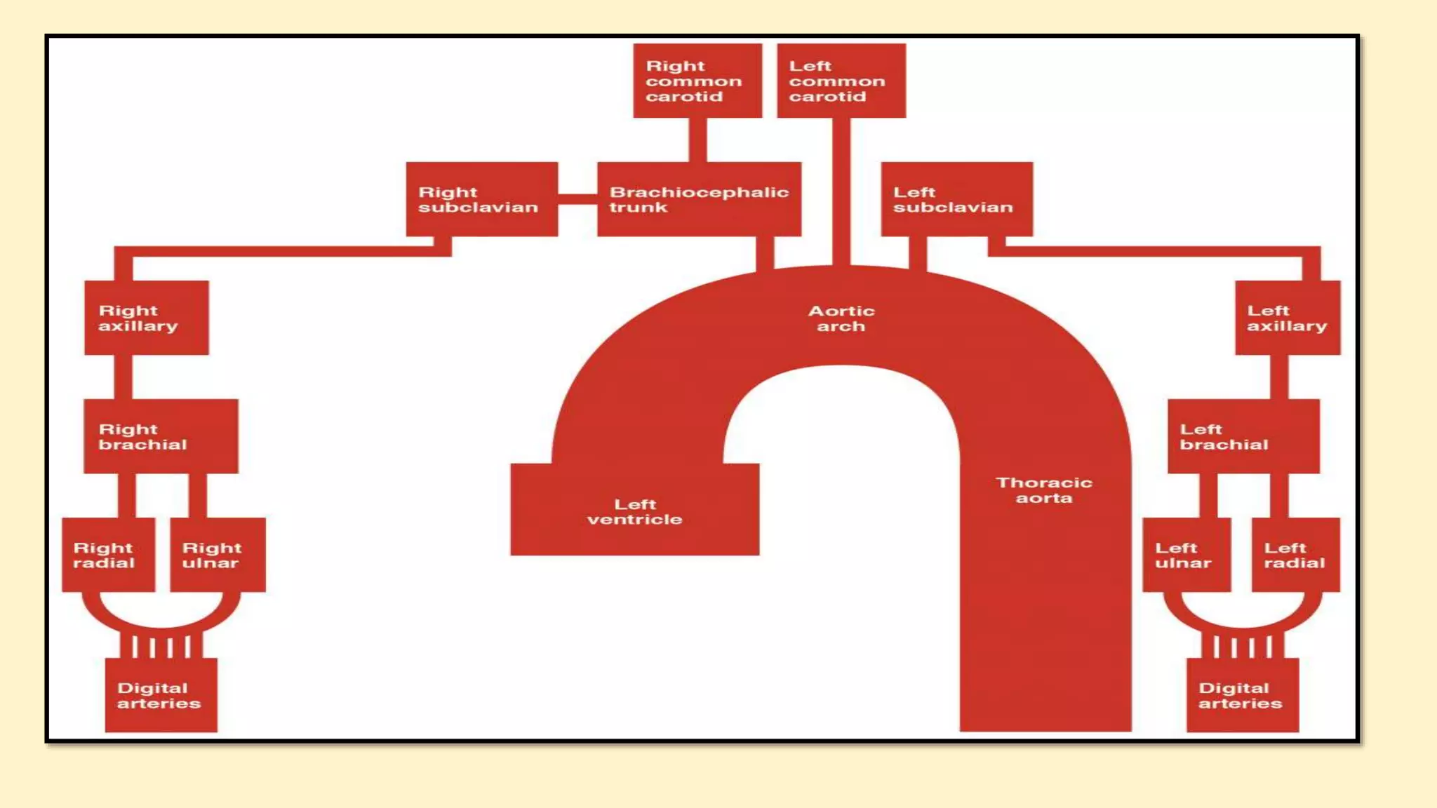
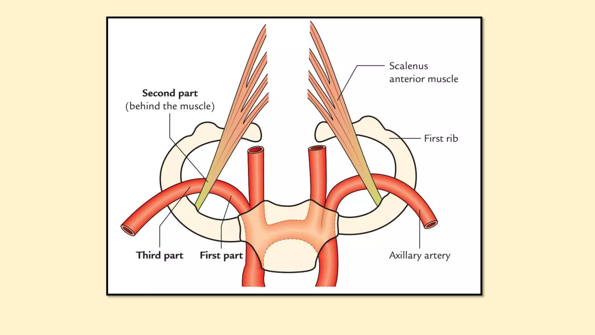
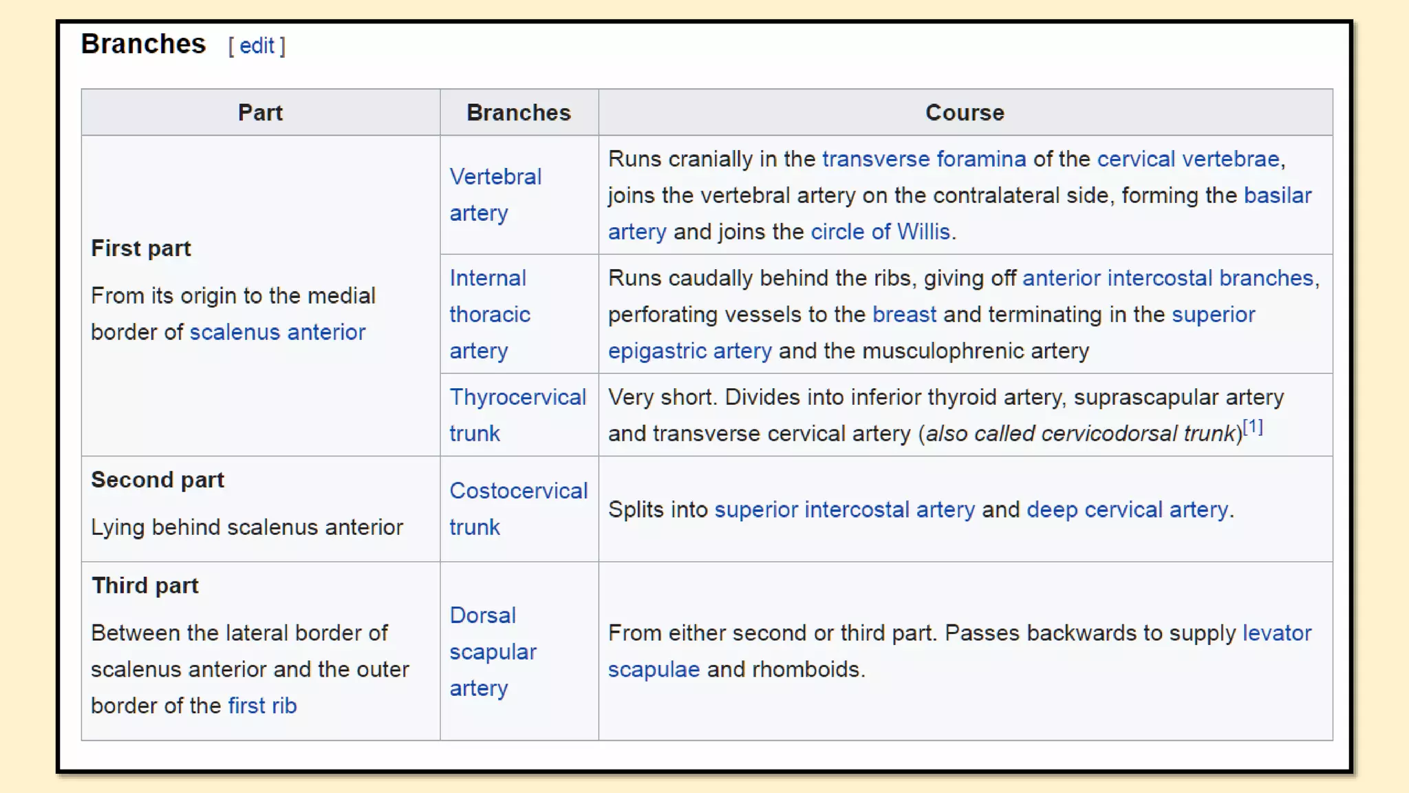
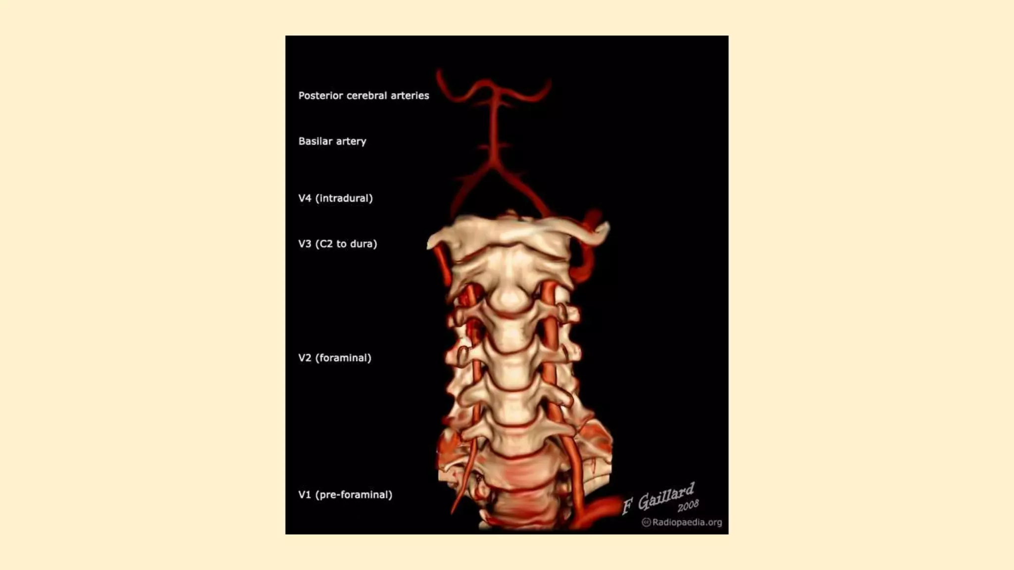
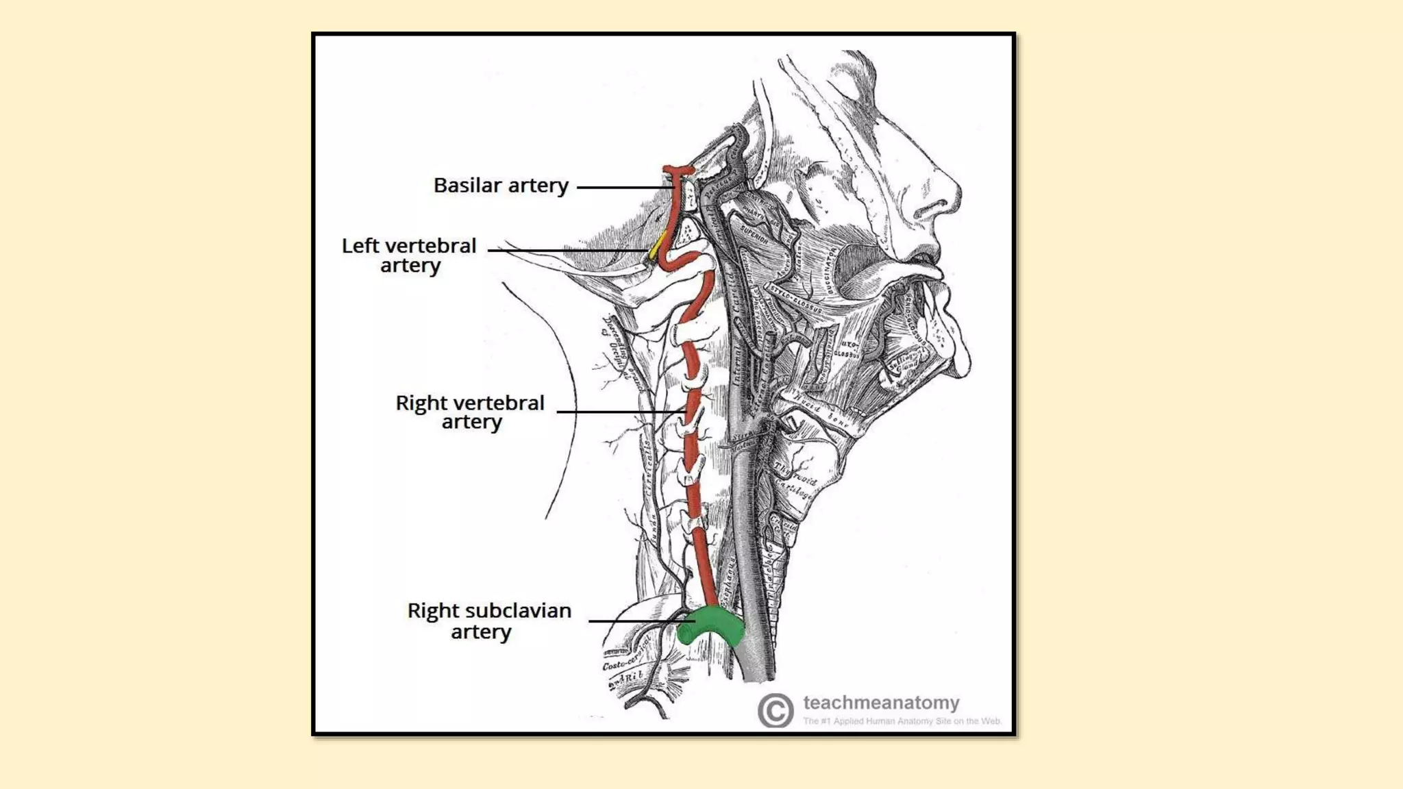
SUPEX
ARTERY
The arterial supply to the upper limb is delivered via five
main vessels (proximal to distal):
1. Subclavian artery
2. Axillary artery
3. Brachial artery
4. Radial artery
5. Ulnar artery
SUBCLAVIAN ARTERY
It is found below the clavicle that is why it is called subcalvian
artery.
It’s the primary source of blood supply to the upper limb and
therefore termed artery of the upper limb.
On the other hand, the subclavian artery also supplies
appreciable part of the thoracic wall, head, neck and brain via its
branches.
OVERVIEW
ORIGIN
The arterial supply to the upper limb
begins as the subclavian artery. On the
right, the subclavian artery arises from
the brachiocephalic trunk. On the left, it
branches directly from the arch of aorta.
COURSE
On every side, the subclavian artery arches
laterally across the anterior surface of the cervical
pleura onto the very first rib posterior to the
scalenus anterior muscle.
At the outer border of 1st rib, it finishes by
becoming axillary artery.
PARTS
On every side, the subclavian artery is split into 3 parts by the
scalenus anterior muscle. These are:
First part-goes from origin to medial border of scalenus anterior.
Second part-is located behind the scalenus anterior muscle.
Third part-goes from the lateral border of scalenus anterior to the
outer border of the very first rib.
BRANCHES OF THE SUBCLAVIAN ARTERY
Very Indignant Tired Individuals Sip Strong Coffee Served
Double DailyVery Indignant Tired Individuals Sip Strong Coffee Served
Double Daily
V: vertebral artery
I: internal thoracic artery
T: thyrocervical trunk
I: inferior thyroid artery
S: superficial cervical artery
S: suprascapular artery
C: costocervical artery
S: superior intercostal artery
D: deep cervical artery
VERTEBRAL ARTERIES
https://www.youtube.com/watch?v=Reo3cVeaKAAThe Vertebral Artery
https://www.youtube.com/watch?v=dmPWJIvHsDAAnatomy of The Vertebral Artery
The right and left vertebral arteries arise from the subclavian arteries, medial to
the anterior scalene muscle. They then ascend the posterior aspect of the neck,
through holes in the transverse processes of the cervical vertebrae, known as
foramen transversarium.
The vertebral arteries enter the cranial cavity via the foramen magnum. Within
the cranial vault, some branches are given off:
Segments
The vertebral artery is typically divided into 4 segments:
V1 (preforaminal): origin to transverse foramen of C6
V2 (foraminal): from the transverse foramen of C6 to the
transverse foramen of C2
V3 (atlantic, extradural or extraspinal): from C2 to the dura
V4 (intradural or intracranial): from the dura to their
confluence to form the basilar artery
V1: segmental cervical muscular and spinal branches
V2: anterior meningeal artery, muscular and spinal branches
V3: posterior meningeal artery
V4: anterior and posterior spinal arteries (ASA and PSA),
perforating branches to medulla, posterior inferior cerebellar
artery
Internal thoracic artery anatomy https://www.youtube.com/watch?v=Wr5PBpk8sfo
INTERNAL THORACIC ARTERY (ITA), previously known as the internal
mammary artery (a name still common among surgeons[citation needed]), is an
artery that supplies the anterior chest wall and the breasts. It is a paired artery,
with one running along each side of the sternum, to continue after its bifurcation
as the superior epigastric and musculophrenic arteries.
Structure
The internal thoracic artery arises from the subclavian artery near its origin.
It travels downward on the inside of the ribcage, approximately a centimeter
from the sides of the sternum, and thus medial to the nipple. It is accompanied
by the internal thoracic vein.
It runs deep to the external oblique, but superficial to the vagus nerve
Branches
1. Mediastinal branches
2. Thymic branches
3. Pericardiacophrenic artery - travels with the phrenic nerve
4. Sternal branches
5. Perforating branches
Twelve anterior intercostal branches, two to each of the top six intercostal
spaces. In a given space, the upper branch travelling laterally along the bottom of
the rib until it anastomoses with its corresponding posterior intercostal artery. The
lower branch of the space anastomoses with a collateral branch of the posterior
intercostal artery.
After passing the sixth intercostal space, the internal thoracic artery splits into the
following two terminal branches:
Musculophrenic artery - roughly follows the costal margin
Superior epigastric artery - continues the course of the internal thoracic
artery, travelling downward into the abdominal wall
Clinical significance
Use in bypass grafts
The internal thoracic artery is the cardiac surgeon's blood vessel of
choice for coronary artery bypass grafting. The left ITA has a
superior long-term patency to saphenous vein graftsand other
arterial grafts (e.g. radial artery, gastroepiploic artery) when grafted
to the left anterior descending coronary artery, generally the most
important vessel, clinically, to revascularize.
Plastic surgeons may use either the left or right internal thoracic
arteries for autologous free flap reconstruction of the breast after
mastectomy. Usually, a microvascular anastomosis is performed at
the second intercostal space to the artery on which the free flap is
based.
The thyrocervical trunk (also called trunk of thyrocervical artery,
latin: truncus thyreocervicalis) is a short wide branch of the
subclavian artery that supplies the thyroid gland and the neck region.
The thyrocervical trunk arises near the origin of the subclavian
artery, ascends vertically and divides into four branches :
1. inferior thyroid artery,
2. suprascapular artery,
3. ascending cervical artery,
4. transverse cervical artery.
The thyrocervical trunk provides blood supply to the viscera of the
neck, brachial plexus, and muscles of the neck.
Costocervical trunk
The costocervical trunk (latin: truncus costocervicalis) is a small
branch of the subclavian artery that supplies blood to the costal and
cervical regions.
The costocervical trunk originates from the posterior surface of the
subclavian artery, runs posteriorly splits into the following branches:
deep cervical artery,
superior intercostal artery.
The costocervical trunk supplies the first two intercostal spaces and
the posterior deep muscles of the neck.
https://www.youtube.com/watch?v=IUIgJ1pegPYSUBCLAVIAN ARTERY Parts Branches Distribution and Clinical
Aspects
Scalene Triangle Syndrome – Subclavian Artery Branches https://www.youtube.com/watch?v=oVIvxRaDG3g
Gross Anatomy: Subclavian Artery and Visceral Section of Neck https://www.youtube.com/watch?v=W_37qEg_bWM
https://www.youtube.com/watch?v=Mcb5BBrmRfwSubclavian Vessels-Brachial Plexus-Scalene Triangle Syndrome
Demonstration
https://www.youtube.com/watch?v=p0DPF-CXbcsAxillary artery with branches and Axillary vein
https://www.youtube.com/watch?v=KNjueT3tU6M
https://www.youtube.com/watch?v=lTH_03MZlDoAxillary – Brachial Artery Full Course Branches and Anatomical
Variation
https://www.ncbi.nlm.nih.gov/books/NBK482174/
https://teachmeanatomy.info/upper-limb/vessels/arteries/

Subclavian artery and it's branches

  • 1.
  • 2.
    The arterial supplyto the upper limb is delivered via five main vessels (proximal to distal): 1. Subclavian artery 2. Axillary artery 3. Brachial artery 4. Radial artery 5. Ulnar artery
  • 4.
  • 5.
    It is foundbelow the clavicle that is why it is called subcalvian artery. It’s the primary source of blood supply to the upper limb and therefore termed artery of the upper limb. On the other hand, the subclavian artery also supplies appreciable part of the thoracic wall, head, neck and brain via its branches. OVERVIEW
  • 7.
    ORIGIN The arterial supplyto the upper limb begins as the subclavian artery. On the right, the subclavian artery arises from the brachiocephalic trunk. On the left, it branches directly from the arch of aorta.
  • 9.
    COURSE On every side,the subclavian artery arches laterally across the anterior surface of the cervical pleura onto the very first rib posterior to the scalenus anterior muscle. At the outer border of 1st rib, it finishes by becoming axillary artery.
  • 11.
    PARTS On every side,the subclavian artery is split into 3 parts by the scalenus anterior muscle. These are: First part-goes from origin to medial border of scalenus anterior. Second part-is located behind the scalenus anterior muscle. Third part-goes from the lateral border of scalenus anterior to the outer border of the very first rib.
  • 12.
    BRANCHES OF THESUBCLAVIAN ARTERY
  • 14.
    Very Indignant TiredIndividuals Sip Strong Coffee Served Double DailyVery Indignant Tired Individuals Sip Strong Coffee Served Double Daily V: vertebral artery I: internal thoracic artery T: thyrocervical trunk I: inferior thyroid artery S: superficial cervical artery S: suprascapular artery C: costocervical artery S: superior intercostal artery D: deep cervical artery
  • 17.
  • 18.
    https://www.youtube.com/watch?v=Reo3cVeaKAAThe Vertebral Artery https://www.youtube.com/watch?v=dmPWJIvHsDAAnatomyof The Vertebral Artery The right and left vertebral arteries arise from the subclavian arteries, medial to the anterior scalene muscle. They then ascend the posterior aspect of the neck, through holes in the transverse processes of the cervical vertebrae, known as foramen transversarium. The vertebral arteries enter the cranial cavity via the foramen magnum. Within the cranial vault, some branches are given off:
  • 19.
    Segments The vertebral arteryis typically divided into 4 segments: V1 (preforaminal): origin to transverse foramen of C6 V2 (foraminal): from the transverse foramen of C6 to the transverse foramen of C2 V3 (atlantic, extradural or extraspinal): from C2 to the dura V4 (intradural or intracranial): from the dura to their confluence to form the basilar artery
  • 20.
    V1: segmental cervicalmuscular and spinal branches V2: anterior meningeal artery, muscular and spinal branches V3: posterior meningeal artery V4: anterior and posterior spinal arteries (ASA and PSA), perforating branches to medulla, posterior inferior cerebellar artery
  • 24.
    Internal thoracic arteryanatomy https://www.youtube.com/watch?v=Wr5PBpk8sfo INTERNAL THORACIC ARTERY (ITA), previously known as the internal mammary artery (a name still common among surgeons[citation needed]), is an artery that supplies the anterior chest wall and the breasts. It is a paired artery, with one running along each side of the sternum, to continue after its bifurcation as the superior epigastric and musculophrenic arteries. Structure The internal thoracic artery arises from the subclavian artery near its origin. It travels downward on the inside of the ribcage, approximately a centimeter from the sides of the sternum, and thus medial to the nipple. It is accompanied by the internal thoracic vein. It runs deep to the external oblique, but superficial to the vagus nerve
  • 25.
    Branches 1. Mediastinal branches 2.Thymic branches 3. Pericardiacophrenic artery - travels with the phrenic nerve 4. Sternal branches 5. Perforating branches Twelve anterior intercostal branches, two to each of the top six intercostal spaces. In a given space, the upper branch travelling laterally along the bottom of the rib until it anastomoses with its corresponding posterior intercostal artery. The lower branch of the space anastomoses with a collateral branch of the posterior intercostal artery. After passing the sixth intercostal space, the internal thoracic artery splits into the following two terminal branches: Musculophrenic artery - roughly follows the costal margin Superior epigastric artery - continues the course of the internal thoracic artery, travelling downward into the abdominal wall
  • 26.
    Clinical significance Use inbypass grafts The internal thoracic artery is the cardiac surgeon's blood vessel of choice for coronary artery bypass grafting. The left ITA has a superior long-term patency to saphenous vein graftsand other arterial grafts (e.g. radial artery, gastroepiploic artery) when grafted to the left anterior descending coronary artery, generally the most important vessel, clinically, to revascularize. Plastic surgeons may use either the left or right internal thoracic arteries for autologous free flap reconstruction of the breast after mastectomy. Usually, a microvascular anastomosis is performed at the second intercostal space to the artery on which the free flap is based.
  • 27.
    The thyrocervical trunk(also called trunk of thyrocervical artery, latin: truncus thyreocervicalis) is a short wide branch of the subclavian artery that supplies the thyroid gland and the neck region. The thyrocervical trunk arises near the origin of the subclavian artery, ascends vertically and divides into four branches : 1. inferior thyroid artery, 2. suprascapular artery, 3. ascending cervical artery, 4. transverse cervical artery. The thyrocervical trunk provides blood supply to the viscera of the neck, brachial plexus, and muscles of the neck.
  • 29.
    Costocervical trunk The costocervicaltrunk (latin: truncus costocervicalis) is a small branch of the subclavian artery that supplies blood to the costal and cervical regions. The costocervical trunk originates from the posterior surface of the subclavian artery, runs posteriorly splits into the following branches: deep cervical artery, superior intercostal artery. The costocervical trunk supplies the first two intercostal spaces and the posterior deep muscles of the neck.
  • 31.
    https://www.youtube.com/watch?v=IUIgJ1pegPYSUBCLAVIAN ARTERY PartsBranches Distribution and Clinical Aspects Scalene Triangle Syndrome – Subclavian Artery Branches https://www.youtube.com/watch?v=oVIvxRaDG3g Gross Anatomy: Subclavian Artery and Visceral Section of Neck https://www.youtube.com/watch?v=W_37qEg_bWM https://www.youtube.com/watch?v=Mcb5BBrmRfwSubclavian Vessels-Brachial Plexus-Scalene Triangle Syndrome Demonstration
  • 32.
    https://www.youtube.com/watch?v=p0DPF-CXbcsAxillary artery withbranches and Axillary vein https://www.youtube.com/watch?v=KNjueT3tU6M https://www.youtube.com/watch?v=lTH_03MZlDoAxillary – Brachial Artery Full Course Branches and Anatomical Variation https://www.ncbi.nlm.nih.gov/books/NBK482174/ https://teachmeanatomy.info/upper-limb/vessels/arteries/