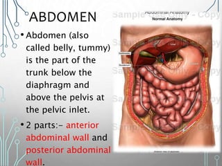
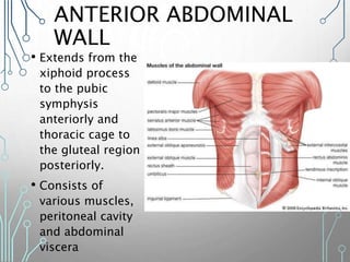
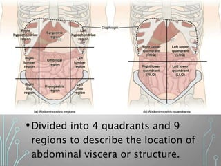
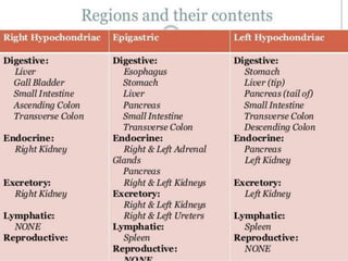
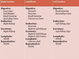
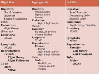
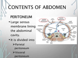
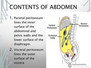
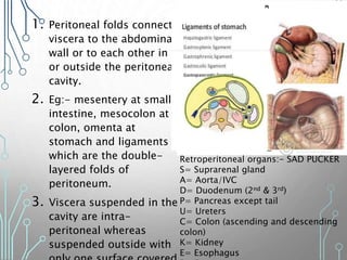
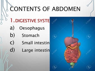
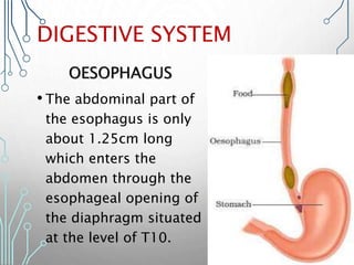
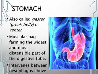
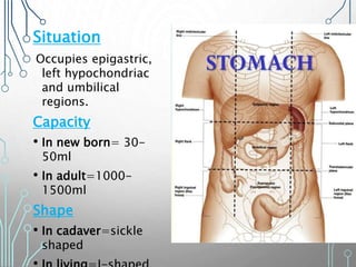
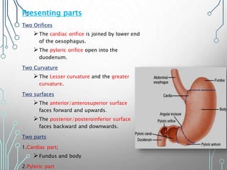
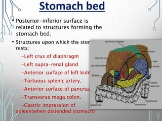
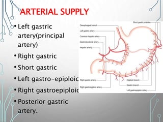
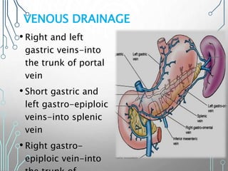
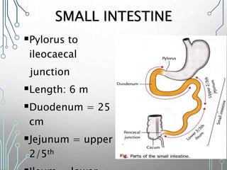
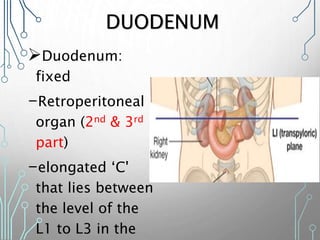
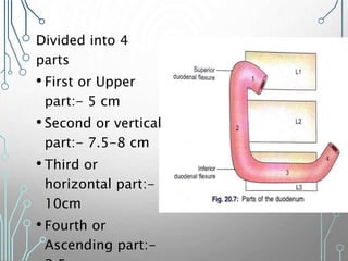
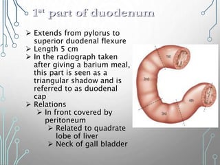
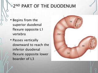
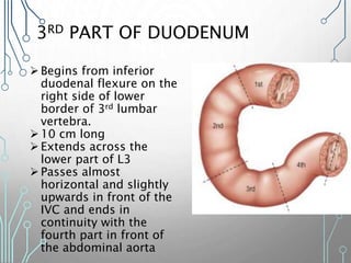
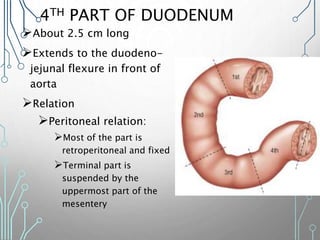
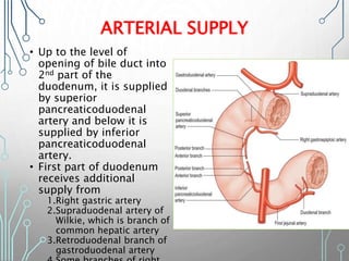
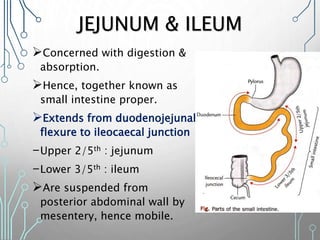
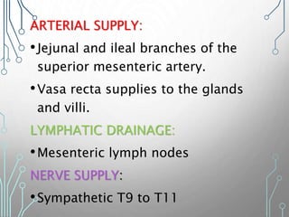
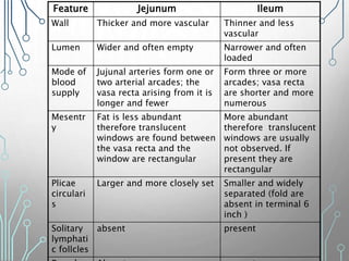
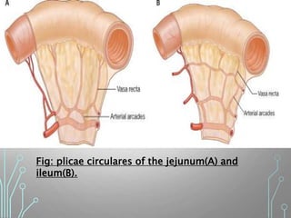
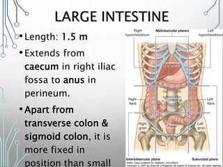
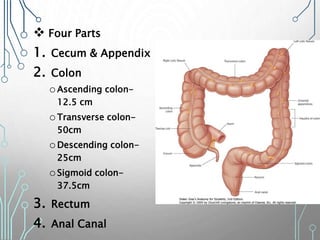
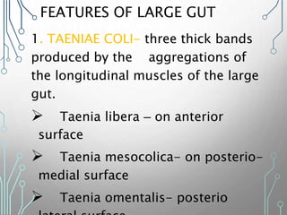
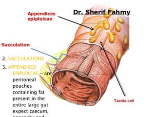
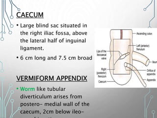
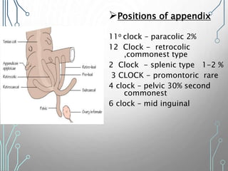
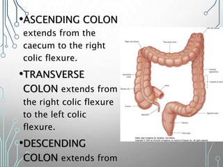
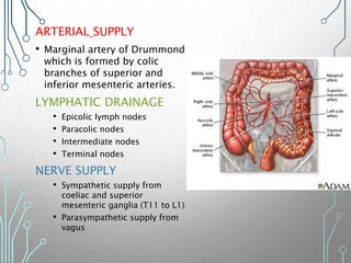
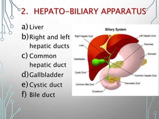
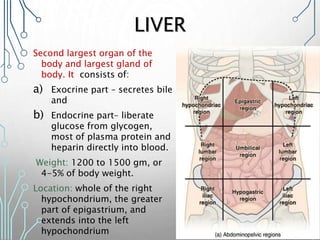
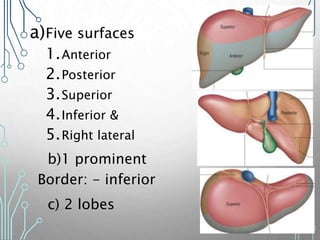
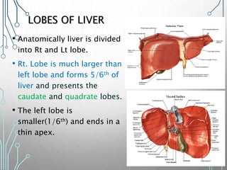
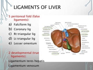
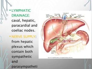
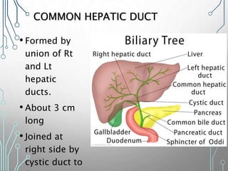
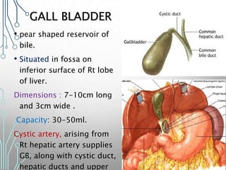
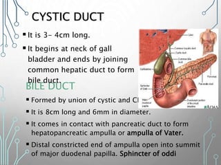
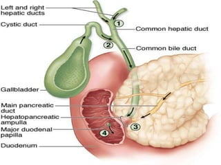
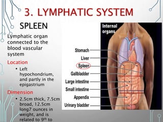
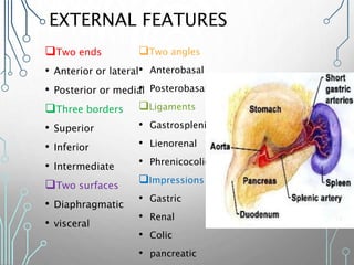
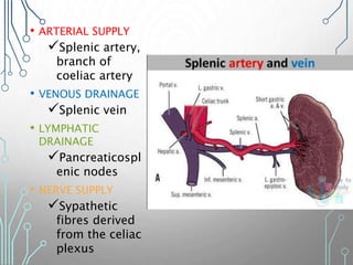
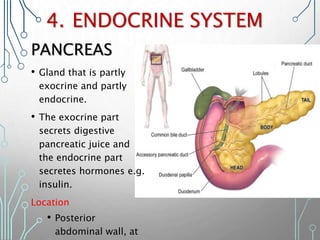
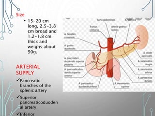
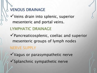
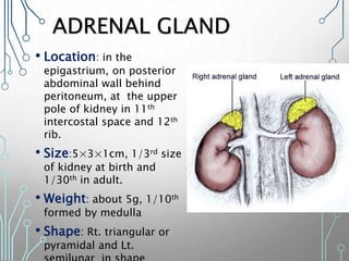
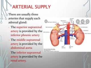
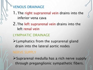
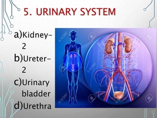
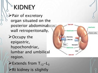
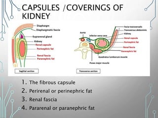
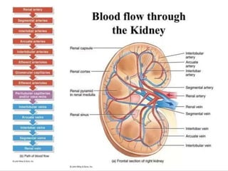
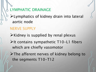
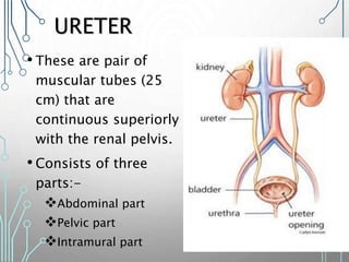
ABDOMEN
& PELVIS
PRESENTED BY:-SARU GOSAIN
B.SC.MIT 2017
MODERATOR:- MR. RANJIT
KUMAR JHA
ASSISTANT
ABDOMEN
• Abdomen (also
called belly, tummy)
is the part of the
trunk below the
diaphragm and
above the pelvis at
the pelvic inlet.
• 2 parts:- anterior
abdominal wall and
posterior abdominal
wall.
ANTERIOR ABDOMINAL
WALL
• Extends from the
xiphoid process
to the pubic
symphysis
anteriorly and
thoracic cage to
the gluteal region
posteriorly.
• Consists of
various muscles,
peritoneal cavity
and abdominal
viscera
•Divided into 4 quadrants and 9
regions to describe the location of
abdominal viscera or structure.
CONTENTS OF ABDOMEN
PERITONEUM
• Large serous
membrane lining
the abdominal
cavity.
• It is divided into
Parietal
peritoneum
Visceral
peritoneum
CONTENTS OF ABDOMEN
1. Parietal peritoneum
lines the inner
surface of the
abdominal and
pelvic walls and the
lower surface of the
diaphragm.
2. Visceral peritoneum
lines the outer
surface of the
viscera
1. Peritoneal folds connect
viscera to the abdominal
wall or to each other in
or outside the peritoneal
cavity.
2. Eg:- mesentery at small
intestine, mesocolon at
colon, omenta at
stomach and ligaments
which are the double-
layered folds of
peritoneum.
3. Viscera suspended in the
cavity are intra-
peritoneal whereas
suspended outside with
Retroperitoneal organs:- SAD PUCKER
S= Suprarenal gland
A= Aorta/IVC
D= Duodenum (2nd & 3rd)
P= Pancreas except tail
U= Ureters
C= Colon (ascending and descending
colon)
K= Kidney
E= Esophagus
CONTENTS OF ABDOMEN
1.DIGESTIVE SYSTEM
a) Oesophagus
b) Stomach
c) Small intestine
d) Large intestine
DIGESTIVE SYSTEM
OESOPHAGUS
• The abdominal part of
the esophagus is only
about 1.25cm long
which enters the
abdomen through the
esophageal opening of
the diaphragm situated
at the level of T10.
STOMACH
•Also called gaster,
(greek belly) or
venter
•Muscular bag
farming the widest
and most
distensible part of
the digestive tube.
•Intervenes between
oesophagus above
Situation
Occupies epigastric,
left hypochondriac
and umbilical
regions.
Capacity
• In new born= 30-
50ml
• In adult=1000-
1500ml
Shape
• In cadaver=sickle
shaped
Presenting parts
Two Orifices
The cardiac orifice is joined by lower end
of the oesophagus.
The pyloric orifice open into the
duodenum.
Two Curvature
The Lesser curvature and the greater
curvature.
Two surfaces
The anterior/anterosuperior surface
faces forward and upwards.
The posterior/posteroinferior surface
faces backward and downwards.
Two parts
1.Cardiac part;
Fundus and body
2.Pyloric part
• Posterior-inferior surface is
related to structures forming the
stomach bed.
• Structures upon which the stomach
rests;
-Left crus of diaphragm
-Left supra-renal gland
-Anterior surface of left kidney
-Tortuous splenic artery.
-Anterior surface of pancreas.
-Transverse mega colon.
-Gastric impression of
spleen(when distended stomach)
Stomach bed
ARTERIAL SUPPLY
• Left gastric
artery(principal
artery)
• Right gastric
• Short gastric
• Left gastro-epiploic
• Right gastroepiploic
• Posterior gastric
artery.
VENOUS DRAINAGE
• Right and left
gastric veins-into
the trunk of portal
vein
• Short gastric and
left gastro-epiploic
veins-into splenic
vein
• Right gastro-
epiploic vein-into
SMALL INTESTINE
Pylorus to
ileocaecal
junction
Length: 6 m
Duodenum = 25
cm
Jejunum = upper
2/5th
Duodenum:
fixed
-Retroperitoneal
organ (2nd & 3rd
part)
-elongated ‘C'
that lies between
the level of the
L1 to L3 in the 20
DUODENUM
Divided into 4
parts
• First or Upper
part:- 5 cm
• Second or vertical
part:- 7.5-8 cm
• Third or
horizontal part:-
10cm
• Fourth or
Ascending part:-
 Extends from pylorus to
superior duodenal flexure
 Length 5 cm
 In the radiograph taken
after giving a barium meal,
this part is seen as a
triangular shadow and is
referred to as duodenal
cap
 Relations
 In front covered by
peritoneum
 Related to quadrate
lobe of liver
 Neck of gall bladder
2ND PART OF THE DUODENUM
• Begins from the
superior duodenal
flexure opposite L1
vertebra
• Passes vertically
downward to reach the
inferior duodenal
flexure opposite lower
boarder of L3
3RD PART OF DUODENUM
Begins from inferior
duodenal flexure on the
right side of lower
border of 3rd lumbar
vertebra.
10 cm long
Extends across the
lower part of L3
Passes almost
horizontal and slightly
upwards in front of the
IVC and ends in
continuity with the
fourth part in front of
the abdominal aorta
4TH PART OF DUODENUM
About 2.5 cm long
Extends to the duodeno-
jejunal flexure in front of
aorta
Relation
Peritoneal relation:
Most of the part is
retroperitoneal and fixed
Terminal part is
suspended by the
uppermost part of the
mesentery
ARTERIAL SUPPLY
• Up to the level of
opening of bile duct into
2nd part of the
duodenum, it is supplied
by superior
pancreaticoduodenal
artery and below it is
supplied by inferior
pancreaticoduodenal
artery.
• First part of duodenum
receives additional
supply from
1.Right gastric artery
2.Supraduodenal artery of
Wilkie, which is branch of
common hepatic artery
3.Retroduodenal branch of
gastroduodenal artery
VENOUS DRAINAGE:
• splenic veins
• Superior mesenteric veins
• portal veins
LYMPHATIC DRAINAGE:
• Pancreatoduodenal nodes
NERVE SUPPLY:
1. Sympathetic nerve: T9 & T10
2. parasympathetic nerve: Vagus nerve
JEJUNUM & ILEUM
Concerned with digestion &
absorption.
Hence, together known as
small intestine proper.
Extends from duodenojejunal
flexure to ileocaecal junction
-Upper 2/5th : jejunum
-Lower 3/5th : ileum
Are suspended from
posterior abdominal wall by
mesentery, hence mobile.
ARTERIAL SUPPLY:
•Jejunal and ileal branches of the
superior mesenteric artery.
•Vasa recta supplies to the glands
and villi.
LYMPHATIC DRAINAGE:
•Mesenteric lymph nodes
NERVE SUPPLY:
•Sympathetic T9 to T11
Feature Jejunum Ileum
Wall Thicker and more vascular Thinner and less
vascular
Lumen Wider and often empty Narrower and often
loaded
Mode of
blood
supply
Jujunal arteries form one or
two arterial arcades; the
vasa recta arising from it is
longer and fewer
Form three or more
arcades; vasa recta
are shorter and more
numerous
Mesentr
y
Fat is less abundant
therefore translucent
windows are found between
the vasa recta and the
window are rectangular
More abundant
therefore translucent
windows are usually
not observed. If
present they are
rectangular
Plicae
circulari
s
Larger and more closely set Smaller and widely
separated (fold are
absent in terminal 6
inch )
Solitary
lymphati
c follcles
absent present
Fig: plicae circulares of the jejunum(A) and
ileum(B).
LARGE INTESTINE
•Length: 1.5 m
•Extends from
caecum in right iliac
fossa to anus in
perineum.
•Apart from
transverse colon &
sigmoid colon, it is
more fixed in
position than small
32
 Four Parts
1. Cecum & Appendix
2. Colon
oAscending colon-
12.5 cm
oTransverse colon-
50cm
oDescending colon-
25cm
oSigmoid colon-
37.5cm
3. Rectum
4. Anal Canal 33
FEATURES OF LARGE GUT
1. TAENIAE COLI- three thick bands
produced by the aggregations of
the longitudinal muscles of the large
gut.
 Taenia libera – on anterior
surface
 Taenia mesocolica- on posterio-
medial surface
 Taenia omentalis- posterio
2. SACCULATIONS
3. APPENDICES
EPIPLOICAE – are
peritoneal
pouches
containing fat
present in the
entire large gut
expect caecum,
CAECUM
• Large blind sac situated in
the right iliac fossa, above
the lateral half of inguinal
ligament.
• 6 cm long and 7.5 cm broad
VERMIFORM APPENDIX
• Worm like tubular
diverticulum arises from
postero- medial wall of the
caecum, 2cm below ileo-
Positions of appendix
11o clock – paracolic 2%
12 Clock - retrocolic
,commonest type
2 Clock - splenic type 1-2 %
3 CLOCK - promontoric rare
4 clock – pelvic 30% second
commonest
6 clock – mid inguinal
•ASCENDING COLON
extends from the
caecum to the right
colic flexure.
•TRANSVERSE
COLON extends from
the right colic flexure
to the left colic
flexure.
•DESCENDING
COLON extends from
ARTERIAL SUPPLY
• Marginal artery of Drummond
which is formed by colic
branches of superior and
inferior mesenteric arteries.
LYMPHATIC DRAINAGE
• Epicolic lymph nodes
• Paracolic nodes
• Intermediate nodes
• Terminal nodes
NERVE SUPPLY
• Sympathetic supply from
coeliac and superior
mesenteric ganglia (T11 to L1)
• Parasympathetic supply from
vagus
2. HEPATO-BILIARY APPARATUS
a) Liver
b)Right and left
hepatic ducts
c) Common
hepatic duct
d)Gallbladder
e)Cystic duct
f) Bile duct
LIVER
Second largest organ of the
body and largest gland of
body. It consists of:
a) Exocrine part – secretes bile
and
b) Endocrine part– liberate
glucose from glycogen,
most of plasma protein and
heparin directly into blood.
Weight: 1200 to 1500 gm, or
4-5% of body weight.
Location: whole of the right
hypochondrium, the greater
part of epigastrium, and
extends into the left
hypochondrium
a)Five surfaces
1.Anterior
2.Posterior
3.Superior
4.Inferior &
5.Right lateral
b)1 prominent
Border: - inferior
c) 2 lobes
LOBES OF LIVER
• Anatomically liver is divided
into Rt and Lt lobe.
• Rt. Lobe is much larger than
left lobe and forms 5/6th of
liver and presents the
caudate and quadrate lobes.
• The left lobe is
smaller(1/6th) and ends in a
thin apex.
LIGAMENTS OF LIVER
5 peritoneal fold (false
ligaments):
a) Falciform lig
b) Coronary lig
c) Rt triangular lig
d) Lt triangular lig
e) Lesser omentum
2 developmental (true
ligaments);
Ligamentum teres hepatis
Ligamentum venosum
PORTA HEPATIS
• deep transverse fissure
(5cm long)
• gateway of liver
• present in inferior
surface of Rt lobe.
• The portal vein, hepatic
artery and hepatic nerve
plexus enters.
• The right and left
hepatic ducts and some
lymph vessels leaves.
PERITONEAL RELATIONS
• Liver is covered by
peritoneum
Except:
-bare area
-groove for IVC
-fossa for gall bladder
-coronary ligaments
-lesser omentum
ARTERIAL SUPPLY
Liver receive dual blood
supply:
1. 20% blood supply from
hepatic artery and
2. 80% through portal vein.
Before entering liver both
divide into Rt and Lt
branches  within liver it
divide into segmental vessels
interlobular vessels which
run along portal canals
further branch and open into
hepatic sinusoid central
VENOUS DRAINAGE
• Hepatic sinusoid  central vein interlobular
veins sublobular veins hepatic veins finally
• LYMPHATIC
DRAINAGE:
caval, hepatic,
paracardial and
coeliac nodes.
• NERVE SUPPLY:
from hepatic
plexus which
contain both
sympathetic
and
parasympatheti
RIGHT AND LEFT HEPATIC DUCTS
•Emerge at
porta
hepatis
from the
right and
left lobes
of the
liver.
COMMON HEPATIC DUCT
• Formed by
union of Rt
and Lt
hepatic
ducts.
• About 3 cm
long
• Joined at
right side by
cystic duct to
GALL BLADDER
• pear shaped reservoir of
bile.
• Situated in fossa on
inferior surface of Rt lobe
of liver.
Dimensions : 7-10cm long
and 3cm wide .
Capacity: 30-50ml.
Cystic artery, arising from
Rt hepatic artery supplies
GB, along with cystic duct,
hepatic ducts and upper
CYSTIC DUCT
BILE DUCT
 Formed by union of cystic and CHD
 It is 8cm long and 6mm in diameter.
 It comes in contact with pancreatic duct to form
hepatopancreatic ampulla or ampulla of Vater.
 Distal constricted end of ampulla open into summit
of major duodenal papilla. Sphincter of oddi
It is 3- 4cm long.
It begins at neck of gall
bladder and ends by joining
common hepatic duct to form
bile duct.
3. LYMPHATIC SYSTEM
Lymphatic organ
connected to the
blood vascular
system
Location
• Left
hypochondrium,
and partly in the
epigastrium
Dimension
• 2.5cm thick, 7.5cm
broad, 12.5cm
long7 ounces in
weight, and is
related to 9th to
SPLEEN
EXTERNAL FEATURES
Two ends
• Anterior or lateral
• Posterior or medial
Three borders
• Superior
• Inferior
• Intermediate
Two surfaces
• Diaphragmatic
• visceral
Two angles
• Anterobasal
• Posterobasal
Ligaments
• Gastrosplenic
• Lienorenal
• Phrenicocolic
Impressions
• Gastric
• Renal
• Colic
• pancreatic
• ARTERIAL SUPPLY
Splenic artery,
branch of
coeliac artery
• VENOUS DRAINAGE
Splenic vein
• LYMPHATIC
DRAINAGE
Pancreaticospl
enic nodes
• NERVE SUPPLY
Sypathetic
fibres derived
from the celiac
plexus
PANCREAS
• Gland that is partly
exocrine and partly
endocrine.
• The exocrine part
secrets digestive
pancreatic juice and
the endocrine part
secretes hormones e.g.
insulin.
Location
• Posterior
abdominal wall, at
4. ENDOCRINE SYSTEM
Size
• 15-20 cm
long, 2.5-3.8
cm broad and
1.2-1.8 cm
thick and
weighs about
90g.
ARTERIAL
SUPPLY
Pancreatic
branches of the
splenic artery
Superior
pancreaticoduoden
al artery
Inferior
VENOUS DRAINAGE
Veins drain into splenic, superior
mesenteric and portal veins.
LYMPHATIC DRAINAGE
Pancreaticosplenic, coeliac and superior
mesenteric groups of lymph nodes
NERVE SUPPLY
Vagus or parasympathetic nerve
Splanchnic sympathetic nerve
ADRENAL GLAND
• Location: in the
epigastrium, on posterior
abdominal wall behind
peritoneum, at the upper
pole of kidney in 11th
intercostal space and 12th
rib.
• Size:5×3×1cm, 1/3rd size
of kidney at birth and
1/30th in adult.
• Weight: about 5g, 1/10th
formed by medulla
• Shape: Rt. triangular or
pyramidal and Lt.
ARTERIAL SUPPLY
There are usually three
arteries that supply each
adrenal gland:
1. The superior suprarenal
artery is provided by the
inferior phrenic artery
2.The middle suprarenal
artery is provided by the
abdominal aorta
3. The inferior suprarenal
artery is provided by the
renal artery.
VENOUS DRAINAGE
1. The right suprarenal vein drains into the
inferior vena cava
2.The left suprarenal vein drains into the
left renal vein
LYMPHATIC DRAINAGE
Lymphatics from the suprarenal gland
drain into the lateral aortic nodes
NERVE SUPPLY
Suprarenal medulla has a rich nerve supply
through preganglionic sympathetic fibers.
5. URINARY SYSTEM
a)Kidney-
2
b)Ureter-
2
c)Urinary
bladder
d)Urethra
KIDNEY
Pair of excretory
organ situated on the
posterior abdominal
wall retroperitonally.
Occupy the
epigastric,
hypochondriac,
lumbar and umbilical
region.
Extends from T12-L3
Rt kidney is slightly
SIZE AND WEIGHT
11cm long, 6cm broad and 3cm thick
Weighs 150 gram in male and 135 gram in female.
Extent:- T12-L3
EXTERNAL FEATURES
• Two poles - upper pole
- lower pole
• Two surfaces –anterior
-posterior
• Two borders- lateral
-medial
RELATIONS OF KIDNEY
• Upper pole of each kidney is related to the corresponding
supranenal gland
• Medial border of each kidney is related to:-
a. The suprarenal gland , above the hilus, and
b. To the ureter below the hilus
• Posterior relations:- related to the following structures:-
a. Diaphragm
b. Medial and lateral arcuate ligaments
c. Psoas major
d. Quardratus lumborum
e. Transversus abdominis
f. Subcoatal vessels
g. Subcostal, iliohypogatric and ilioinguinal nerves
OTHER RELATIONS OF RIGHT
KIDNEY
1. 12th rib
2. Right suprarenal
gland
3. Liver
4. Second part of
duodenum
5. Hepatic flexure of
colon
6. Small intestine
OTHER RELATIONS OF LEFT
KIDNEY
1. 11th and 12th ribs
2. Left suprarenal gland
3. Spleen
4. Stomach
5. Pancreas
6. Splenic vessels
7. Splenic flexure and
descending colon
8. Jejunum
CAPSULES /COVERINGS OF
KIDNEY
1. The fibrous capsule
2. Perirenal or perinephric fat
3. Renal fascia
4. Pararenal or paranephric fat
LYMPHATIC DRAINAGE
Lymphatics of kidney drain into lateral
aortic node
NERVE SUPPLY
Kidney is supplied by renal plexus
It contains sympathetic T10-L1 fibers
which are chiefly vasomotor
The afferent nerves of kidney belong to
the segments T10-T12
URETER
• These are pair of
muscular tubes (25
cm) that are
continuous superiorly
with the renal pelvis.
• Consists of three
parts:-
Abdominal part
Pelvic part
Intramural part
DIMENSION
10 inch long of which upper half
5 inch lies in the abdomen and
lower half 5 inch in the pelvis
measures about 3mm in diameter
but it is slightly constricted at 5
places
1. At the pelviuretric junction
2. At the brim of lesser pelvis
3. Point of crossing of ureter by
ductus deference or broad
ligament of uterus.
4. Intravesical course and
5. At the trigone of urinary
BLOOD SUPPLY
• Upper part- renal
artery
• Middle part- from
aorta
• Pelvic part- vesical,
middle rectal or
uterine vessels
NERVE SUPPLY
Sympathetic- T10-
L1
POSTERIOR ABDOMINAL
WALL
• Includes following
1. Abdominal aorta
2. Inferior vena cava
3. Abdominal parts of the azygos and
hemiazygos veins
4. Lymph nodes of posterior abdominal wall
and cisterna chili
5. Muscles of the posterior abdominal wall and
thoracolumbar fascia
6. Nerves of the posterior abdominal wall
including lumbar plexus and the abdominal
part of autonomic nervous system
BONES IN RELATION TO
ABDOMEN
• Lumbar
vertebrae-5
• Sacrum-1
(formed by
fusion of 5
bones)
• Coccyx-1
(formed by
fusion of 4
bones)
LUMBAR VERTEBRAE
• There are 5 lumbar
vertebrae, of which the
first four are typical, and
the fifth is atypical
• Lumbar vertebrae is
identified by
1. Its large size
2. The absence of costal
facets on the body and
3. The absence of
foramen in transverse
process
TYPICAL FEATURES OF LUMBAR
VERTEBRAE
• The body is large, kidney-shaped
• The vertebral foramen is triangular in
shape, and is larger than in the
thoracic region but is smaller than in
cervical region
• The pedicles are short and strong
• The alminae are short, thick and
broad
• The spine forms a vertical
quadrilateral plate, directed almost
backwards and only slightly
downwards.
• The transverse process are thin and
tapering
• The superior articular processes lie
ATYPICAL FEATURES OF LUMBAR
VERTEBRAE
1. The transverse processes are thick,
short and pyramidal in shape.
2. The distance between the inferior
articular processes is equal to or
more than the distance between the
superior articular processes.
3. The spine is small, short and rounded
at the tip
4. The body is the largest of all the
lumbar vertebrae.
5. The pedicles are directed backwards
and laterally.
6. The superior articular facets look
more backwards than medially, and
the inferior articular facets look more
forwards than laterally, as compared
to other lumbar vertebrae
SACRUM
• Large, flattened, triangular bone formed from
the fusion of five sacral vertebrae
Male sacrum Female sacrum
Longer and narrower, i.e., the average
sacral index is 105
Shorter and wider, i.e., the average
sacral index is 115
The width of the body of the first
sacral vertebrae is greater than that of
each ala in the male
The two are equal
The dorsal concavity of auricular
surface is less marked in male
More marked
The concavity on the ventral aspect of
sacrum is more uniform, and is
shallower in males
The concavity is irregular especially
between S1 and S2 and between S3
and S4
The sacrovertebral angle and the size
of pelvic cavity is less
More
COCCYX
• Small
triangular
bone formed
by fusion of
four
rudimentary
coccygeal
vertebrae
Abdomen & pelvis part I

Abdomen & pelvis part I

  • 1.
    ABDOMEN & PELVIS PRESENTED BY:-SARUGOSAIN B.SC.MIT 2017 MODERATOR:- MR. RANJIT KUMAR JHA ASSISTANT
  • 2.
    ABDOMEN • Abdomen (also calledbelly, tummy) is the part of the trunk below the diaphragm and above the pelvis at the pelvic inlet. • 2 parts:- anterior abdominal wall and posterior abdominal wall.
  • 3.
    ANTERIOR ABDOMINAL WALL • Extendsfrom the xiphoid process to the pubic symphysis anteriorly and thoracic cage to the gluteal region posteriorly. • Consists of various muscles, peritoneal cavity and abdominal viscera
  • 4.
    •Divided into 4quadrants and 9 regions to describe the location of abdominal viscera or structure.
  • 8.
    CONTENTS OF ABDOMEN PERITONEUM •Large serous membrane lining the abdominal cavity. • It is divided into Parietal peritoneum Visceral peritoneum
  • 9.
    CONTENTS OF ABDOMEN 1.Parietal peritoneum lines the inner surface of the abdominal and pelvic walls and the lower surface of the diaphragm. 2. Visceral peritoneum lines the outer surface of the viscera
  • 10.
    1. Peritoneal foldsconnect viscera to the abdominal wall or to each other in or outside the peritoneal cavity. 2. Eg:- mesentery at small intestine, mesocolon at colon, omenta at stomach and ligaments which are the double- layered folds of peritoneum. 3. Viscera suspended in the cavity are intra- peritoneal whereas suspended outside with Retroperitoneal organs:- SAD PUCKER S= Suprarenal gland A= Aorta/IVC D= Duodenum (2nd & 3rd) P= Pancreas except tail U= Ureters C= Colon (ascending and descending colon) K= Kidney E= Esophagus
  • 11.
    CONTENTS OF ABDOMEN 1.DIGESTIVESYSTEM a) Oesophagus b) Stomach c) Small intestine d) Large intestine
  • 12.
    DIGESTIVE SYSTEM OESOPHAGUS • Theabdominal part of the esophagus is only about 1.25cm long which enters the abdomen through the esophageal opening of the diaphragm situated at the level of T10.
  • 13.
    STOMACH •Also called gaster, (greekbelly) or venter •Muscular bag farming the widest and most distensible part of the digestive tube. •Intervenes between oesophagus above
  • 14.
    Situation Occupies epigastric, left hypochondriac andumbilical regions. Capacity • In new born= 30- 50ml • In adult=1000- 1500ml Shape • In cadaver=sickle shaped
  • 15.
    Presenting parts Two Orifices Thecardiac orifice is joined by lower end of the oesophagus. The pyloric orifice open into the duodenum. Two Curvature The Lesser curvature and the greater curvature. Two surfaces The anterior/anterosuperior surface faces forward and upwards. The posterior/posteroinferior surface faces backward and downwards. Two parts 1.Cardiac part; Fundus and body 2.Pyloric part
  • 16.
    • Posterior-inferior surfaceis related to structures forming the stomach bed. • Structures upon which the stomach rests; -Left crus of diaphragm -Left supra-renal gland -Anterior surface of left kidney -Tortuous splenic artery. -Anterior surface of pancreas. -Transverse mega colon. -Gastric impression of spleen(when distended stomach) Stomach bed
  • 17.
    ARTERIAL SUPPLY • Leftgastric artery(principal artery) • Right gastric • Short gastric • Left gastro-epiploic • Right gastroepiploic • Posterior gastric artery.
  • 18.
    VENOUS DRAINAGE • Rightand left gastric veins-into the trunk of portal vein • Short gastric and left gastro-epiploic veins-into splenic vein • Right gastro- epiploic vein-into
  • 19.
    SMALL INTESTINE Pylorus to ileocaecal junction Length:6 m Duodenum = 25 cm Jejunum = upper 2/5th
  • 20.
    Duodenum: fixed -Retroperitoneal organ (2nd &3rd part) -elongated ‘C' that lies between the level of the L1 to L3 in the 20 DUODENUM
  • 21.
    Divided into 4 parts •First or Upper part:- 5 cm • Second or vertical part:- 7.5-8 cm • Third or horizontal part:- 10cm • Fourth or Ascending part:-
  • 22.
     Extends frompylorus to superior duodenal flexure  Length 5 cm  In the radiograph taken after giving a barium meal, this part is seen as a triangular shadow and is referred to as duodenal cap  Relations  In front covered by peritoneum  Related to quadrate lobe of liver  Neck of gall bladder
  • 23.
    2ND PART OFTHE DUODENUM • Begins from the superior duodenal flexure opposite L1 vertebra • Passes vertically downward to reach the inferior duodenal flexure opposite lower boarder of L3
  • 24.
    3RD PART OFDUODENUM Begins from inferior duodenal flexure on the right side of lower border of 3rd lumbar vertebra. 10 cm long Extends across the lower part of L3 Passes almost horizontal and slightly upwards in front of the IVC and ends in continuity with the fourth part in front of the abdominal aorta
  • 25.
    4TH PART OFDUODENUM About 2.5 cm long Extends to the duodeno- jejunal flexure in front of aorta Relation Peritoneal relation: Most of the part is retroperitoneal and fixed Terminal part is suspended by the uppermost part of the mesentery
  • 26.
    ARTERIAL SUPPLY • Upto the level of opening of bile duct into 2nd part of the duodenum, it is supplied by superior pancreaticoduodenal artery and below it is supplied by inferior pancreaticoduodenal artery. • First part of duodenum receives additional supply from 1.Right gastric artery 2.Supraduodenal artery of Wilkie, which is branch of common hepatic artery 3.Retroduodenal branch of gastroduodenal artery
  • 27.
    VENOUS DRAINAGE: • splenicveins • Superior mesenteric veins • portal veins LYMPHATIC DRAINAGE: • Pancreatoduodenal nodes NERVE SUPPLY: 1. Sympathetic nerve: T9 & T10 2. parasympathetic nerve: Vagus nerve
  • 28.
    JEJUNUM & ILEUM Concernedwith digestion & absorption. Hence, together known as small intestine proper. Extends from duodenojejunal flexure to ileocaecal junction -Upper 2/5th : jejunum -Lower 3/5th : ileum Are suspended from posterior abdominal wall by mesentery, hence mobile.
  • 29.
    ARTERIAL SUPPLY: •Jejunal andileal branches of the superior mesenteric artery. •Vasa recta supplies to the glands and villi. LYMPHATIC DRAINAGE: •Mesenteric lymph nodes NERVE SUPPLY: •Sympathetic T9 to T11
  • 30.
    Feature Jejunum Ileum WallThicker and more vascular Thinner and less vascular Lumen Wider and often empty Narrower and often loaded Mode of blood supply Jujunal arteries form one or two arterial arcades; the vasa recta arising from it is longer and fewer Form three or more arcades; vasa recta are shorter and more numerous Mesentr y Fat is less abundant therefore translucent windows are found between the vasa recta and the window are rectangular More abundant therefore translucent windows are usually not observed. If present they are rectangular Plicae circulari s Larger and more closely set Smaller and widely separated (fold are absent in terminal 6 inch ) Solitary lymphati c follcles absent present
  • 31.
    Fig: plicae circularesof the jejunum(A) and ileum(B).
  • 32.
    LARGE INTESTINE •Length: 1.5m •Extends from caecum in right iliac fossa to anus in perineum. •Apart from transverse colon & sigmoid colon, it is more fixed in position than small 32
  • 33.
     Four Parts 1.Cecum & Appendix 2. Colon oAscending colon- 12.5 cm oTransverse colon- 50cm oDescending colon- 25cm oSigmoid colon- 37.5cm 3. Rectum 4. Anal Canal 33
  • 34.
    FEATURES OF LARGEGUT 1. TAENIAE COLI- three thick bands produced by the aggregations of the longitudinal muscles of the large gut.  Taenia libera – on anterior surface  Taenia mesocolica- on posterio- medial surface  Taenia omentalis- posterio
  • 35.
    2. SACCULATIONS 3. APPENDICES EPIPLOICAE– are peritoneal pouches containing fat present in the entire large gut expect caecum,
  • 36.
    CAECUM • Large blindsac situated in the right iliac fossa, above the lateral half of inguinal ligament. • 6 cm long and 7.5 cm broad VERMIFORM APPENDIX • Worm like tubular diverticulum arises from postero- medial wall of the caecum, 2cm below ileo-
  • 37.
    Positions of appendix 11oclock – paracolic 2% 12 Clock - retrocolic ,commonest type 2 Clock - splenic type 1-2 % 3 CLOCK - promontoric rare 4 clock – pelvic 30% second commonest 6 clock – mid inguinal
  • 38.
    •ASCENDING COLON extends fromthe caecum to the right colic flexure. •TRANSVERSE COLON extends from the right colic flexure to the left colic flexure. •DESCENDING COLON extends from
  • 39.
    ARTERIAL SUPPLY • Marginalartery of Drummond which is formed by colic branches of superior and inferior mesenteric arteries. LYMPHATIC DRAINAGE • Epicolic lymph nodes • Paracolic nodes • Intermediate nodes • Terminal nodes NERVE SUPPLY • Sympathetic supply from coeliac and superior mesenteric ganglia (T11 to L1) • Parasympathetic supply from vagus
  • 40.
    2. HEPATO-BILIARY APPARATUS a)Liver b)Right and left hepatic ducts c) Common hepatic duct d)Gallbladder e)Cystic duct f) Bile duct
  • 41.
    LIVER Second largest organof the body and largest gland of body. It consists of: a) Exocrine part – secretes bile and b) Endocrine part– liberate glucose from glycogen, most of plasma protein and heparin directly into blood. Weight: 1200 to 1500 gm, or 4-5% of body weight. Location: whole of the right hypochondrium, the greater part of epigastrium, and extends into the left hypochondrium
  • 42.
    a)Five surfaces 1.Anterior 2.Posterior 3.Superior 4.Inferior & 5.Rightlateral b)1 prominent Border: - inferior c) 2 lobes
  • 43.
    LOBES OF LIVER •Anatomically liver is divided into Rt and Lt lobe. • Rt. Lobe is much larger than left lobe and forms 5/6th of liver and presents the caudate and quadrate lobes. • The left lobe is smaller(1/6th) and ends in a thin apex.
  • 44.
    LIGAMENTS OF LIVER 5peritoneal fold (false ligaments): a) Falciform lig b) Coronary lig c) Rt triangular lig d) Lt triangular lig e) Lesser omentum 2 developmental (true ligaments); Ligamentum teres hepatis Ligamentum venosum
  • 45.
    PORTA HEPATIS • deeptransverse fissure (5cm long) • gateway of liver • present in inferior surface of Rt lobe. • The portal vein, hepatic artery and hepatic nerve plexus enters. • The right and left hepatic ducts and some lymph vessels leaves.
  • 46.
    PERITONEAL RELATIONS • Liveris covered by peritoneum Except: -bare area -groove for IVC -fossa for gall bladder -coronary ligaments -lesser omentum
  • 47.
    ARTERIAL SUPPLY Liver receivedual blood supply: 1. 20% blood supply from hepatic artery and 2. 80% through portal vein. Before entering liver both divide into Rt and Lt branches  within liver it divide into segmental vessels interlobular vessels which run along portal canals further branch and open into hepatic sinusoid central
  • 48.
    VENOUS DRAINAGE • Hepaticsinusoid  central vein interlobular veins sublobular veins hepatic veins finally
  • 49.
    • LYMPHATIC DRAINAGE: caval, hepatic, paracardialand coeliac nodes. • NERVE SUPPLY: from hepatic plexus which contain both sympathetic and parasympatheti
  • 50.
    RIGHT AND LEFTHEPATIC DUCTS •Emerge at porta hepatis from the right and left lobes of the liver.
  • 51.
    COMMON HEPATIC DUCT •Formed by union of Rt and Lt hepatic ducts. • About 3 cm long • Joined at right side by cystic duct to
  • 52.
    GALL BLADDER • pearshaped reservoir of bile. • Situated in fossa on inferior surface of Rt lobe of liver. Dimensions : 7-10cm long and 3cm wide . Capacity: 30-50ml. Cystic artery, arising from Rt hepatic artery supplies GB, along with cystic duct, hepatic ducts and upper
  • 53.
    CYSTIC DUCT BILE DUCT Formed by union of cystic and CHD  It is 8cm long and 6mm in diameter.  It comes in contact with pancreatic duct to form hepatopancreatic ampulla or ampulla of Vater.  Distal constricted end of ampulla open into summit of major duodenal papilla. Sphincter of oddi It is 3- 4cm long. It begins at neck of gall bladder and ends by joining common hepatic duct to form bile duct.
  • 55.
    3. LYMPHATIC SYSTEM Lymphaticorgan connected to the blood vascular system Location • Left hypochondrium, and partly in the epigastrium Dimension • 2.5cm thick, 7.5cm broad, 12.5cm long7 ounces in weight, and is related to 9th to SPLEEN
  • 56.
    EXTERNAL FEATURES Two ends •Anterior or lateral • Posterior or medial Three borders • Superior • Inferior • Intermediate Two surfaces • Diaphragmatic • visceral Two angles • Anterobasal • Posterobasal Ligaments • Gastrosplenic • Lienorenal • Phrenicocolic Impressions • Gastric • Renal • Colic • pancreatic
  • 57.
    • ARTERIAL SUPPLY Splenicartery, branch of coeliac artery • VENOUS DRAINAGE Splenic vein • LYMPHATIC DRAINAGE Pancreaticospl enic nodes • NERVE SUPPLY Sypathetic fibres derived from the celiac plexus
  • 58.
    PANCREAS • Gland thatis partly exocrine and partly endocrine. • The exocrine part secrets digestive pancreatic juice and the endocrine part secretes hormones e.g. insulin. Location • Posterior abdominal wall, at 4. ENDOCRINE SYSTEM
  • 59.
    Size • 15-20 cm long,2.5-3.8 cm broad and 1.2-1.8 cm thick and weighs about 90g. ARTERIAL SUPPLY Pancreatic branches of the splenic artery Superior pancreaticoduoden al artery Inferior
  • 60.
    VENOUS DRAINAGE Veins draininto splenic, superior mesenteric and portal veins. LYMPHATIC DRAINAGE Pancreaticosplenic, coeliac and superior mesenteric groups of lymph nodes NERVE SUPPLY Vagus or parasympathetic nerve Splanchnic sympathetic nerve
  • 61.
    ADRENAL GLAND • Location:in the epigastrium, on posterior abdominal wall behind peritoneum, at the upper pole of kidney in 11th intercostal space and 12th rib. • Size:5×3×1cm, 1/3rd size of kidney at birth and 1/30th in adult. • Weight: about 5g, 1/10th formed by medulla • Shape: Rt. triangular or pyramidal and Lt.
  • 62.
    ARTERIAL SUPPLY There areusually three arteries that supply each adrenal gland: 1. The superior suprarenal artery is provided by the inferior phrenic artery 2.The middle suprarenal artery is provided by the abdominal aorta 3. The inferior suprarenal artery is provided by the renal artery.
  • 63.
    VENOUS DRAINAGE 1. Theright suprarenal vein drains into the inferior vena cava 2.The left suprarenal vein drains into the left renal vein LYMPHATIC DRAINAGE Lymphatics from the suprarenal gland drain into the lateral aortic nodes NERVE SUPPLY Suprarenal medulla has a rich nerve supply through preganglionic sympathetic fibers.
  • 64.
  • 65.
    KIDNEY Pair of excretory organsituated on the posterior abdominal wall retroperitonally. Occupy the epigastric, hypochondriac, lumbar and umbilical region. Extends from T12-L3 Rt kidney is slightly
  • 66.
    SIZE AND WEIGHT 11cmlong, 6cm broad and 3cm thick Weighs 150 gram in male and 135 gram in female. Extent:- T12-L3 EXTERNAL FEATURES • Two poles - upper pole - lower pole • Two surfaces –anterior -posterior • Two borders- lateral -medial
  • 67.
    RELATIONS OF KIDNEY •Upper pole of each kidney is related to the corresponding supranenal gland • Medial border of each kidney is related to:- a. The suprarenal gland , above the hilus, and b. To the ureter below the hilus • Posterior relations:- related to the following structures:- a. Diaphragm b. Medial and lateral arcuate ligaments c. Psoas major d. Quardratus lumborum e. Transversus abdominis f. Subcoatal vessels g. Subcostal, iliohypogatric and ilioinguinal nerves
  • 68.
    OTHER RELATIONS OFRIGHT KIDNEY 1. 12th rib 2. Right suprarenal gland 3. Liver 4. Second part of duodenum 5. Hepatic flexure of colon 6. Small intestine
  • 69.
    OTHER RELATIONS OFLEFT KIDNEY 1. 11th and 12th ribs 2. Left suprarenal gland 3. Spleen 4. Stomach 5. Pancreas 6. Splenic vessels 7. Splenic flexure and descending colon 8. Jejunum
  • 70.
    CAPSULES /COVERINGS OF KIDNEY 1.The fibrous capsule 2. Perirenal or perinephric fat 3. Renal fascia 4. Pararenal or paranephric fat
  • 72.
    LYMPHATIC DRAINAGE Lymphatics ofkidney drain into lateral aortic node NERVE SUPPLY Kidney is supplied by renal plexus It contains sympathetic T10-L1 fibers which are chiefly vasomotor The afferent nerves of kidney belong to the segments T10-T12
  • 73.
    URETER • These arepair of muscular tubes (25 cm) that are continuous superiorly with the renal pelvis. • Consists of three parts:- Abdominal part Pelvic part Intramural part
  • 74.
    DIMENSION 10 inch longof which upper half 5 inch lies in the abdomen and lower half 5 inch in the pelvis measures about 3mm in diameter but it is slightly constricted at 5 places 1. At the pelviuretric junction 2. At the brim of lesser pelvis 3. Point of crossing of ureter by ductus deference or broad ligament of uterus. 4. Intravesical course and 5. At the trigone of urinary
  • 75.
    BLOOD SUPPLY • Upperpart- renal artery • Middle part- from aorta • Pelvic part- vesical, middle rectal or uterine vessels NERVE SUPPLY Sympathetic- T10- L1
  • 76.
    POSTERIOR ABDOMINAL WALL • Includesfollowing 1. Abdominal aorta 2. Inferior vena cava 3. Abdominal parts of the azygos and hemiazygos veins 4. Lymph nodes of posterior abdominal wall and cisterna chili 5. Muscles of the posterior abdominal wall and thoracolumbar fascia 6. Nerves of the posterior abdominal wall including lumbar plexus and the abdominal part of autonomic nervous system
  • 77.
    BONES IN RELATIONTO ABDOMEN • Lumbar vertebrae-5 • Sacrum-1 (formed by fusion of 5 bones) • Coccyx-1 (formed by fusion of 4 bones)
  • 78.
    LUMBAR VERTEBRAE • Thereare 5 lumbar vertebrae, of which the first four are typical, and the fifth is atypical • Lumbar vertebrae is identified by 1. Its large size 2. The absence of costal facets on the body and 3. The absence of foramen in transverse process
  • 79.
    TYPICAL FEATURES OFLUMBAR VERTEBRAE • The body is large, kidney-shaped • The vertebral foramen is triangular in shape, and is larger than in the thoracic region but is smaller than in cervical region • The pedicles are short and strong • The alminae are short, thick and broad • The spine forms a vertical quadrilateral plate, directed almost backwards and only slightly downwards. • The transverse process are thin and tapering • The superior articular processes lie
  • 80.
    ATYPICAL FEATURES OFLUMBAR VERTEBRAE 1. The transverse processes are thick, short and pyramidal in shape. 2. The distance between the inferior articular processes is equal to or more than the distance between the superior articular processes. 3. The spine is small, short and rounded at the tip 4. The body is the largest of all the lumbar vertebrae. 5. The pedicles are directed backwards and laterally. 6. The superior articular facets look more backwards than medially, and the inferior articular facets look more forwards than laterally, as compared to other lumbar vertebrae
  • 81.
    SACRUM • Large, flattened,triangular bone formed from the fusion of five sacral vertebrae Male sacrum Female sacrum Longer and narrower, i.e., the average sacral index is 105 Shorter and wider, i.e., the average sacral index is 115 The width of the body of the first sacral vertebrae is greater than that of each ala in the male The two are equal The dorsal concavity of auricular surface is less marked in male More marked The concavity on the ventral aspect of sacrum is more uniform, and is shallower in males The concavity is irregular especially between S1 and S2 and between S3 and S4 The sacrovertebral angle and the size of pelvic cavity is less More
  • 82.
    COCCYX • Small triangular bone formed byfusion of four rudimentary coccygeal vertebrae

Editor's Notes

  • #5 9 regions are divided by the two horizontal planes(trans pyloric plane and transtubercular plane) and two vertical planes (right and left lateral planes).
  • #11 Some organs may be covered by peritoneum only on one side which are known as retroperitoneal organs. Like duodenum, kidney, pancreas, ureter and suprarenal. Retro peritoenal organs: @SAD PUCKER suprarenal gland Aorta/ivc Duodenum(2nd & 3rd) Pancrease except tail Ureters Colon (acending & decending colon) Kidney Esophagus rectum
  • #21 Vasa recta; straight arteries coming off from arcades in mesentry Fixation of duodenum is essential because mobile duodenum will cause twisting & kinking of CBD & pancreatic duct
  • #41 The system collects bile from the liver, stores in the gall bladder and transmits it to the second part of duodenum.
  • #48 Portal vein is about 8cm long which is formed by the union of the superior mesenteric vein and the splenic vein at the level of L2. It conveys blood from the abdominal part of the alimentary tract, the gallbladder, the spleen, and the pancreas and conveys it to the liver. Hepatic arterial blood mixes with the portal venous blood in the sinusoids.