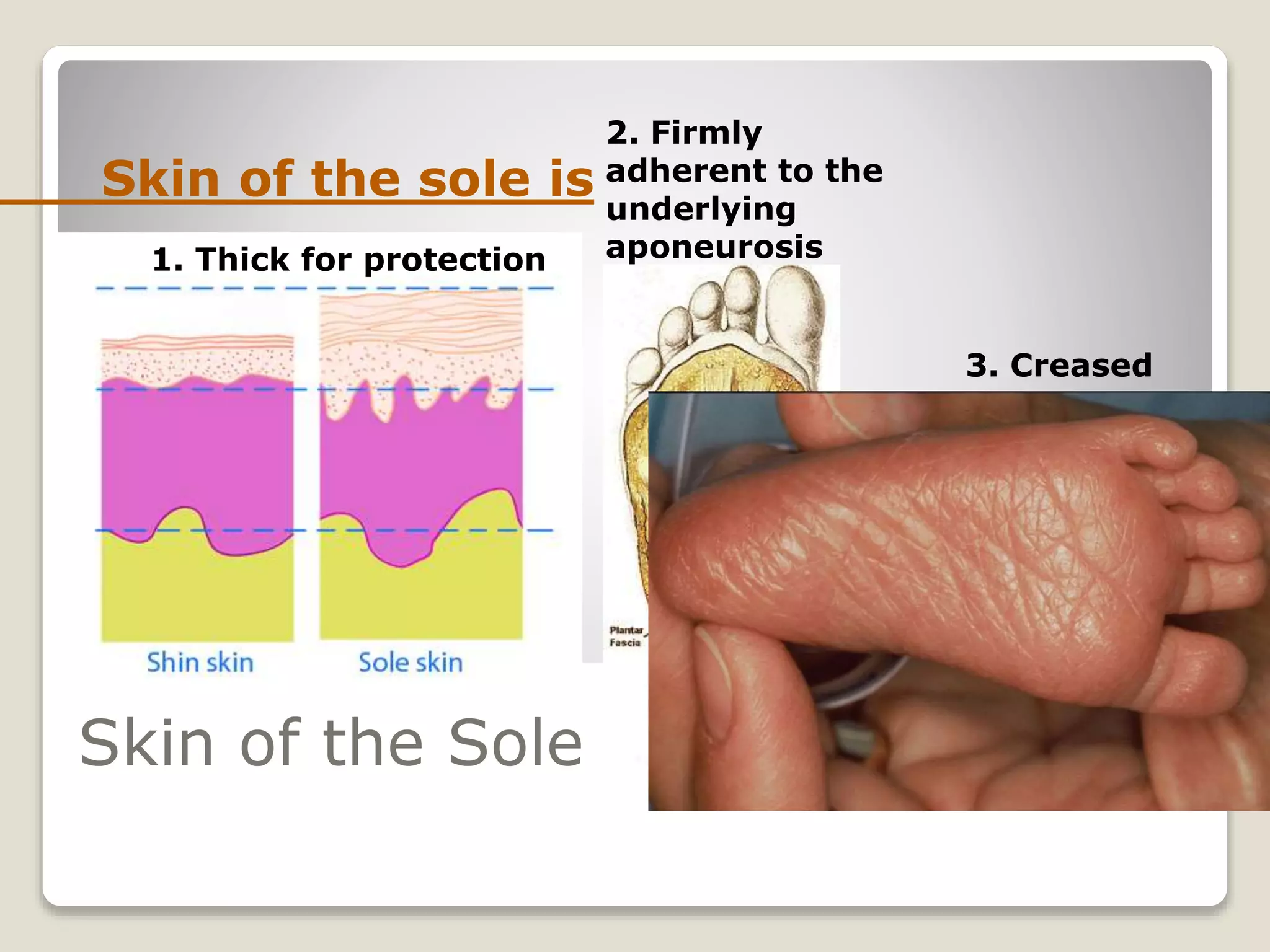
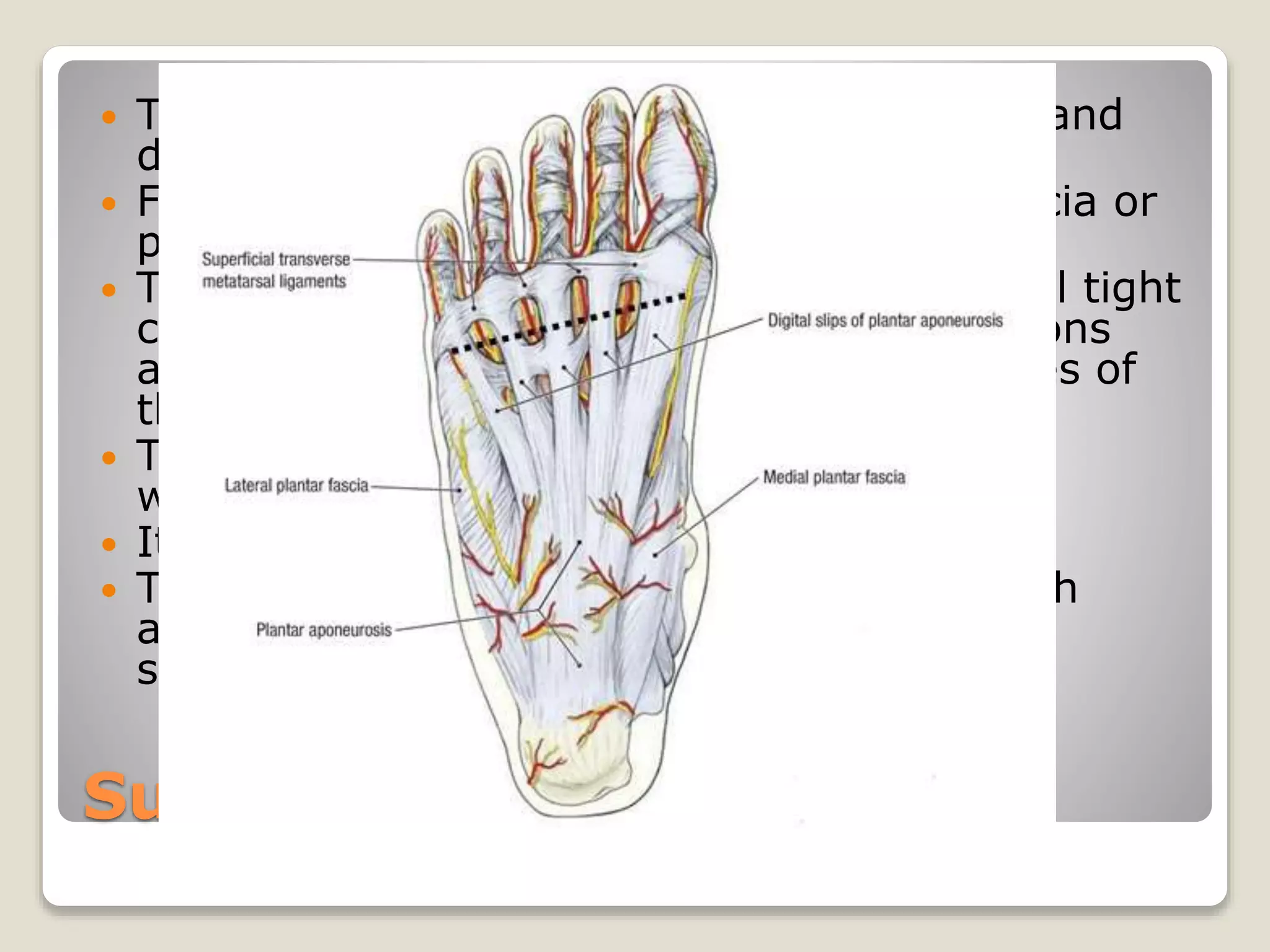
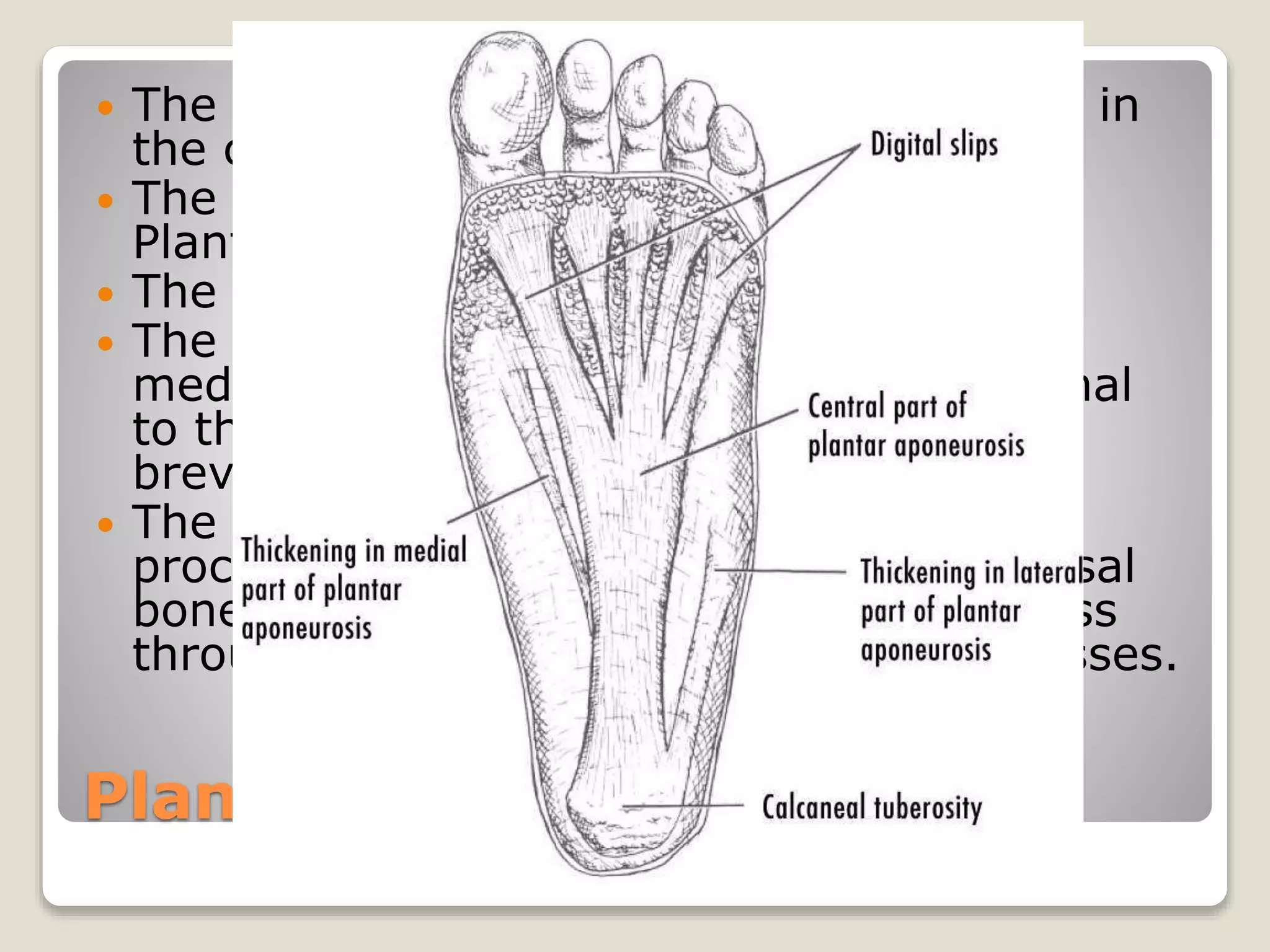
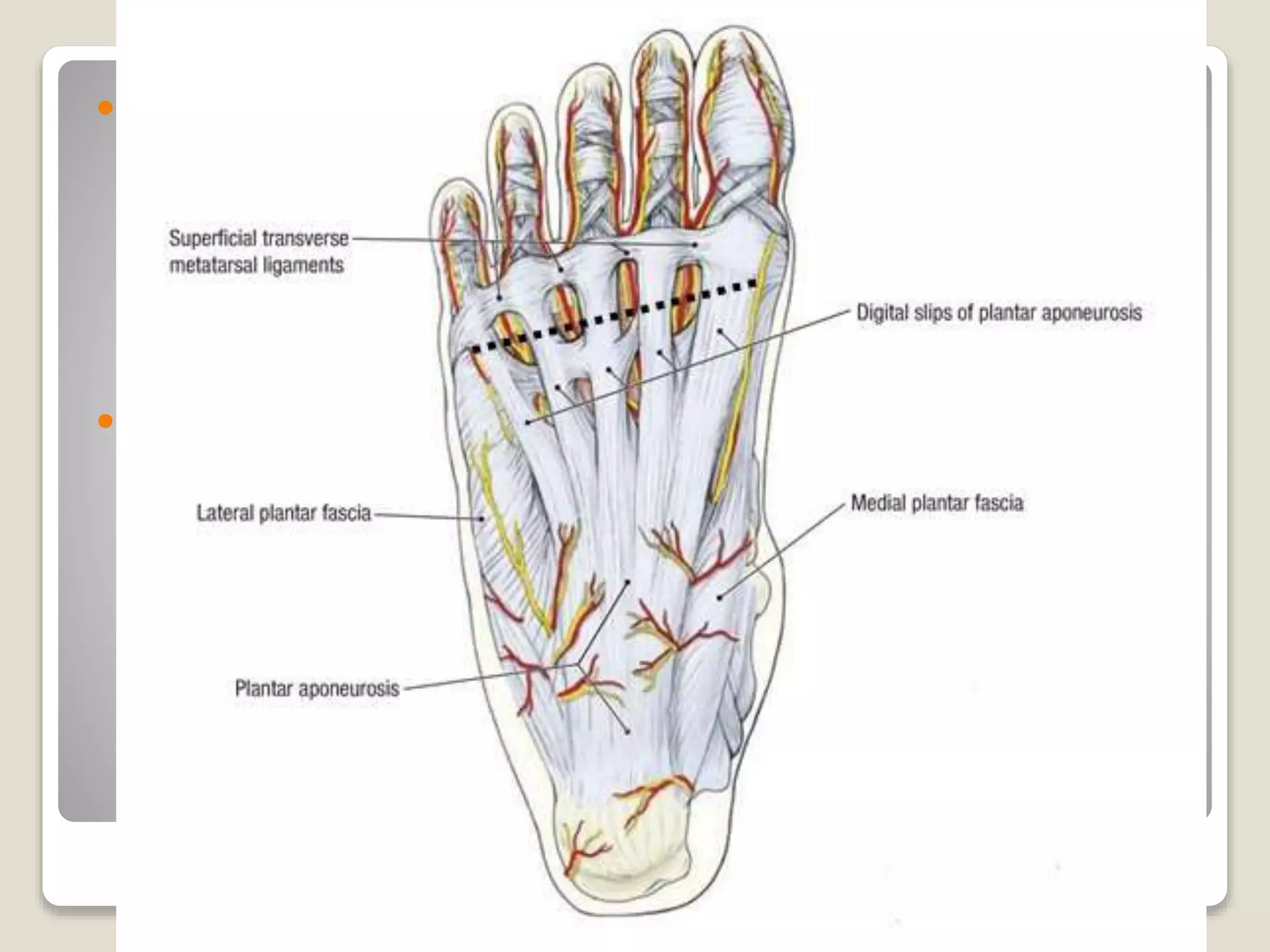
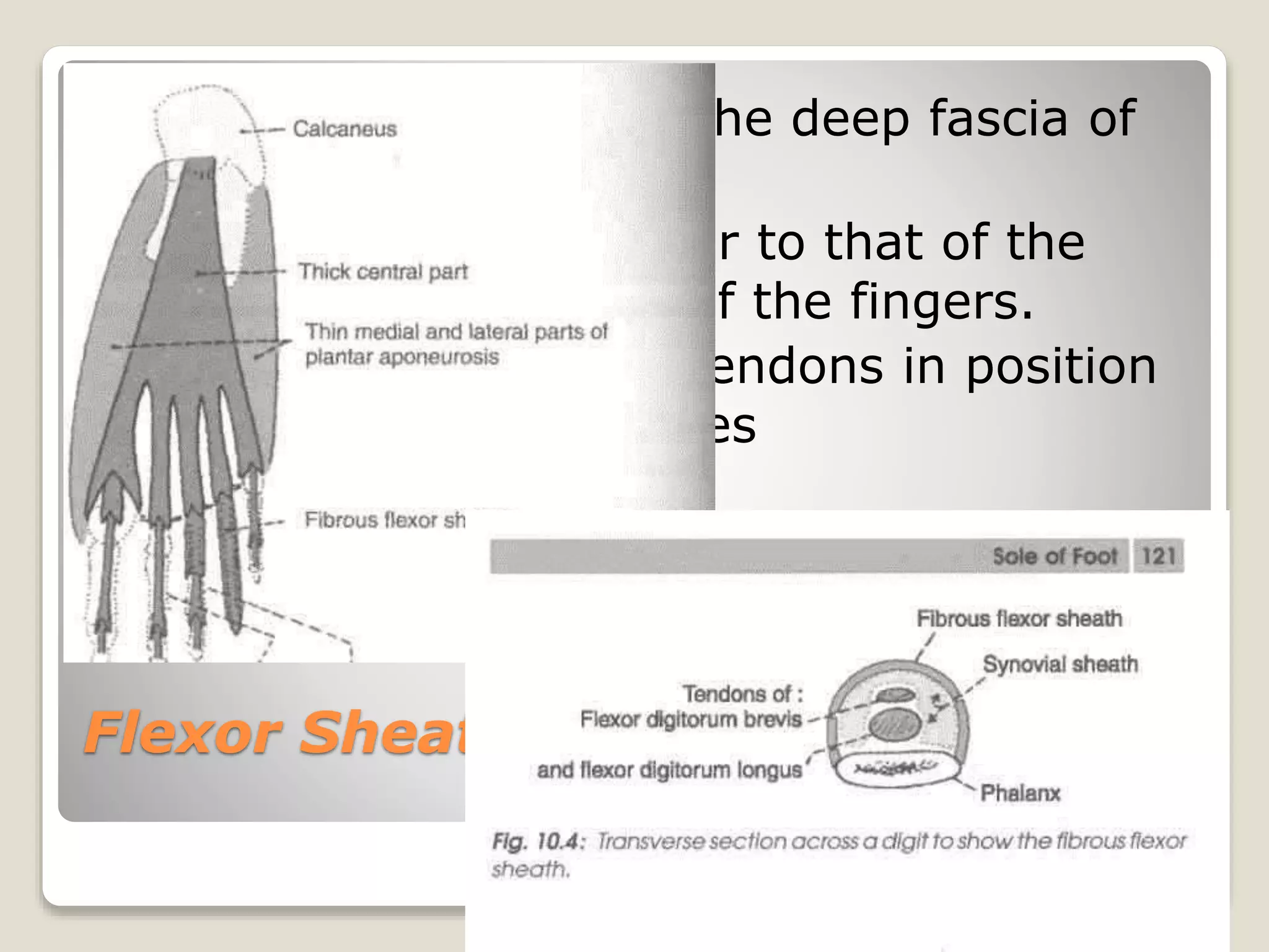
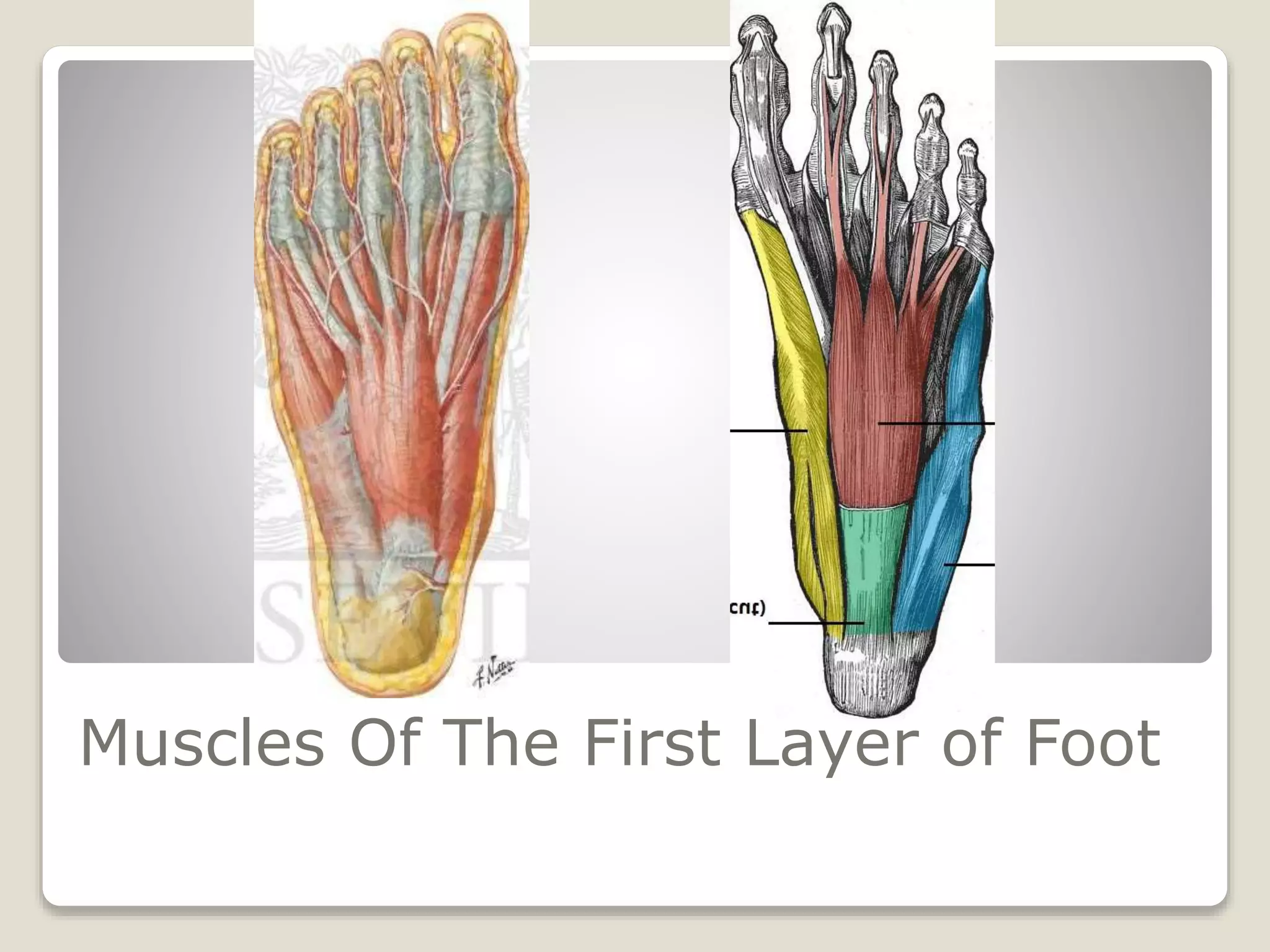
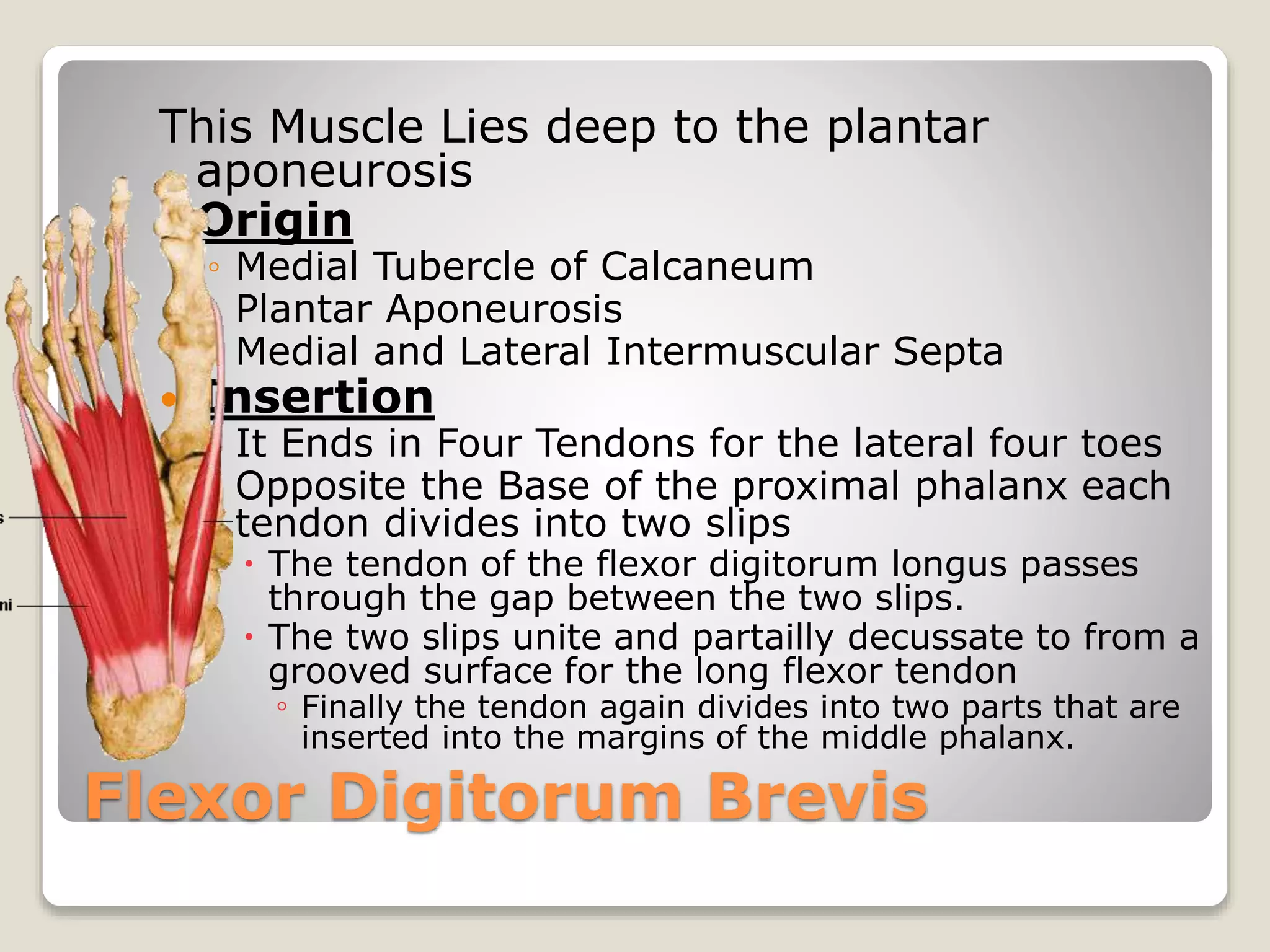
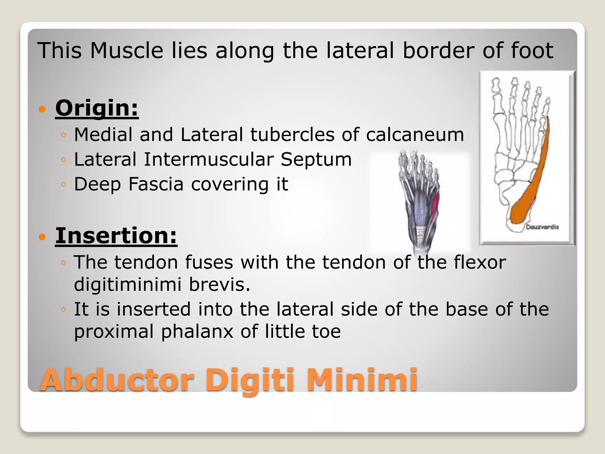
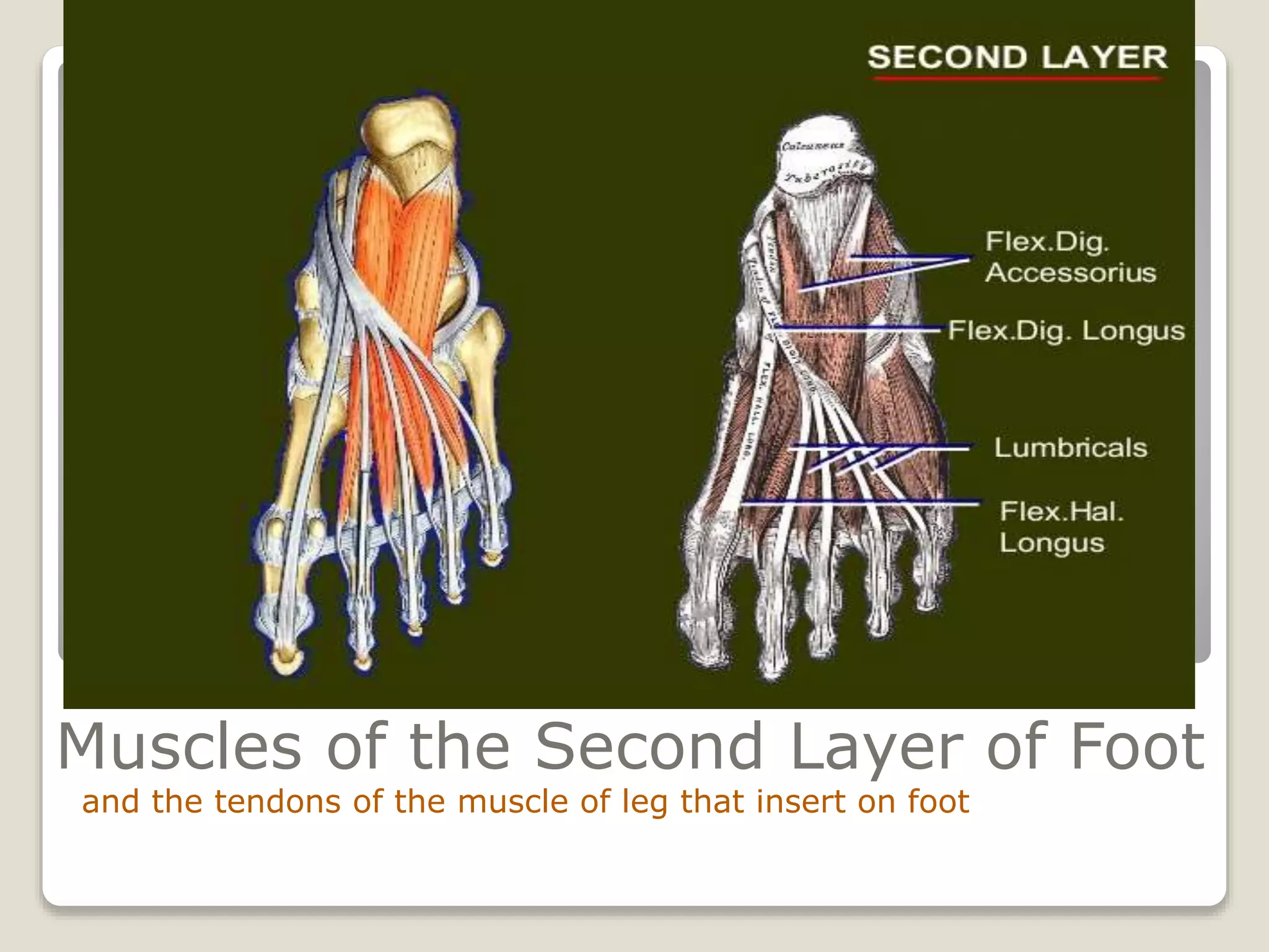
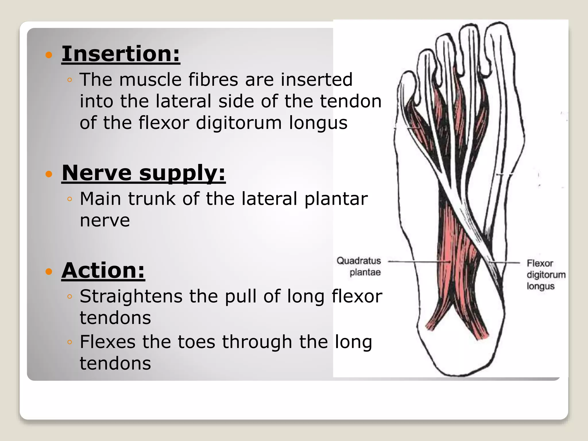
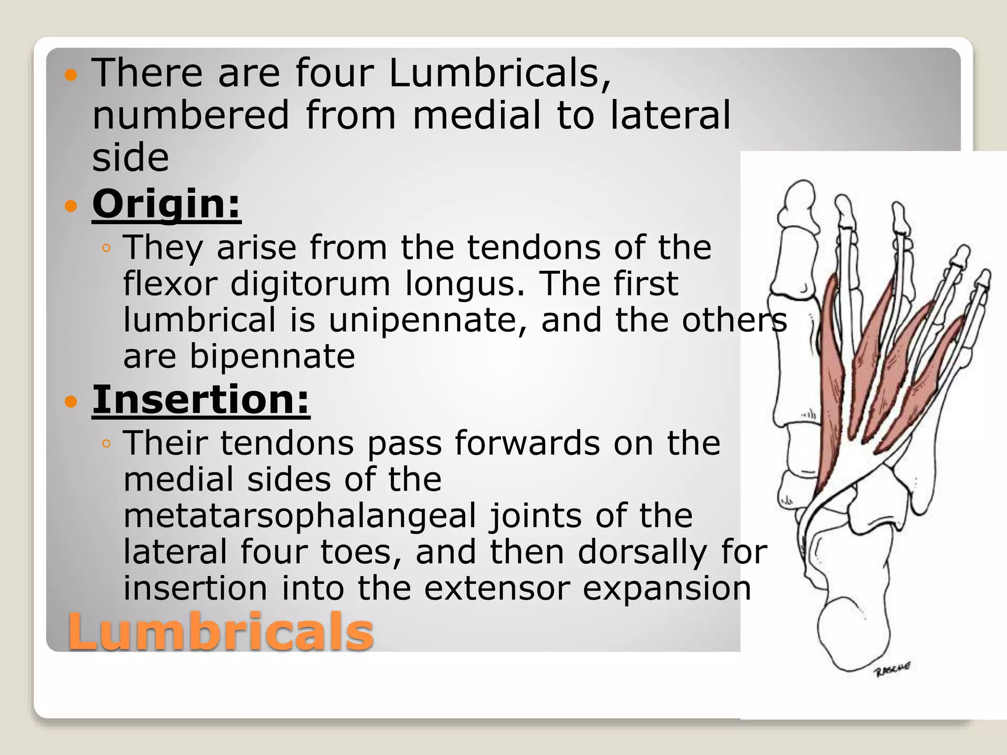
Sole Of Foot
Introduction
Skin of the Sole
Superficial Fascia
Deep Fascia
Muscles of the 1st Layer
Muscles of the 2nd Layer
Prepared By: Dr. Mateen
Reference: BD Chaurasia’s Human Anatomy
Intoduction
The Homologous Structures
Introduction
 Foot and Hand are homologous Parts
 However Foot is an organ of support and
locomotion so structures of the foot are
modified accordingly. For Example
◦ The great toe has lost its mobility and its
power of prehension (gripping)
◦ The Lesser four toes are markedly reduced
in size
◦ And the Tarsal Bones and the first
metatarsal are enlarged to form a broad
base for better support
Skin of the Sole
Skin of the sole is
1. Thick for protection
2. Firmly
adherent to the
underlying
aponeurosis
3. Creased
Skin of the Sole
Cutaneous nerve supply
a) The Medial Calcanean Branches of Tibial
Nerve supply the posterior and medial
portion
b) Branches from the medial plantar nerve
supply the larger antero-medial portion
including the medial three and a half
digits
c) Branches from the Lateral Plantar
Nerve supply the smaller antero-lateral
portion including the lateral one and a
half digits.
 These nerves are derived from spinal
nerves L4, L5 and S1.
 In eliciting the plantar reflex, the area
supplied by segment S1 is stimulated.
Superficial Fascia
Superficial Fascia
 The superficial fascia of the sole is fibrous and
dense.
 Fibrous band bind the skin to the deep fascia or
plantar Aponeurosis
 They divide the subcutaneous fat into small tight
compartments which serve as water-cushions
and reinforce the spring-effect of the arches of
the foot
 The fascia is very thick and dense over the
weight-bearing points.
 It contains cutaneous nerves and vessels.
 Thickened bands of superficial fascia stretch
across the roots of the toes forming the
superficial transverse metatarsal ligaments
Deep Fascia
The deep fascia of the sole is
specialized to form
 The Plantar Aponeurosis in the sole
 The Deep Transverse Metatarsal
Ligaments.
 The Fibrous Flexor Sheaths in the toes.
Plantar Aponeurosis
 The deep fascia covering the sole is thick in
the centre and thin at the sides.
 The thickened central part known as the
Plantar Aponeurosis
 The Aponeurosis is triangular in shape.
 The apex is proximal and attached to the
medial tubercle of the calcaneum, proximal
to the attachment of the flexor digitorum
brevis.
 The base is distal. It divides into five
processes near the heads of the metatarsal
bones. The digital nerves and vessels pass
through the intervals between the processes.
 Each process splits, opposite the metatarso-
phalangeal joints, into a superficial and a
deep slip. The superficial slip is attached to
skin. The deep slip divides into two parts
which embrace the flexor tendons, and blend
with the fibrous flexor sheaths and with the
deep transverse metatarsal ligaments.
 From the margins of the aponeurosis, lateral
and medial vertical intermuscular septa pass
deeply, and divide the sole into three
compartments. Thinner transverse septa
arise from the vertical septa and divide the
muscles of the sole into four layers.
 Functions
 1. It fixes the skin of the sole.
 2. It protects deeper structures.
 3. It helps in maintaining the longitudinal
arches of the foot.
 4. It gives origin to muscles of the first layer
of the sole.
 The plantar aponeurosis gives off an
additional process to the great toe, which
restricts its movement.
Deep Transverse Metatarsal
Ligaments
 These are four short, flat bands which
connect the plantar ligaments of the
adjoining metatars-o-phalangeal joints.
 They are related dorsally to the interossei,
and ventrally to the lumbricals and the
digital vessels and nerves.
Flexor Sheaths
 These are made up of the deep fascia of
the toes.
 Their structure is similar to that of the
fibrous flexor sheaths of the fingers.
 They retain the flexor tendons in position
during flexion of the toes
Muscles Of The First Layer of Foot
MUSCLES OF THE FIRST LAYER OF
THE SOLE
The muscles of the sole are arranged in
four layers, which will be considered one
by one.
◦The muscles of the first layer are
the
 Flexor Digitorum Brevis
 The Abductor Hallucis and
 The Abductor Digiti Minimi
Flexor Digitorum Brevis
This Muscle Lies deep to the plantar
aponeurosis
 Origin
◦ Medial Tubercle of Calcaneum
◦ Plantar Aponeurosis
◦ Medial and Lateral Intermuscular Septa
 Insertion
◦ It Ends in Four Tendons for the lateral four toes
◦ Opposite the Base of the proximal phalanx each
tendon divides into two slips
 The tendon of the flexor digitorum longus passes
through the gap between the two slips.
 The two slips unite and partailly decussate to from a
grooved surface for the long flexor tendon
◦ Finally the tendon again divides into two parts that are
inserted into the margins of the middle phalanx.
 Nerve Supply
◦ Medial Plantar Nerve
 Actions
◦ Flexion of the toes at
the proximal
interphalangeal joints
and
metatarsophalangeal
joints.
Abductor Hallucis
This Muscle Lies Along the Medial border of
the foot and covers the origin of the
plantar vessels and nerves
 Origin:
 Medial Tubercle of Calcanium
 Flexor Retinaculum and its deep fascia
 Medial Intermuscular Septum
 Insertion:
 The Tendon Fuses with the Medial
Portion of the flexor hallucis brevis
 It is inserted on to the medial side of
the base of the great toe
Abductor Hallucis
 Nerve Supply:
◦ Medial Plantar
Nerve
 Action:
◦ Abduction of the
great toe away from
the second toe
Trigger Point
Abductor Digiti Minimi
This Muscle lies along the lateral border of foot
 Origin:
◦ Medial and Lateral tubercles of calcaneum
◦ Lateral Intermuscular Septum
◦ Deep Fascia covering it
 Insertion:
◦ The tendon fuses with the tendon of the flexor
digitiminimi brevis.
◦ It is inserted into the lateral side of the base of the
proximal phalanx of little toe
Abductor Digiti Minimi
 Nevrve Supply:
◦ Main Trunk of the
Lateral Plantar
Nerve
 Action:
◦ Abduction of little
toe
Muscles of the Second Layer of Foot
and the tendons of the muscle of leg that insert on foot
Quadratus Plantae
It is also called as “flexor digitorum
accessorius” as it is accessory to the
flexor digitorum longus.
 Origin: It arises by two heads
◦ Medial head is large and fleshy; It arises
from the medial concave surface of the
calcaneum and from its medial tubercle.
◦ Lateral head is smaller and tendinous; it
arises form the calcaneum in front of the
lateral tubercle and from the long plantar
ligament.
◦ The two heads unite at an acute angle.
 Insertion:
◦ The muscle fibres are inserted
into the lateral side of the tendon
of the flexor digitorum longus
 Nerve supply:
◦ Main trunk of the lateral plantar
nerve
 Action:
◦ Straightens the pull of long flexor
tendons
◦ Flexes the toes through the long
tendons
Lumbricals
 There are four Lumbricals,
numbered from medial to lateral
side
 Origin:
◦ They arise from the tendons of the
flexor digitorum longus. The first
lumbrical is unipennate, and the others
are bipennate
 Insertion:
◦ Their tendons pass forwards on the
medial sides of the
metatarsophalangeal joints of the
lateral four toes, and then dorsally for
insertion into the extensor expansion
 Nerve Supply:
◦ The first muscle by the medial plantar nerve;
and the other three by the deep branch of
lateral plantar nerve
 Actions:
◦ They maintain extension of the digits at the
interphalangeal joints so that in walking and
running the toes do not buckle under
Introduction contd.
 The Arches of the Foot serve as elastic
springs for efficient walking, running,
jumping and supporting of body
 CLASSIFICATION OF ARCHES
◦I A. Longitudinal
 1. Medial
 2. Lateral
◦B. Transverse
 1. Anterior
 2. Posterior

Sole of foot

  • 1.
    Sole Of Foot Introduction Skinof the Sole Superficial Fascia Deep Fascia Muscles of the 1st Layer Muscles of the 2nd Layer Prepared By: Dr. Mateen Reference: BD Chaurasia’s Human Anatomy
  • 2.
  • 3.
    Introduction  Foot andHand are homologous Parts  However Foot is an organ of support and locomotion so structures of the foot are modified accordingly. For Example ◦ The great toe has lost its mobility and its power of prehension (gripping) ◦ The Lesser four toes are markedly reduced in size ◦ And the Tarsal Bones and the first metatarsal are enlarged to form a broad base for better support
  • 4.
    Skin of theSole Skin of the sole is 1. Thick for protection 2. Firmly adherent to the underlying aponeurosis 3. Creased
  • 5.
    Skin of theSole Cutaneous nerve supply a) The Medial Calcanean Branches of Tibial Nerve supply the posterior and medial portion b) Branches from the medial plantar nerve supply the larger antero-medial portion including the medial three and a half digits c) Branches from the Lateral Plantar Nerve supply the smaller antero-lateral portion including the lateral one and a half digits.
  • 6.
     These nervesare derived from spinal nerves L4, L5 and S1.  In eliciting the plantar reflex, the area supplied by segment S1 is stimulated.
  • 7.
  • 8.
    Superficial Fascia  Thesuperficial fascia of the sole is fibrous and dense.  Fibrous band bind the skin to the deep fascia or plantar Aponeurosis  They divide the subcutaneous fat into small tight compartments which serve as water-cushions and reinforce the spring-effect of the arches of the foot  The fascia is very thick and dense over the weight-bearing points.  It contains cutaneous nerves and vessels.  Thickened bands of superficial fascia stretch across the roots of the toes forming the superficial transverse metatarsal ligaments
  • 9.
    Deep Fascia The deepfascia of the sole is specialized to form  The Plantar Aponeurosis in the sole  The Deep Transverse Metatarsal Ligaments.  The Fibrous Flexor Sheaths in the toes.
  • 10.
    Plantar Aponeurosis  Thedeep fascia covering the sole is thick in the centre and thin at the sides.  The thickened central part known as the Plantar Aponeurosis  The Aponeurosis is triangular in shape.  The apex is proximal and attached to the medial tubercle of the calcaneum, proximal to the attachment of the flexor digitorum brevis.  The base is distal. It divides into five processes near the heads of the metatarsal bones. The digital nerves and vessels pass through the intervals between the processes.
  • 11.
     Each processsplits, opposite the metatarso- phalangeal joints, into a superficial and a deep slip. The superficial slip is attached to skin. The deep slip divides into two parts which embrace the flexor tendons, and blend with the fibrous flexor sheaths and with the deep transverse metatarsal ligaments.  From the margins of the aponeurosis, lateral and medial vertical intermuscular septa pass deeply, and divide the sole into three compartments. Thinner transverse septa arise from the vertical septa and divide the muscles of the sole into four layers.
  • 12.
     Functions  1.It fixes the skin of the sole.  2. It protects deeper structures.  3. It helps in maintaining the longitudinal arches of the foot.  4. It gives origin to muscles of the first layer of the sole.  The plantar aponeurosis gives off an additional process to the great toe, which restricts its movement.
  • 13.
    Deep Transverse Metatarsal Ligaments These are four short, flat bands which connect the plantar ligaments of the adjoining metatars-o-phalangeal joints.  They are related dorsally to the interossei, and ventrally to the lumbricals and the digital vessels and nerves.
  • 14.
    Flexor Sheaths  Theseare made up of the deep fascia of the toes.  Their structure is similar to that of the fibrous flexor sheaths of the fingers.  They retain the flexor tendons in position during flexion of the toes
  • 15.
    Muscles Of TheFirst Layer of Foot
  • 16.
    MUSCLES OF THEFIRST LAYER OF THE SOLE The muscles of the sole are arranged in four layers, which will be considered one by one. ◦The muscles of the first layer are the  Flexor Digitorum Brevis  The Abductor Hallucis and  The Abductor Digiti Minimi
  • 17.
    Flexor Digitorum Brevis ThisMuscle Lies deep to the plantar aponeurosis  Origin ◦ Medial Tubercle of Calcaneum ◦ Plantar Aponeurosis ◦ Medial and Lateral Intermuscular Septa  Insertion ◦ It Ends in Four Tendons for the lateral four toes ◦ Opposite the Base of the proximal phalanx each tendon divides into two slips  The tendon of the flexor digitorum longus passes through the gap between the two slips.  The two slips unite and partailly decussate to from a grooved surface for the long flexor tendon ◦ Finally the tendon again divides into two parts that are inserted into the margins of the middle phalanx.
  • 18.
     Nerve Supply ◦Medial Plantar Nerve  Actions ◦ Flexion of the toes at the proximal interphalangeal joints and metatarsophalangeal joints.
  • 19.
    Abductor Hallucis This MuscleLies Along the Medial border of the foot and covers the origin of the plantar vessels and nerves  Origin:  Medial Tubercle of Calcanium  Flexor Retinaculum and its deep fascia  Medial Intermuscular Septum  Insertion:  The Tendon Fuses with the Medial Portion of the flexor hallucis brevis  It is inserted on to the medial side of the base of the great toe
  • 20.
    Abductor Hallucis  NerveSupply: ◦ Medial Plantar Nerve  Action: ◦ Abduction of the great toe away from the second toe Trigger Point
  • 21.
    Abductor Digiti Minimi ThisMuscle lies along the lateral border of foot  Origin: ◦ Medial and Lateral tubercles of calcaneum ◦ Lateral Intermuscular Septum ◦ Deep Fascia covering it  Insertion: ◦ The tendon fuses with the tendon of the flexor digitiminimi brevis. ◦ It is inserted into the lateral side of the base of the proximal phalanx of little toe
  • 22.
    Abductor Digiti Minimi Nevrve Supply: ◦ Main Trunk of the Lateral Plantar Nerve  Action: ◦ Abduction of little toe
  • 23.
    Muscles of theSecond Layer of Foot and the tendons of the muscle of leg that insert on foot
  • 24.
    Quadratus Plantae It isalso called as “flexor digitorum accessorius” as it is accessory to the flexor digitorum longus.  Origin: It arises by two heads ◦ Medial head is large and fleshy; It arises from the medial concave surface of the calcaneum and from its medial tubercle. ◦ Lateral head is smaller and tendinous; it arises form the calcaneum in front of the lateral tubercle and from the long plantar ligament. ◦ The two heads unite at an acute angle.
  • 25.
     Insertion: ◦ Themuscle fibres are inserted into the lateral side of the tendon of the flexor digitorum longus  Nerve supply: ◦ Main trunk of the lateral plantar nerve  Action: ◦ Straightens the pull of long flexor tendons ◦ Flexes the toes through the long tendons
  • 26.
    Lumbricals  There arefour Lumbricals, numbered from medial to lateral side  Origin: ◦ They arise from the tendons of the flexor digitorum longus. The first lumbrical is unipennate, and the others are bipennate  Insertion: ◦ Their tendons pass forwards on the medial sides of the metatarsophalangeal joints of the lateral four toes, and then dorsally for insertion into the extensor expansion
  • 27.
     Nerve Supply: ◦The first muscle by the medial plantar nerve; and the other three by the deep branch of lateral plantar nerve  Actions: ◦ They maintain extension of the digits at the interphalangeal joints so that in walking and running the toes do not buckle under
  • 29.
    Introduction contd.  TheArches of the Foot serve as elastic springs for efficient walking, running, jumping and supporting of body  CLASSIFICATION OF ARCHES ◦I A. Longitudinal  1. Medial  2. Lateral ◦B. Transverse  1. Anterior  2. Posterior

Editor's Notes

  • #7 Dermatome: an area of the skin supplied by nerves from a single spinal root.