Downloaded 110 times


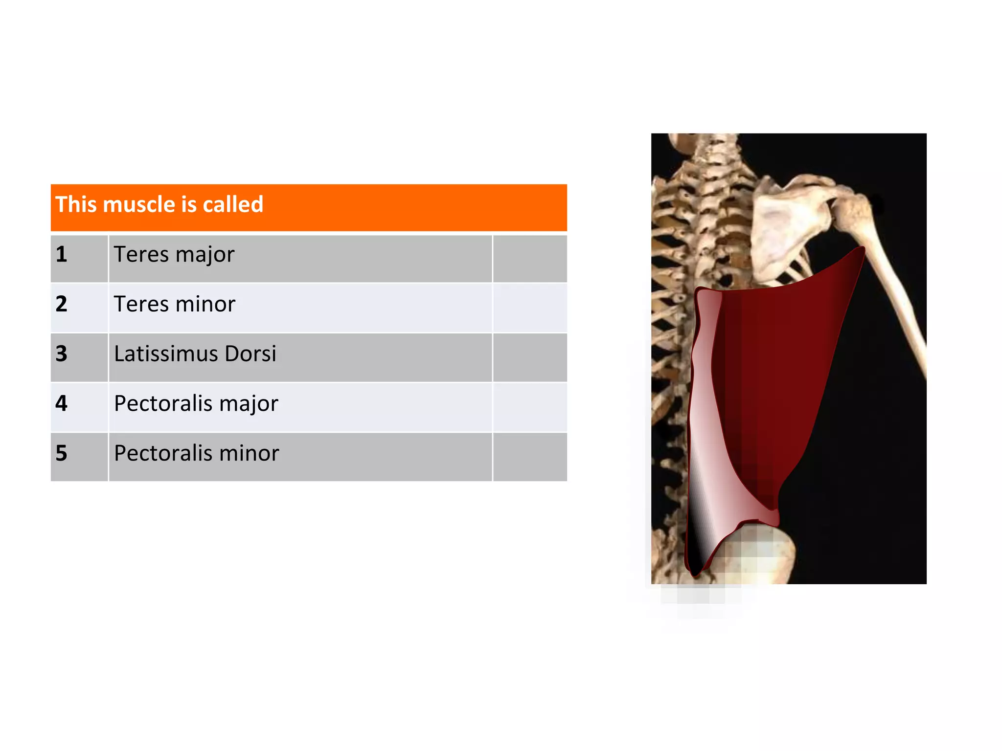


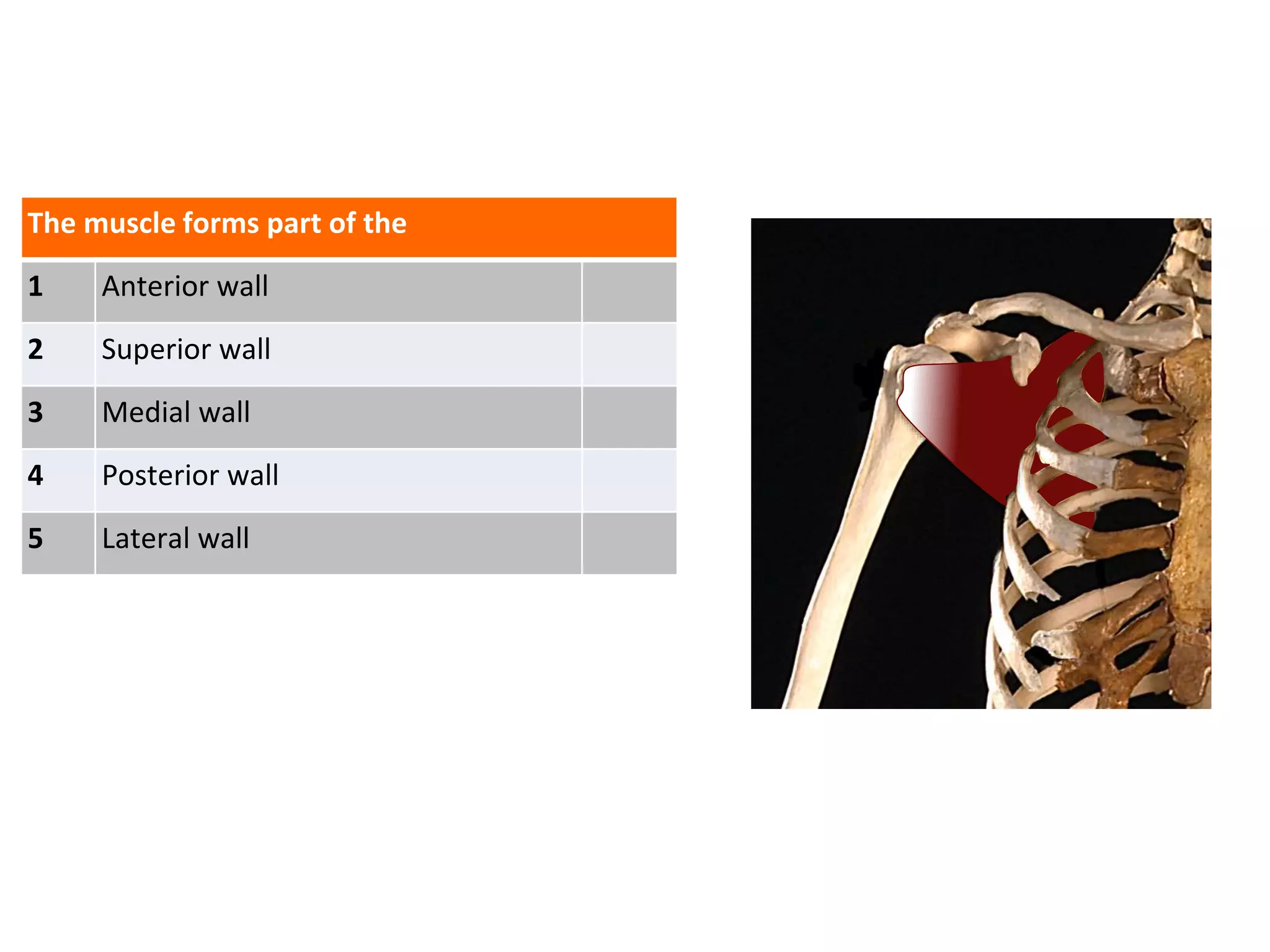


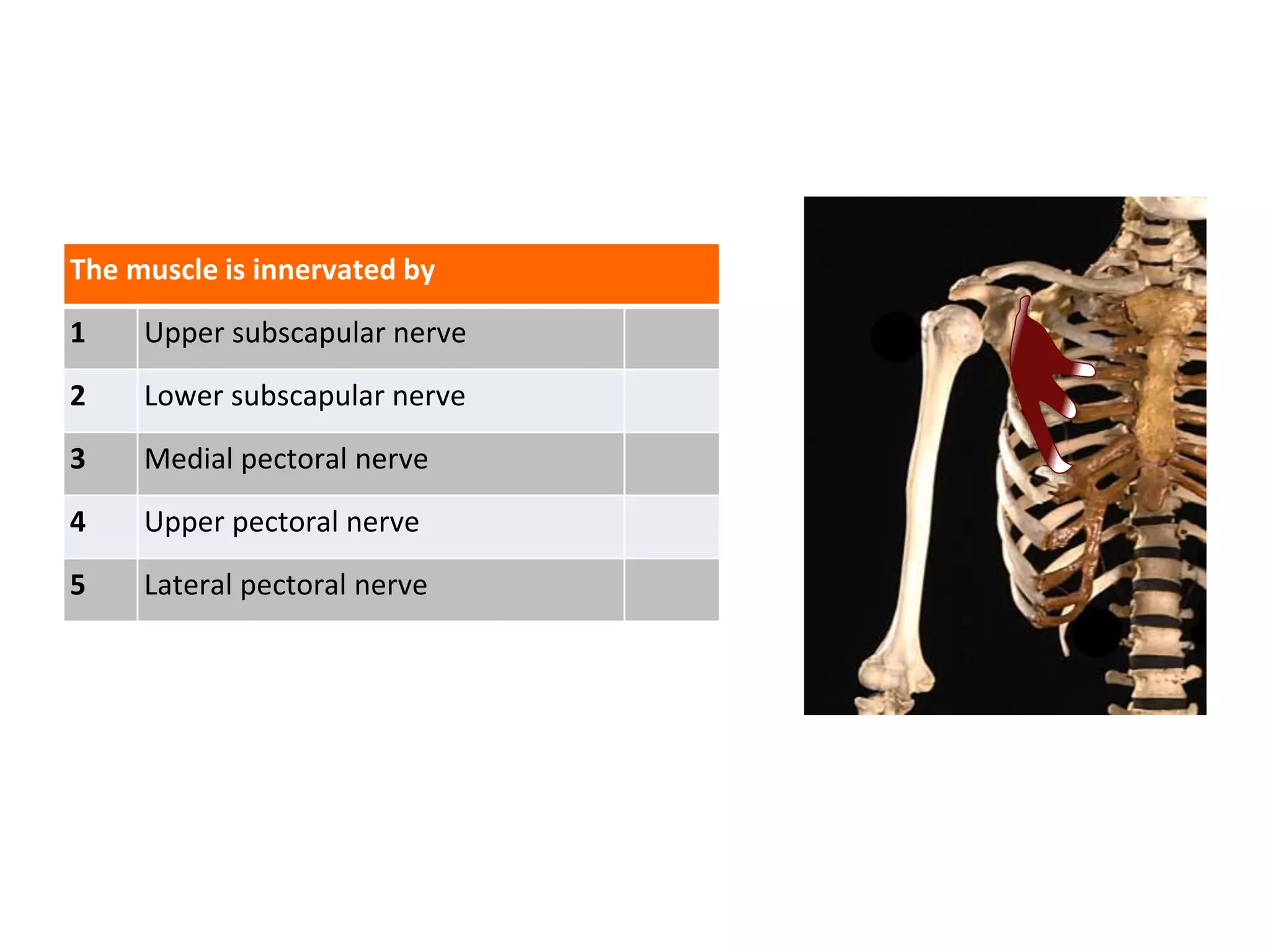


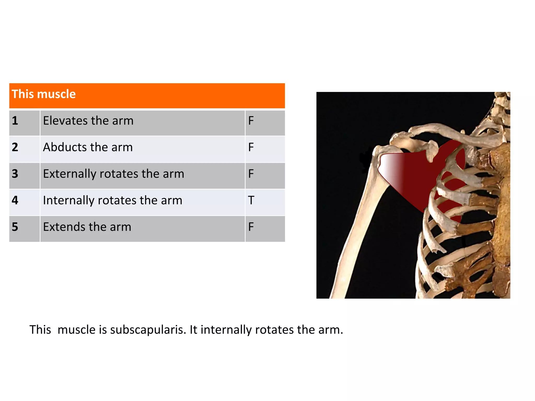


The document provides a detailed anatomical overview of the axilla, including the structures that make up its walls, the muscles involved, their innervation, and vascular connections. It elaborates on the relationships between various components such as the axillary artery and nerves of the brachial plexus. Key muscles like pectoralis major and minor, teres major, and latissimus dorsi are discussed in terms of their functional roles and anatomical locations.


