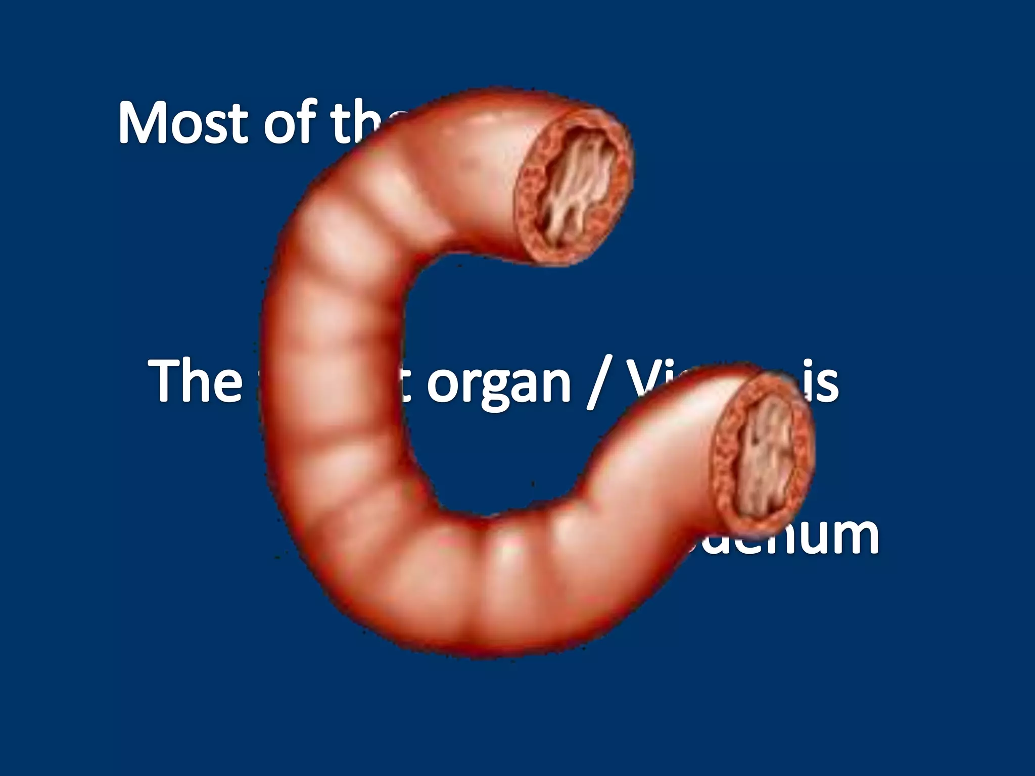
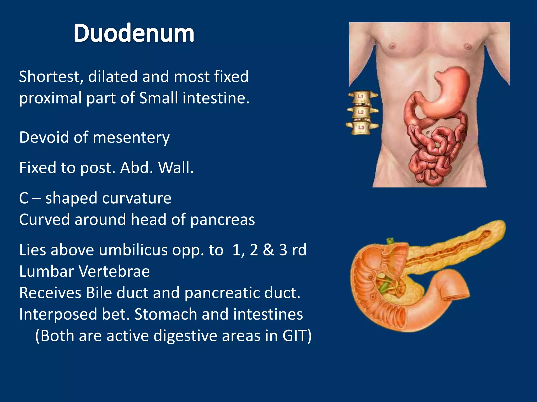
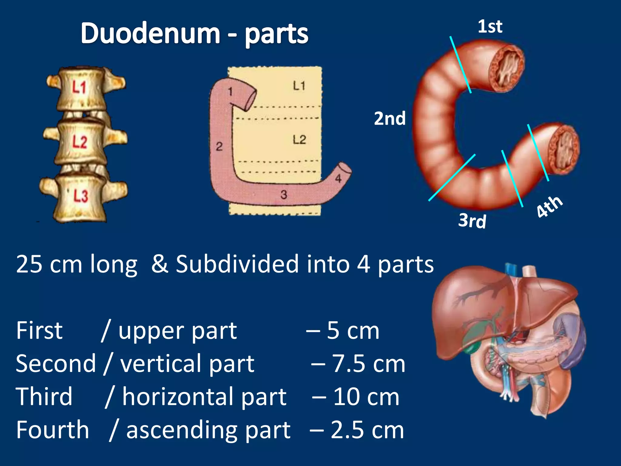
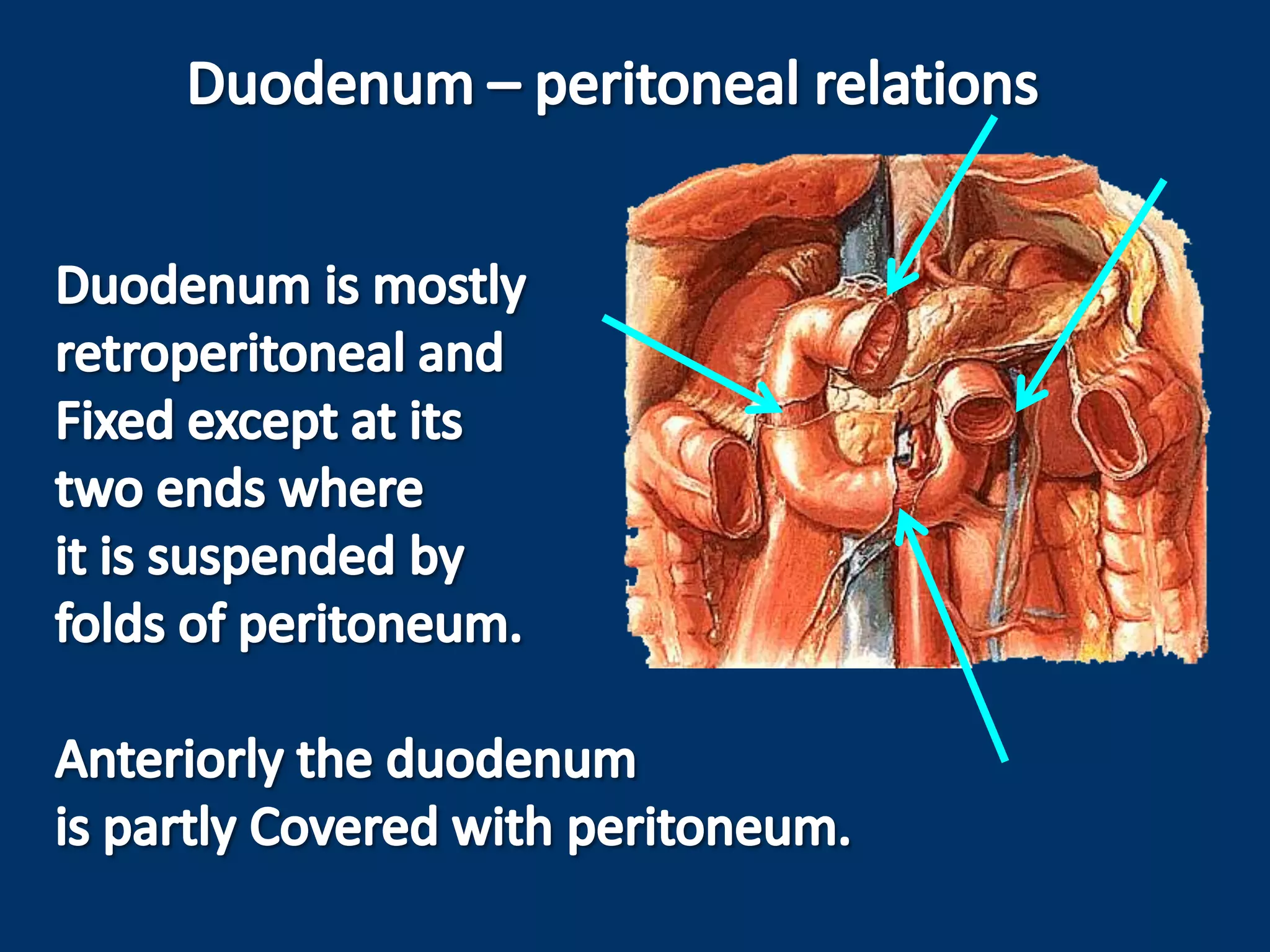
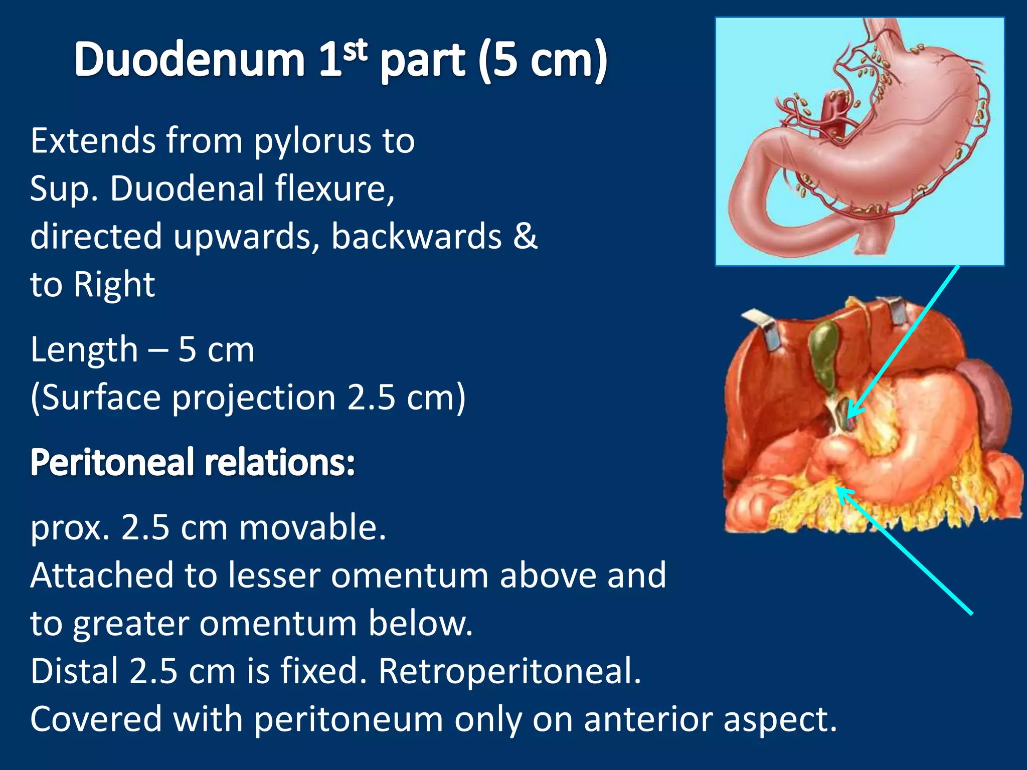
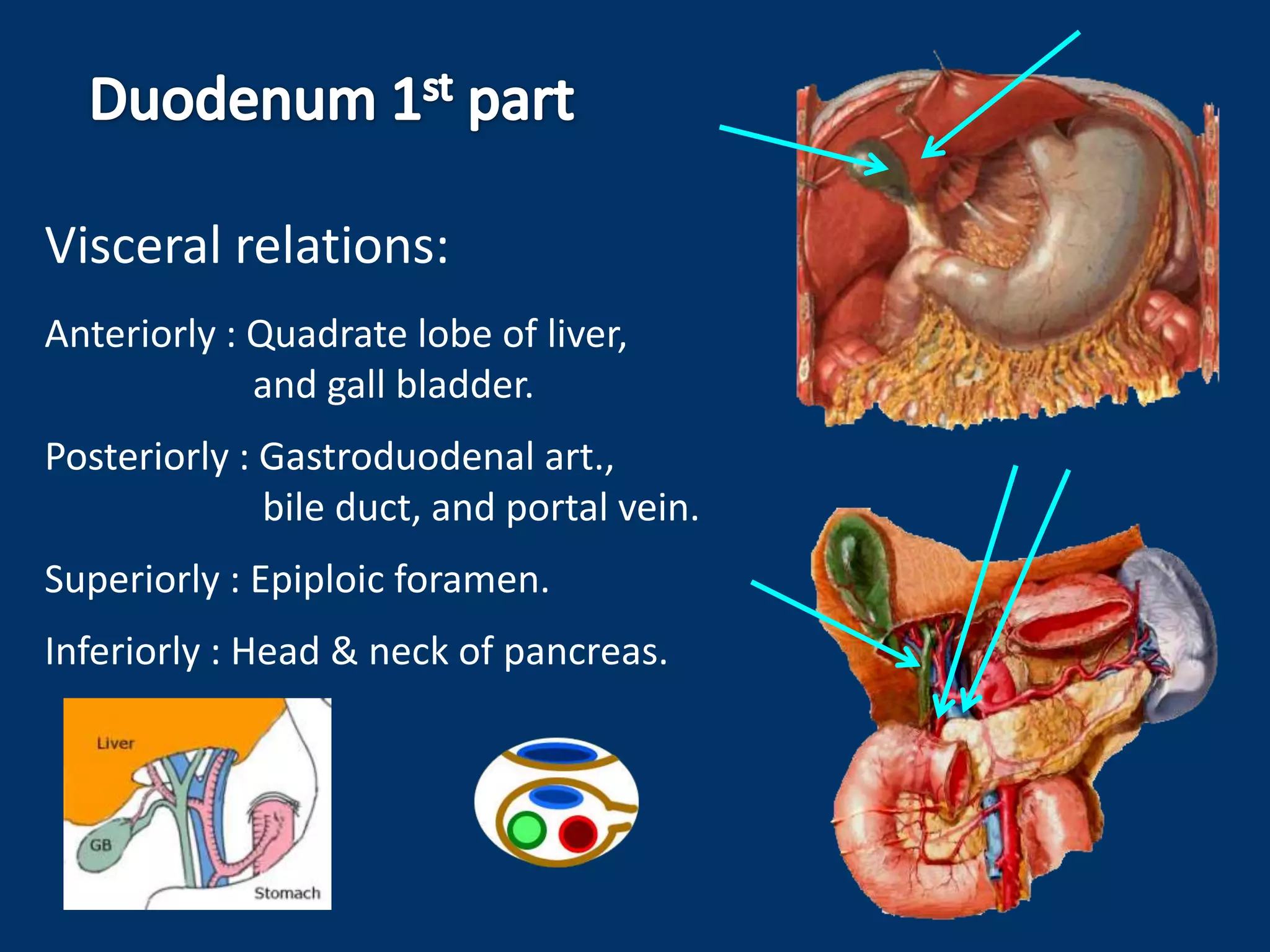
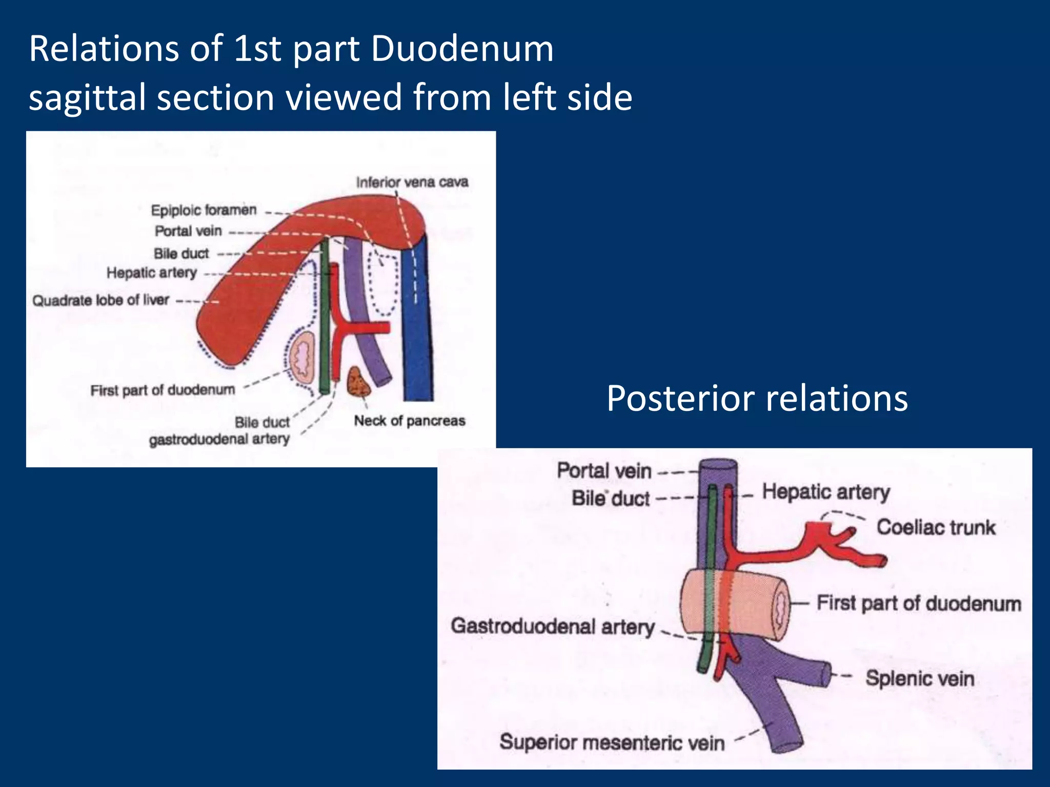
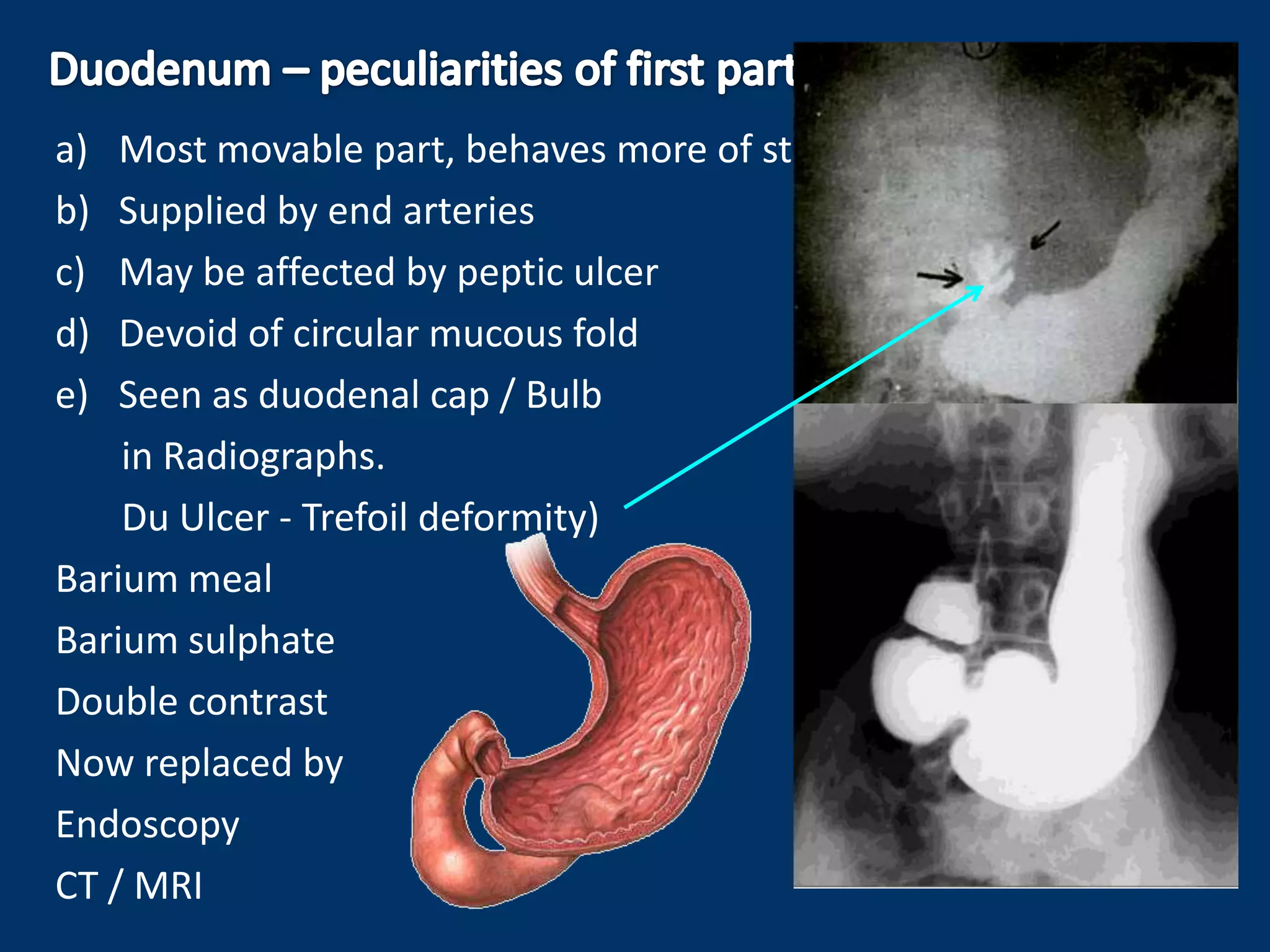
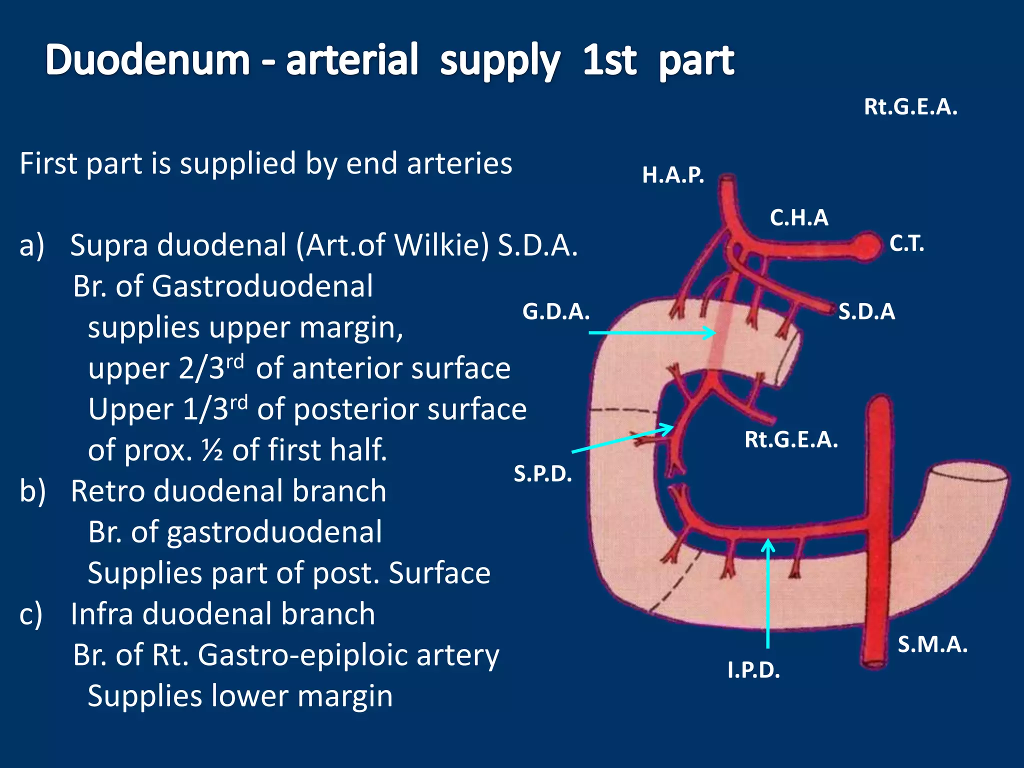
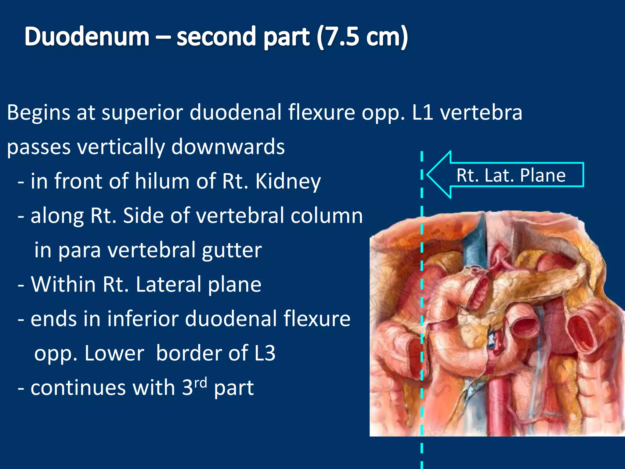
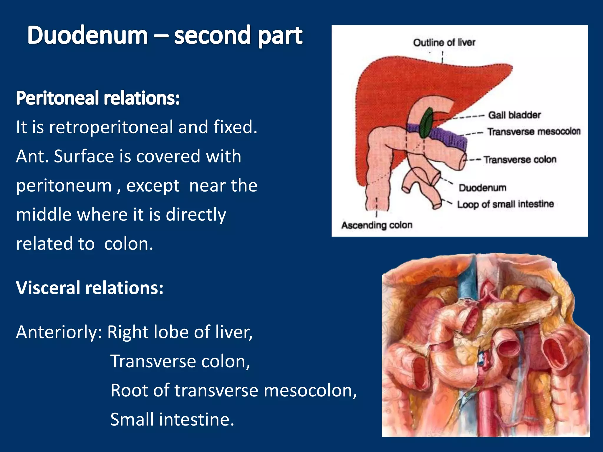
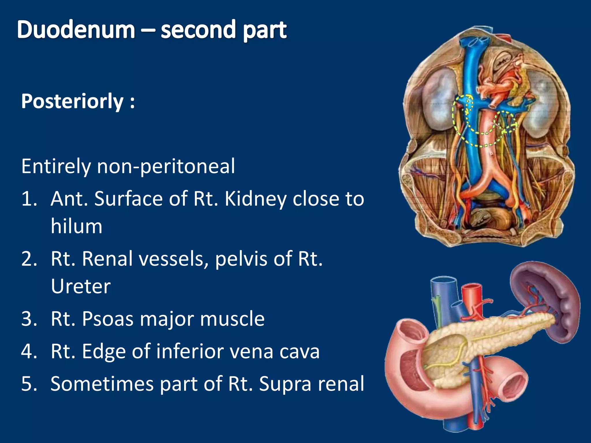
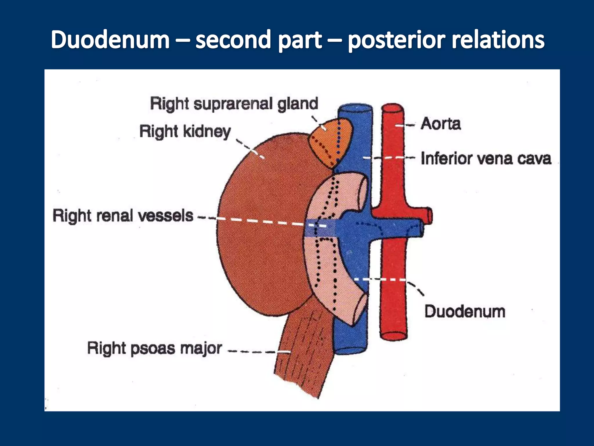
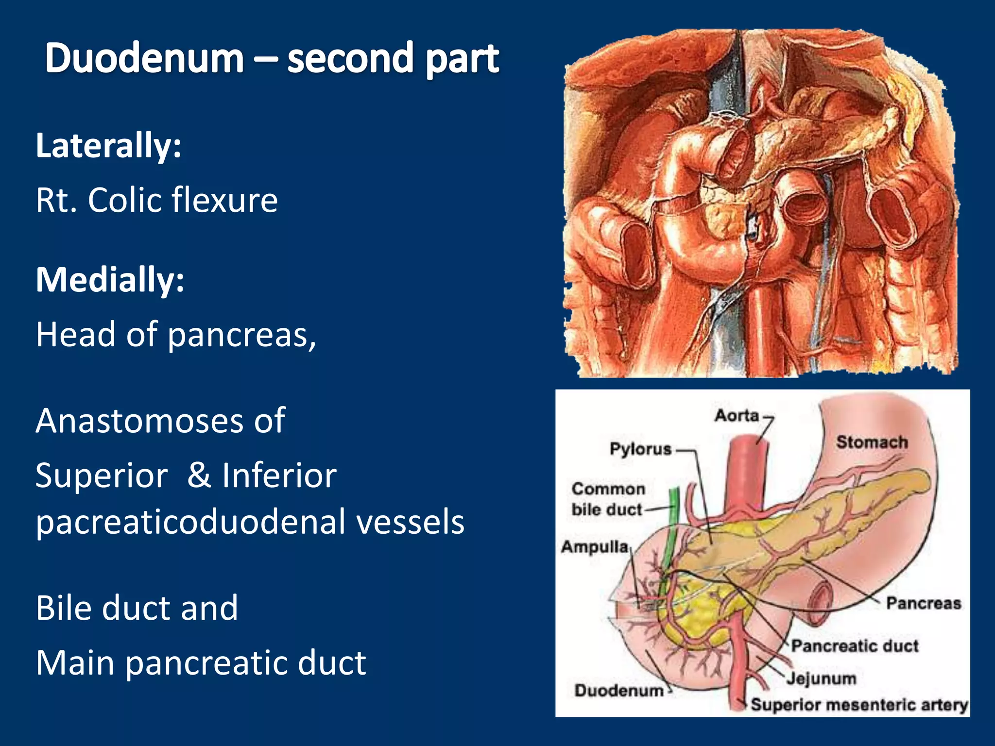
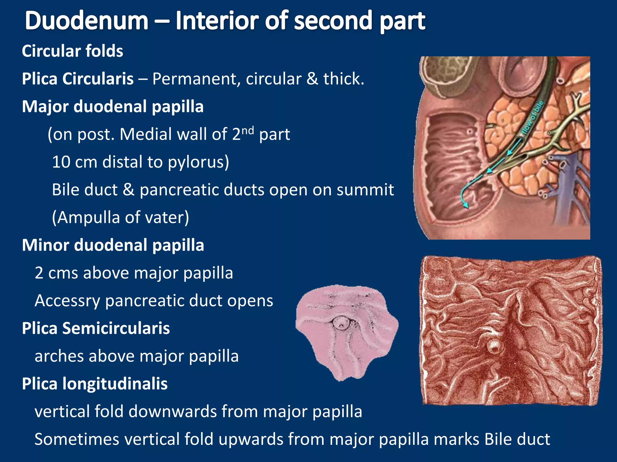
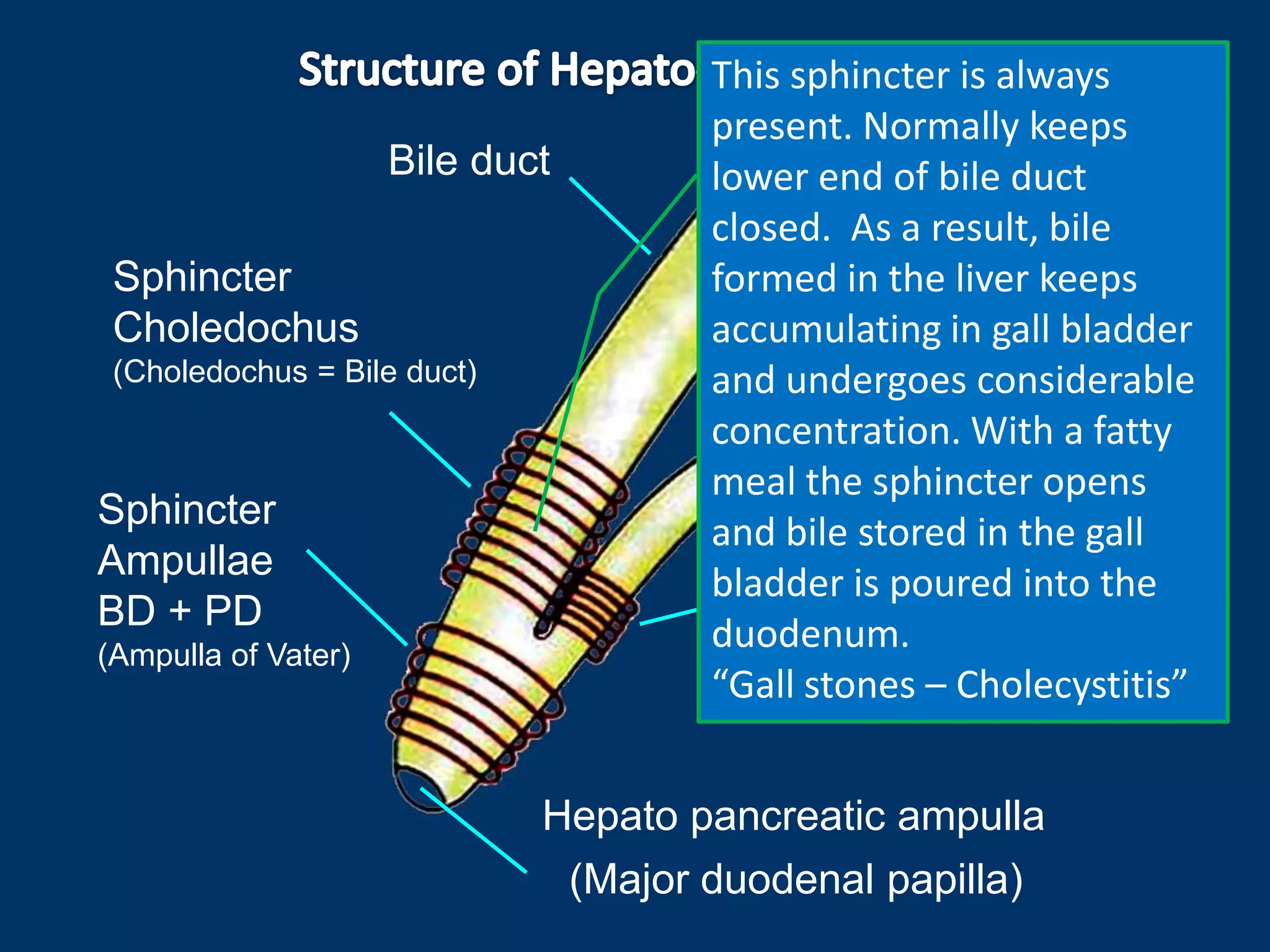
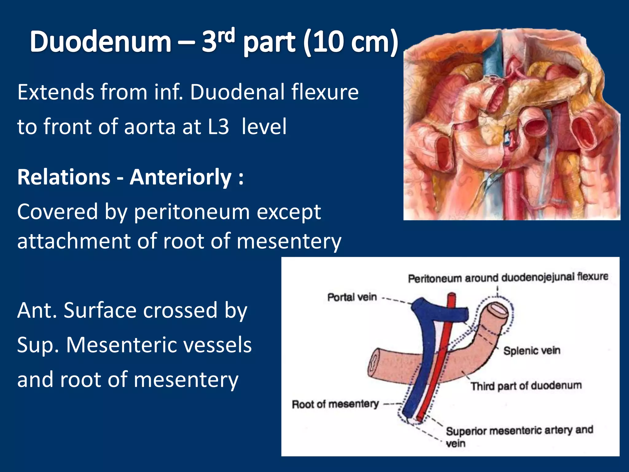
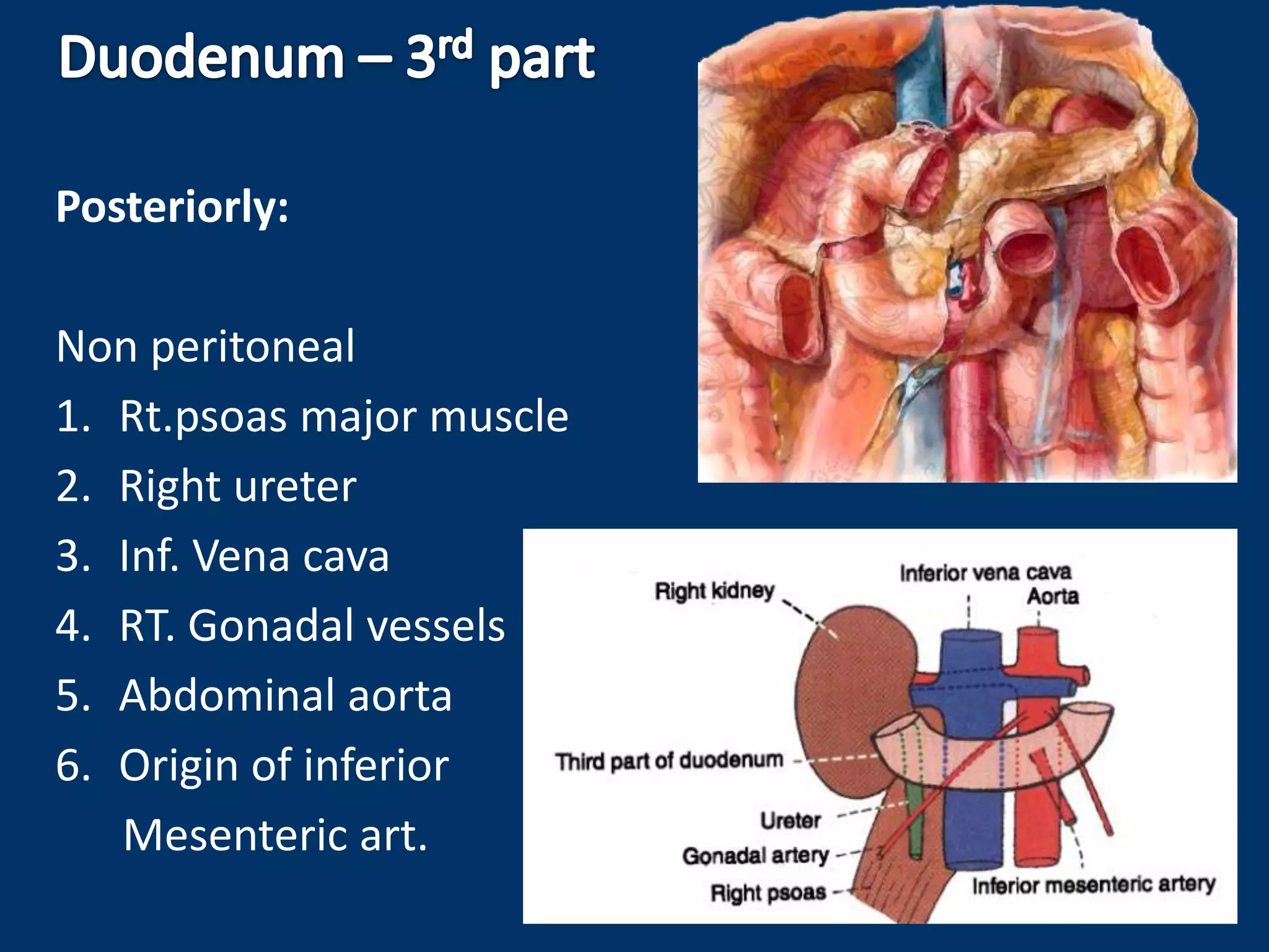
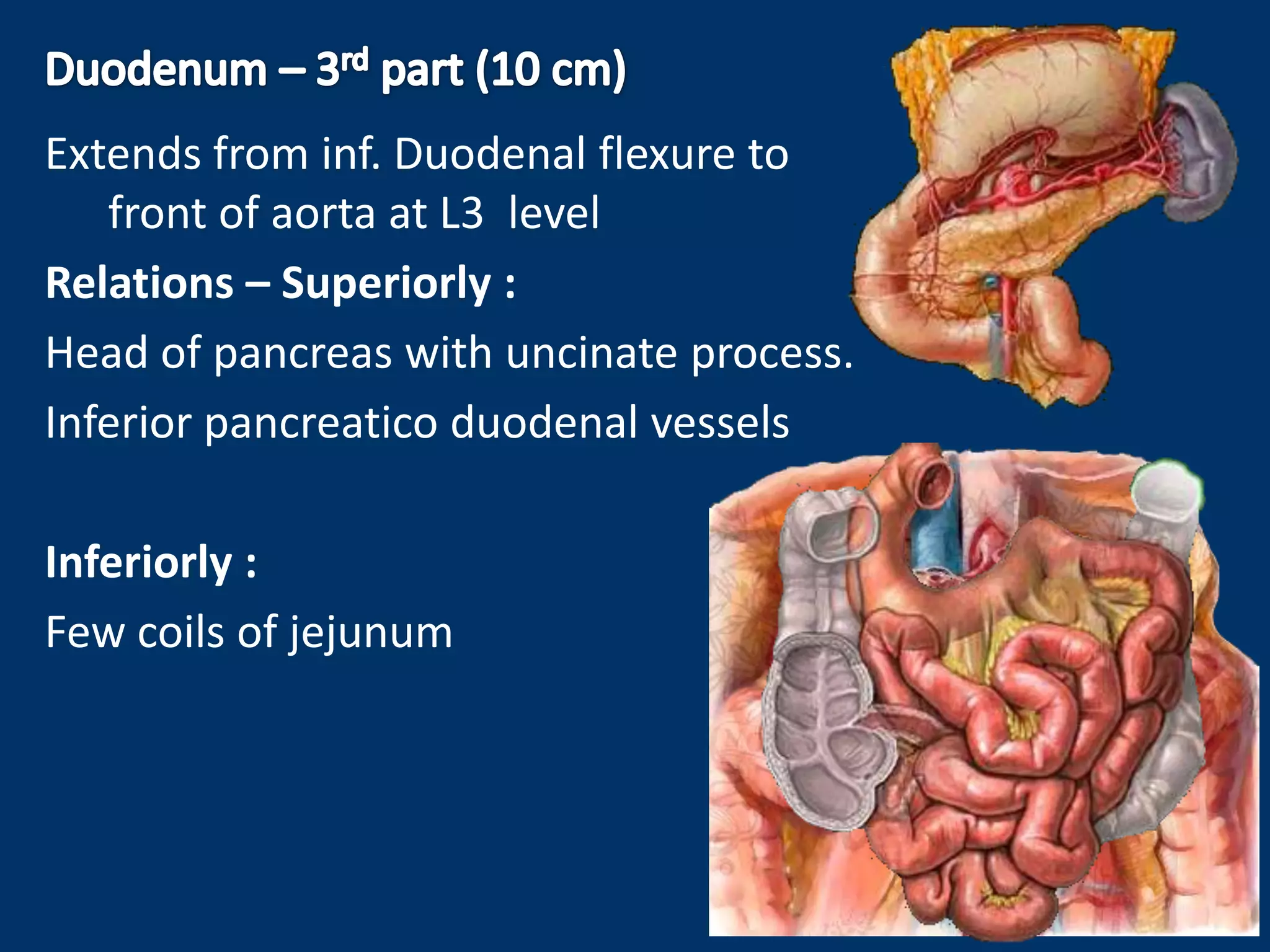
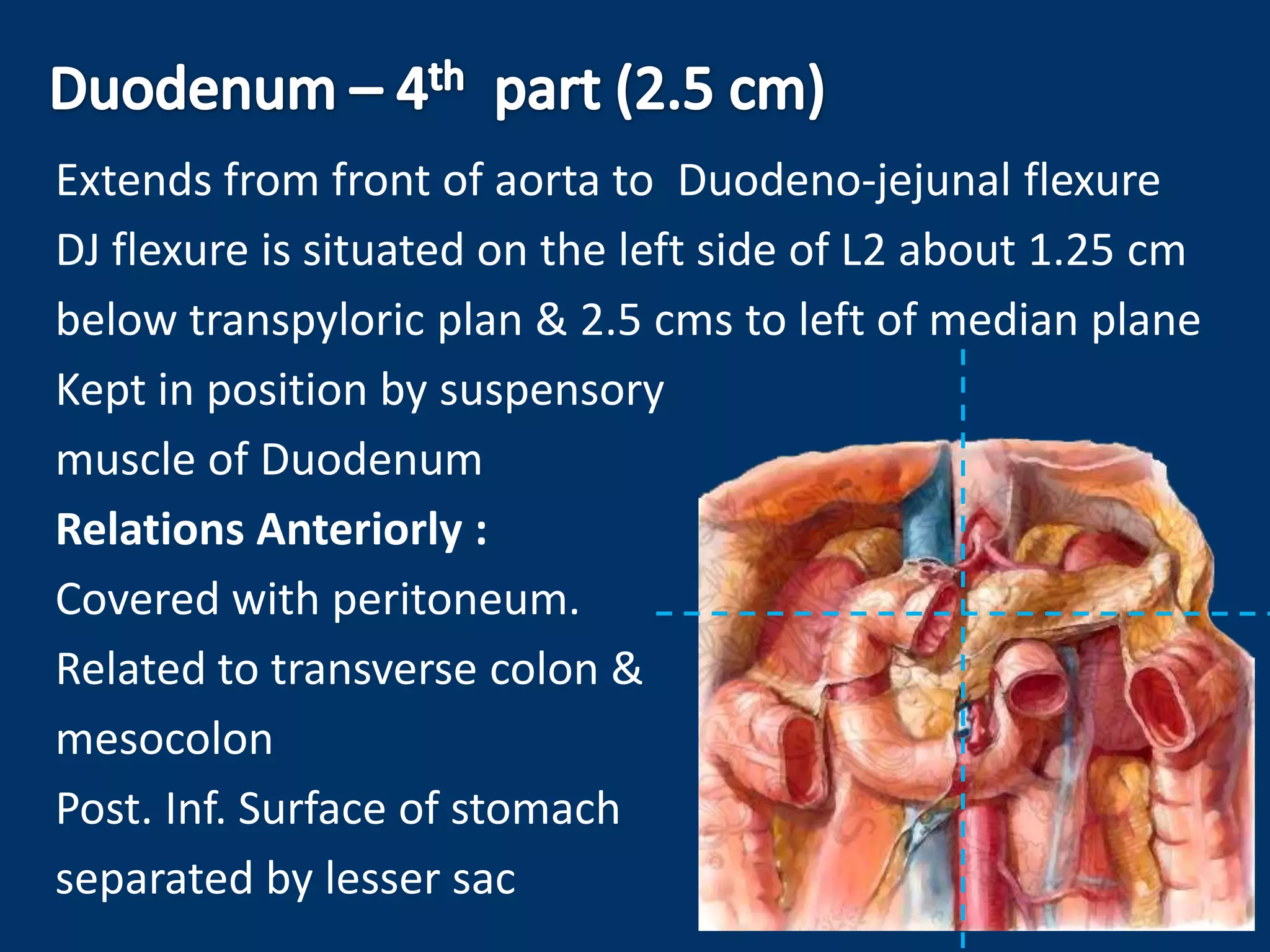
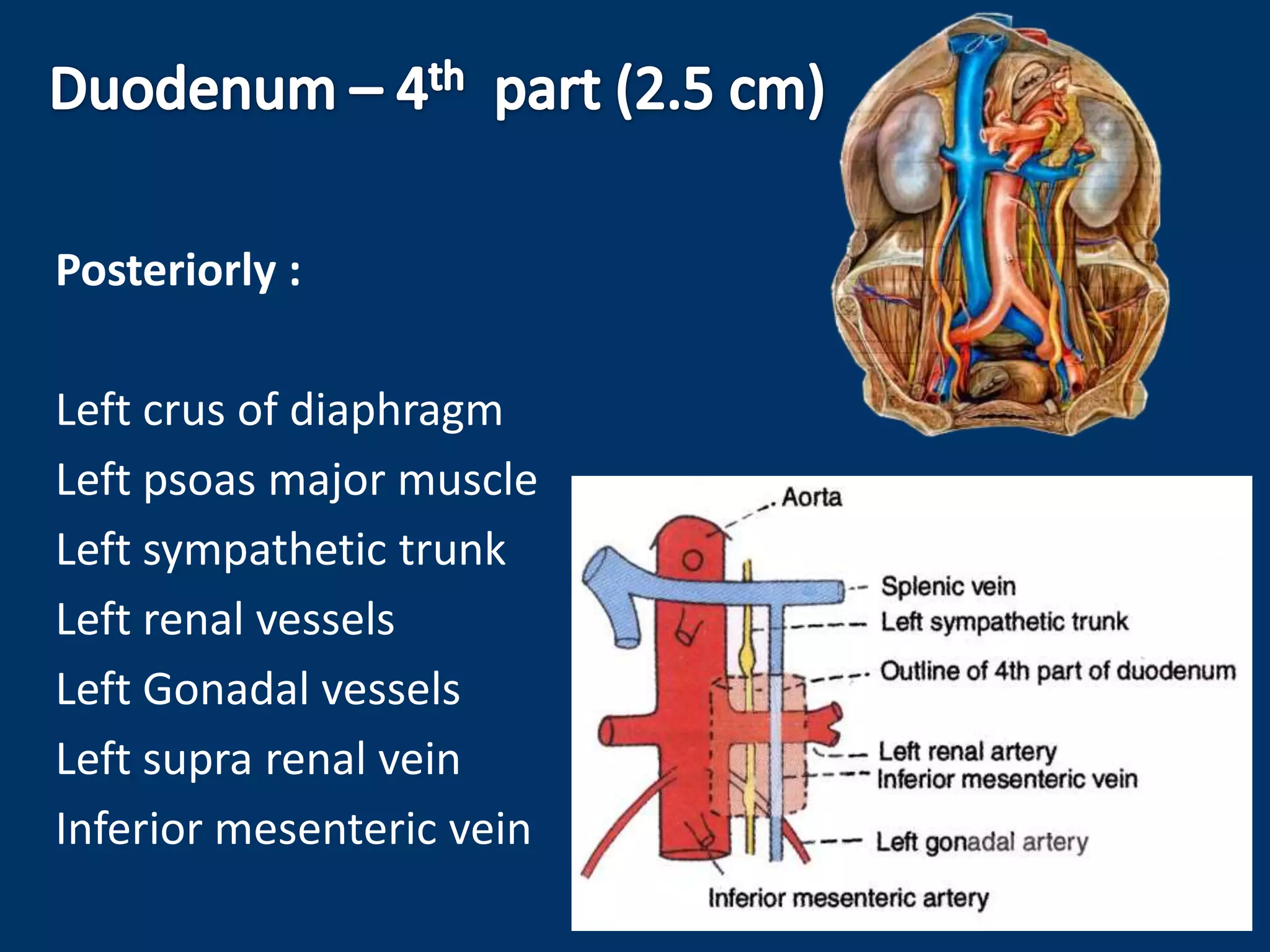
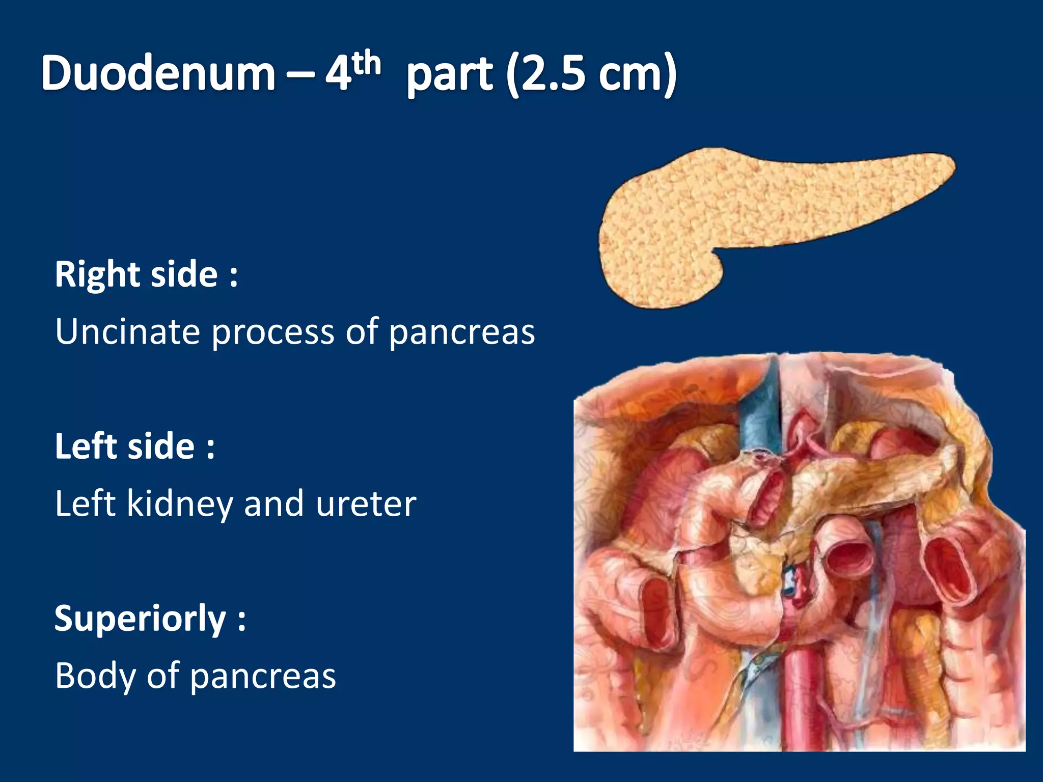
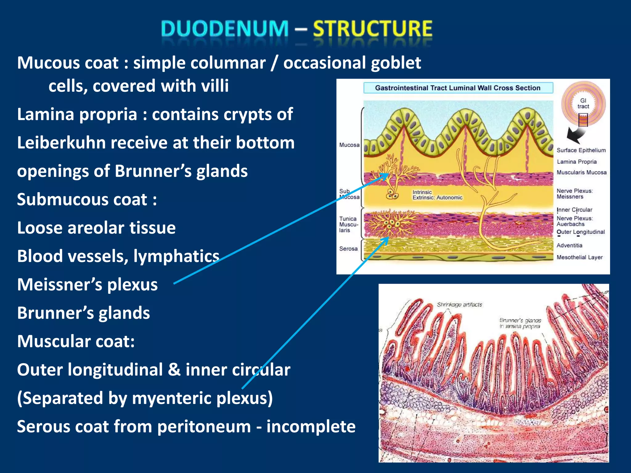
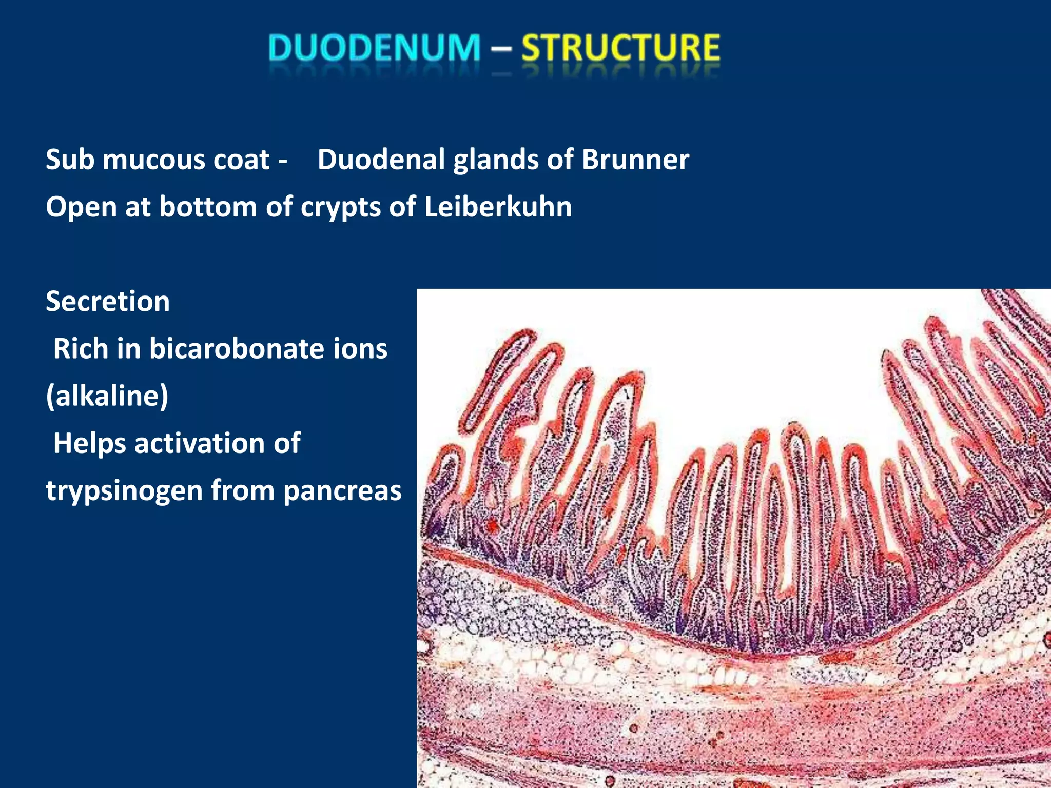
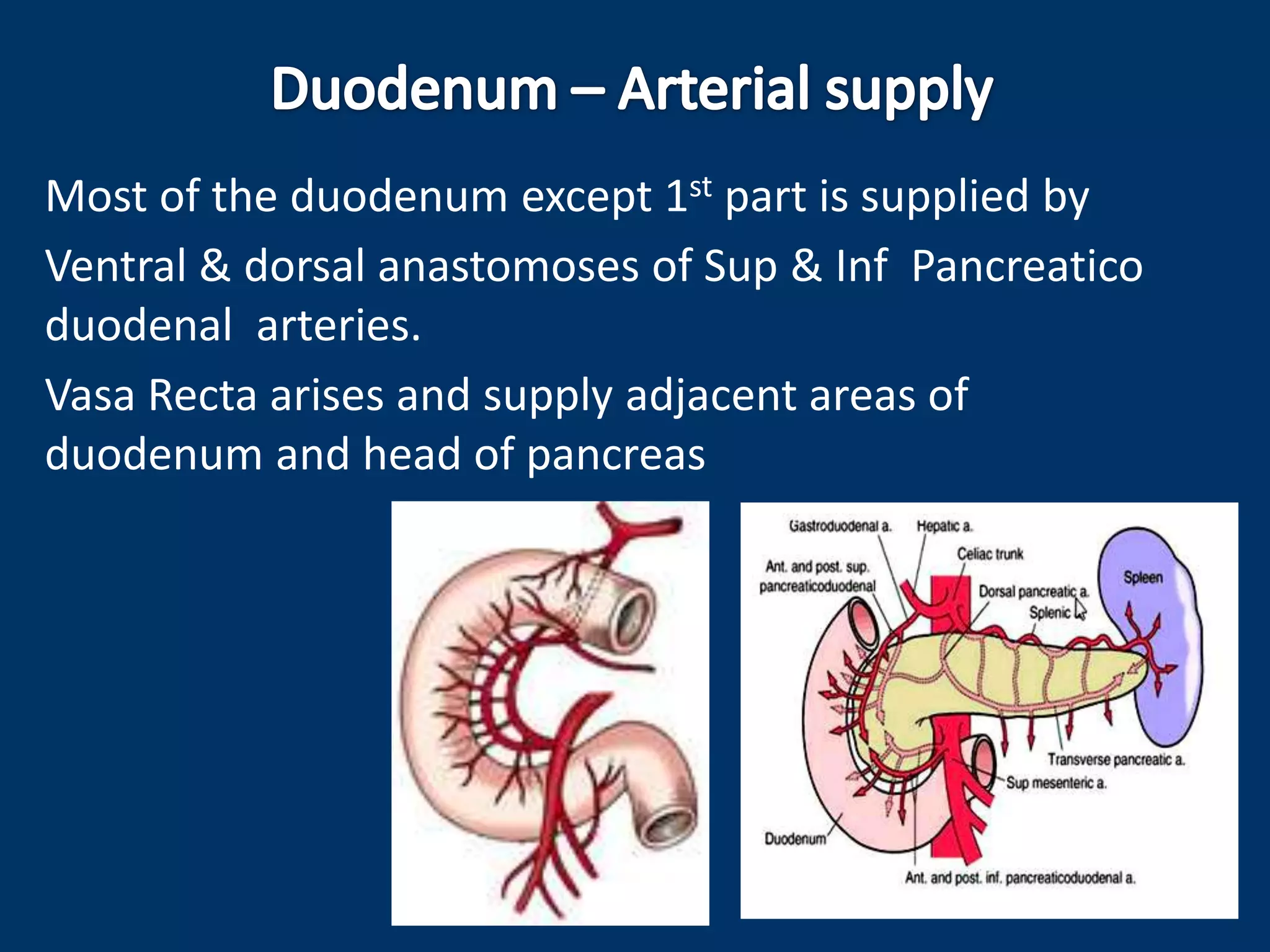
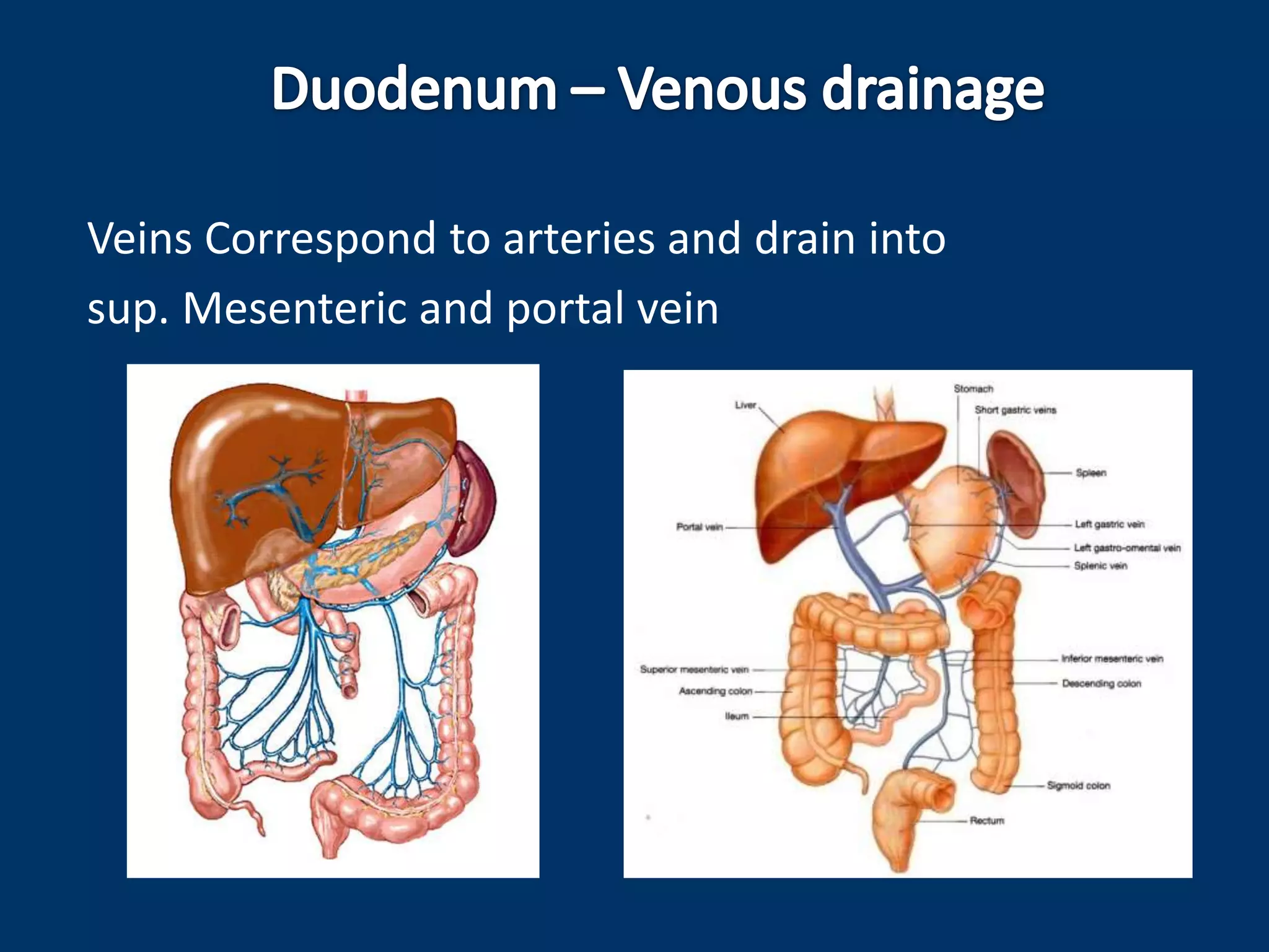
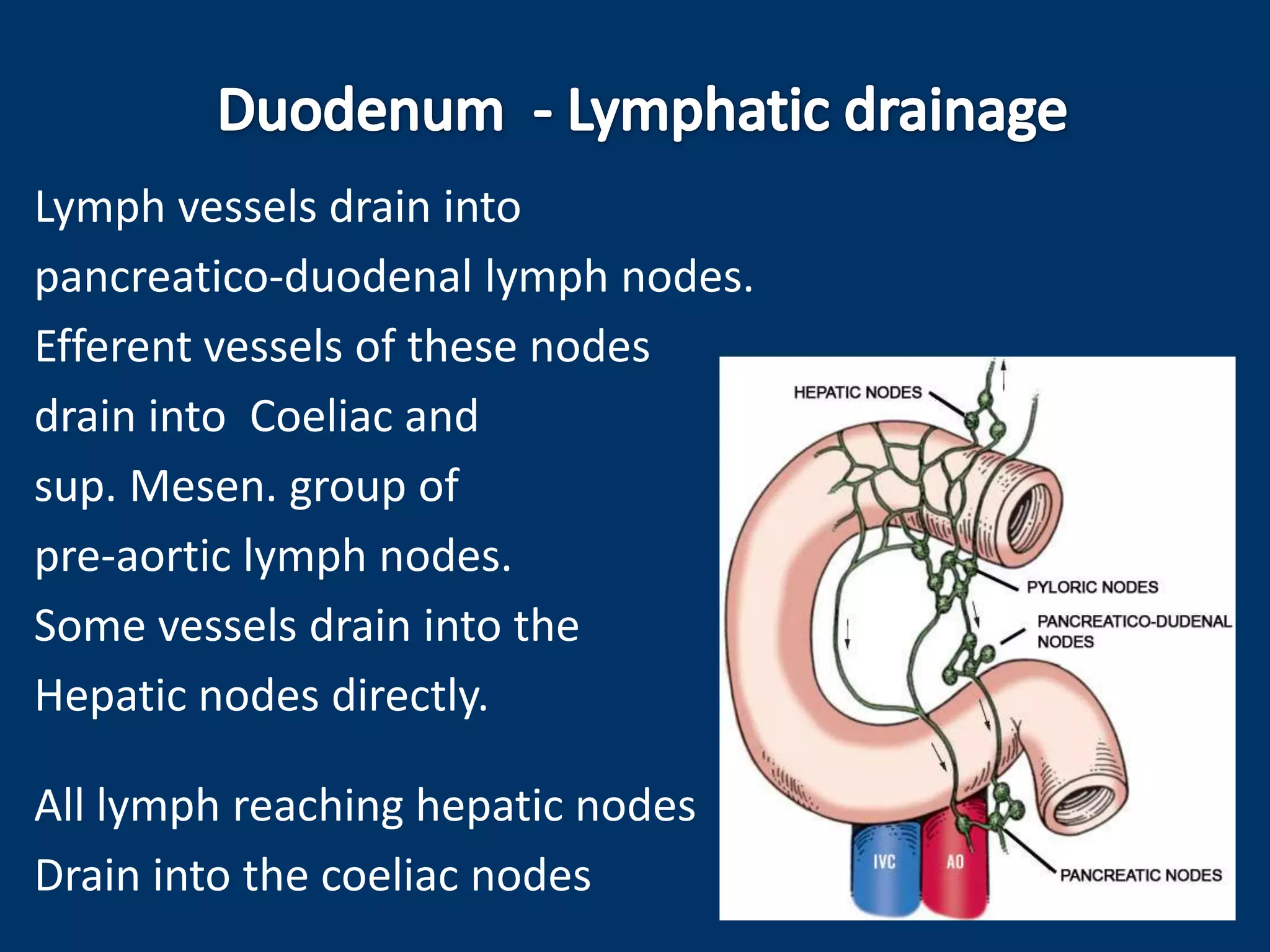
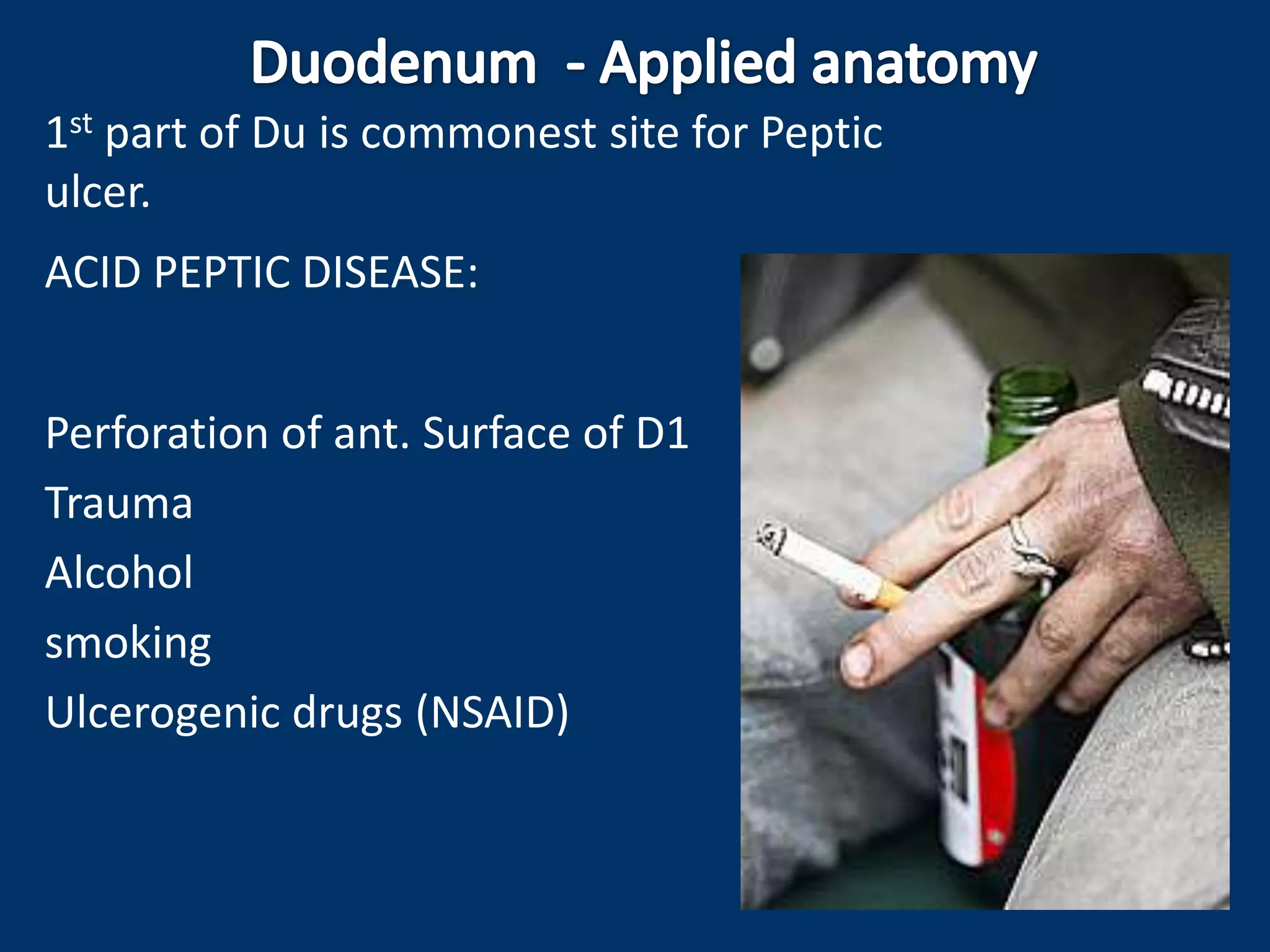
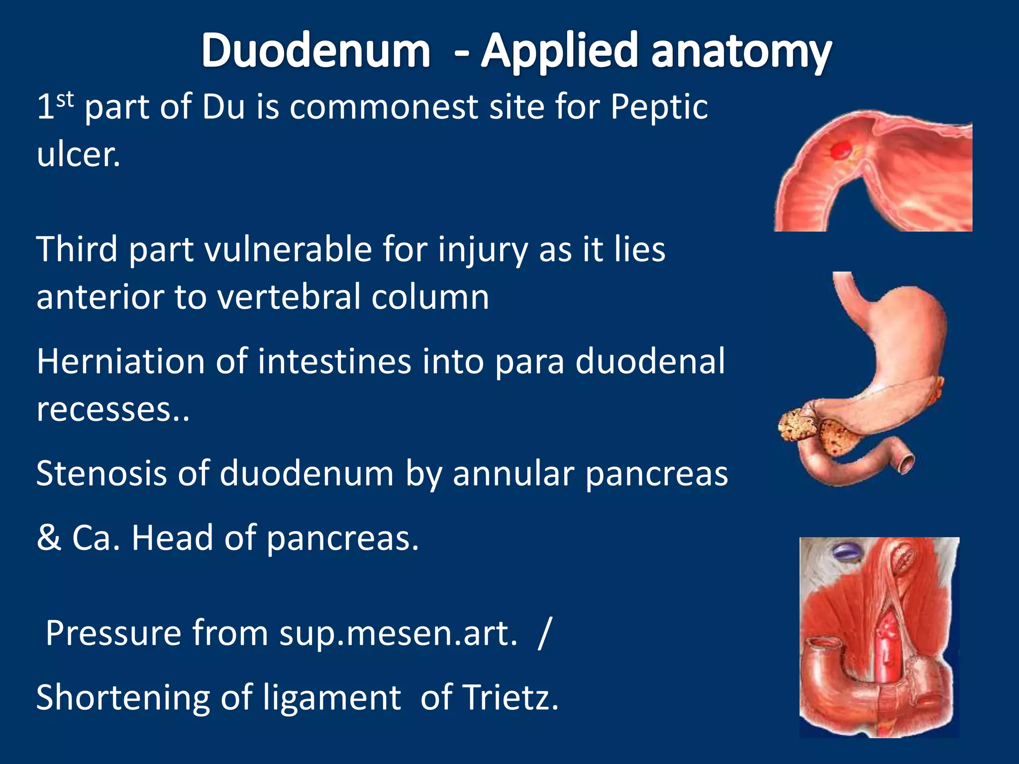
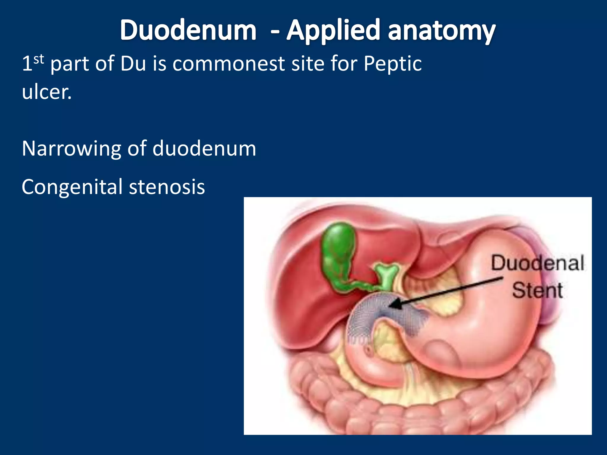
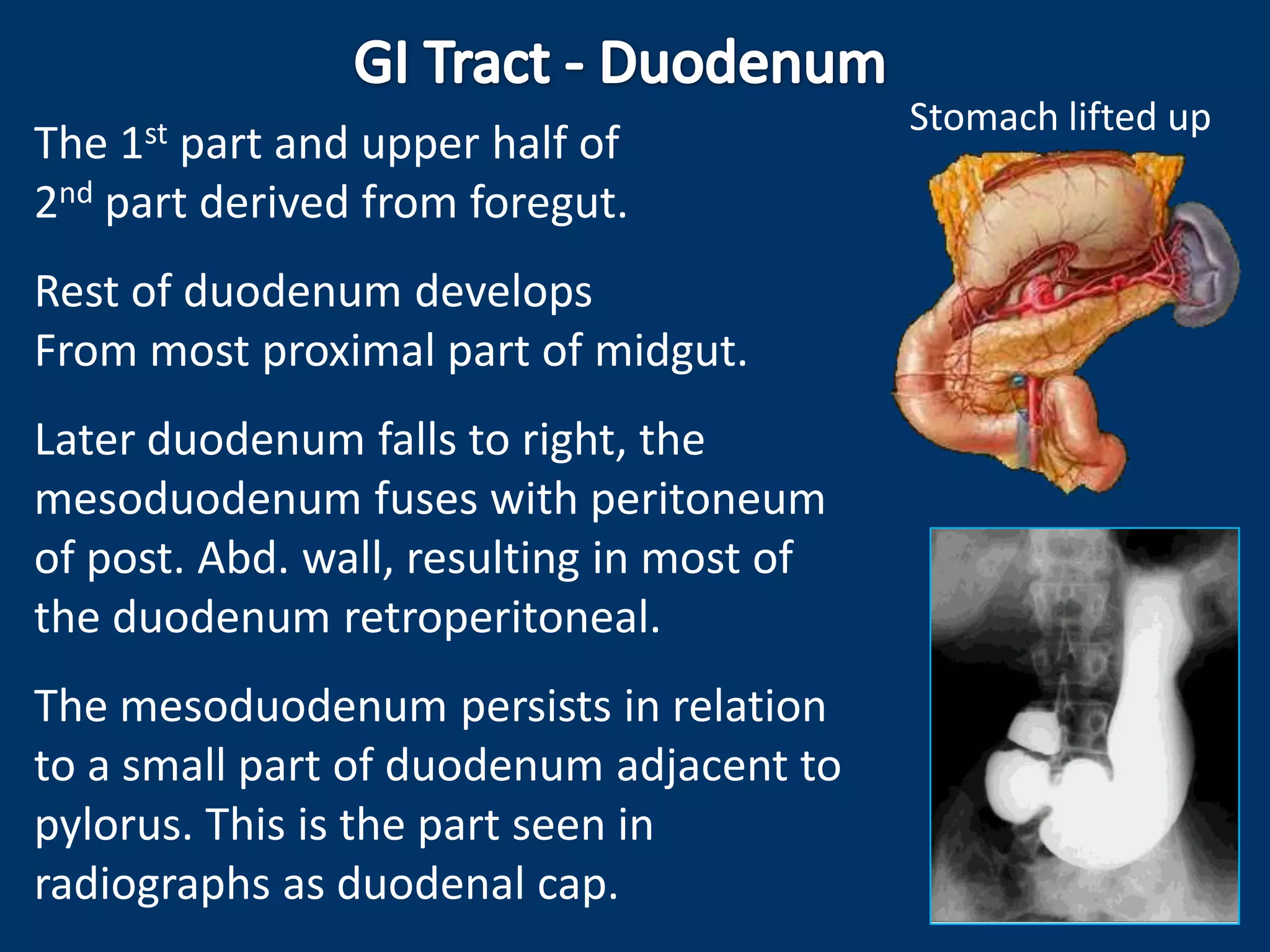
5. Shortest, dilated and most fixed
proximal part of Small intestine.
Devoid of mesentery
Fixed to post. Abd. Wall.
C – shaped curvature
Curved around head of pancreas
Lies above umbilicus opp. to 1, 2 & 3 rd
Lumbar Vertebrae
Receives Bile duct and pancreatic duct.
Interposed bet. Stomach and intestines
(Both are active digestive areas in GIT)
7. 25 cm long & Subdivided into 4 parts
First / upper part – 5 cm
Second / vertical part – 7.5 cm
Third / horizontal part – 10 cm
Fourth / ascending part – 2.5 cm
1st
2nd
8.
9. Extends from pylorus to
Sup. Duodenal flexure,
directed upwards, backwards &
to Right
Length – 5 cm
(Surface projection 2.5 cm)
prox. 2.5 cm movable.
Attached to lesser omentum above and
to greater omentum below.
Distal 2.5 cm is fixed. Retroperitoneal.
Covered with peritoneum only on anterior aspect.
10. Visceral relations:
Anteriorly : Quadrate lobe of liver,
and gall bladder.
Posteriorly : Gastroduodenal art.,
bile duct, and portal vein.
Superiorly : Epiploic foramen.
Inferiorly : Head & neck of pancreas.
11. Relations of 1st part Duodenum
sagittal section viewed from left side
Posterior relations
12. a) Most movable part, behaves more of stomach than duodenum
b) Supplied by end arteries
c) May be affected by peptic ulcer
d) Devoid of circular mucous fold
e) Seen as duodenal cap / Bulb
in Radiographs.
Du Ulcer - Trefoil deformity)
Barium meal
Barium sulphate
Double contrast
Now replaced by
Endoscopy
CT / MRI
13. First part is supplied by end arteries
a) Supra duodenal (Art.of Wilkie) S.D.A.
Br. of Gastroduodenal
supplies upper margin,
upper 2/3rd of anterior surface
Upper 1/3rd of posterior surface
of prox. ½ of first half.
b) Retro duodenal branch
Br. of gastroduodenal
Supplies part of post. Surface
c) Infra duodenal branch
Br. of Rt. Gastro-epiploic artery
Supplies lower margin
C.T.
S.M.A.
I.P.D.
S.D.A
Rt.G.E.A.
C.H.A
H.A.P.
G.D.A.
S.P.D.
Rt.G.E.A.
14. Begins at superior duodenal flexure opp. L1 vertebra
passes vertically downwards
- in front of hilum of Rt. Kidney
- along Rt. Side of vertebral column
in para vertebral gutter
- Within Rt. Lateral plane
- ends in inferior duodenal flexure
opp. Lower border of L3
- continues with 3rd part
Rt. Lat. Plane
15. It is retroperitoneal and fixed.
Ant. Surface is covered with
peritoneum , except near the
middle where it is directly
related to colon.
Visceral relations:
Anteriorly: Right lobe of liver,
Transverse colon,
Root of transverse mesocolon,
Small intestine.
16. Posteriorly :
Entirely non-peritoneal
1. Ant. Surface of Rt. Kidney close to
hilum
2. Rt. Renal vessels, pelvis of Rt.
Ureter
3. Rt. Psoas major muscle
4. Rt. Edge of inferior vena cava
5. Sometimes part of Rt. Supra renal
19. Circular folds
Plica Circularis – Permanent, circular & thick.
Major duodenal papilla
(on post. Medial wall of 2nd part
10 cm distal to pylorus)
Bile duct & pancreatic ducts open on summit
(Ampulla of vater)
Minor duodenal papilla
2 cms above major papilla
Accessry pancreatic duct opens
Plica Semicircularis
arches above major papilla
Plica longitudinalis
vertical fold downwards from major papilla
Sometimes vertical fold upwards from major papilla marks Bile duct
20. Bile duct
Pancreatic duct
Sphincter pancreaticus
Sphincter
Ampullae
BD + PD
(Ampulla of Vater)
Hepato pancreatic ampulla
Sphincter
Choledochus
(Choledochus = Bile duct)
This sphincter is always
present. Normally keeps
lower end of bile duct
closed. As a result, bile
formed in the liver keeps
accumulating in gall bladder
and undergoes considerable
concentration. With a fatty
meal the sphincter opens
and bile stored in the gall
bladder is poured into the
duodenum.
“Gall stones – Cholecystitis”
(Major duodenal papilla)
21. Extends from inf. Duodenal flexure
to front of aorta at L3 level
Relations - Anteriorly :
Covered by peritoneum except
attachment of root of mesentery
Ant. Surface crossed by
Sup. Mesenteric vessels
and root of mesentery
22. Posteriorly:
Non peritoneal
1. Rt.psoas major muscle
2. Right ureter
3. Inf. Vena cava
4. RT. Gonadal vessels
5. Abdominal aorta
6. Origin of inferior
Mesenteric art.
23. Extends from inf. Duodenal flexure to
front of aorta at L3 level
Relations – Superiorly :
Head of pancreas with uncinate process.
Inferior pancreatico duodenal vessels
Inferiorly :
Few coils of jejunum
24. Extends from front of aorta to Duodeno-jejunal flexure
DJ flexure is situated on the left side of L2 about 1.25 cm
below transpyloric plan & 2.5 cms to left of median plane
Kept in position by suspensory
muscle of Duodenum
Relations Anteriorly :
Covered with peritoneum.
Related to transverse colon &
mesocolon
Post. Inf. Surface of stomach
separated by lesser sac
25. Posteriorly :
Left crus of diaphragm
Left psoas major muscle
Left sympathetic trunk
Left renal vessels
Left Gonadal vessels
Left supra renal vein
Inferior mesenteric vein
26. Right side :
Uncinate process of pancreas
Left side :
Left kidney and ureter
Superiorly :
Body of pancreas
27. Fibro muscular band, which suspends and
supports the duodenojejunal flexure.
Arises from the right crus of the diaphragm;
close to the right side of the esophagus,
passes downwards behind pancreas,
and is attached to the posterior surface of the
duodenojejunal flexure and the 3rd & 4th
parts of duodenum.
Made up of stripped muscle fibers in its upper part,
Elastic fibers in its middle part,
and plain muscle fibers in its lower part.
28. Mucous coat : simple columnar / occasional goblet
cells, covered with villi
Lamina propria : contains crypts of
Leiberkuhn receive at their bottom
openings of Brunner’s glands
Submucous coat :
Loose areolar tissue
Blood vessels, lymphatics
Meissner’s plexus
Brunner’s glands
Muscular coat:
Outer longitudinal & inner circular
(Separated by myenteric plexus)
Serous coat from peritoneum - incomplete
29. Sub mucous coat - Duodenal glands of Brunner
Open at bottom of crypts of Leiberkuhn
Secretion
Rich in bicarobonate ions
(alkaline)
Helps activation of
trypsinogen from pancreas
30. Most of the duodenum except 1st part is supplied by
Ventral & dorsal anastomoses of Sup & Inf Pancreatico
duodenal arteries.
Vasa Recta arises and supply adjacent areas of
duodenum and head of pancreas
32. Lymph vessels drain into
pancreatico-duodenal lymph nodes.
Efferent vessels of these nodes
drain into Coeliac and
sup. Mesen. group of
pre-aortic lymph nodes.
Some vessels drain into the
Hepatic nodes directly.
All lymph reaching hepatic nodes
Drain into the coeliac nodes
33. Sympathetic nerves are derived from coeliac and sup.
Mesen. Plexuses.
Preganglionic fibres come from T6 to T9 segments of
spinal cord.
Parasympathetic are derived from both vagus nerves .
The myenteric (Auerbach’s plexus) & Meisner’s plexuses
Act as post ganglionic neurons for parasypathetic fibres
only.
34. 1st part of Du is commonest site for Peptic
ulcer.
ACID PEPTIC DISEASE:
Perforation of ant. Surface of D1
Trauma
Alcohol
smoking
Ulcerogenic drugs (NSAID)
35. 1st part of Du is commonest site for Peptic
ulcer.
Third part vulnerable for injury as it lies
anterior to vertebral column
Herniation of intestines into para duodenal
recesses..
Stenosis of duodenum by annular pancreas
& Ca. Head of pancreas.
Pressure from sup.mesen.art. /
Shortening of ligament of Trietz.
36. Acid peptic disease and Duodenal trefoil appearance:
Besides a genetic predisposition to acid peptic disease
abnormal high secretion of acid and pepsin, reflux of bile
and pancreatic juice to stomach, reduced amount of
mucus or structurally deficient mucus, reduced
submucosal blood flow, (stress and alcohol ingestion),
reduced cell renewal or bicarbonate secretion are all
contributory singly or in combination in the causation of
acid peptic disease, Diet has a limited role in both
causation and cure of the ailment. Emotional stress,
delayed gastric emptying, cigarette smoking, use of
ulcerogenic drugs
37. 1st part of Du is commonest site for Peptic
ulcer.
Narrowing of duodenum
Congenital stenosis
39. The 1st part and upper half of
2nd part derived from foregut.
Rest of duodenum develops
From most proximal part of midgut.
Later duodenum falls to right, the
mesoduodenum fuses with peritoneum
of post. Abd. wall, resulting in most of
the duodenum retroperitoneal.
The mesoduodenum persists in relation
to a small part of duodenum adjacent to
pylorus. This is the part seen in
radiographs as duodenal cap.
Stomach lifted up