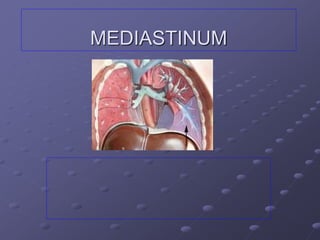
3. MEDIASTINUM
• Mediastinum is the middle
space in thooracic cavity
between lungs.
• It is a median septum .
• Covered by mediastinal pleura
Contents: all thoracic viscera
EXCEPT lungs
Extent:
Superior - thoracic inlet
Inferior - diaphragm
Anterior - sternum & costal
cartilages
Posterior- bodies of thoracic
vertebrae.
On each sides – mediastinal
pleura.
4. MEDIASTINUM
• Mediastinum is divided
into 2 divisions.
• By imaginary plane
passing from
manubriosternal joint to
disc between
t4&t5(sternal angle).
• Above –superior
mediastinum.
• Below – inferior
mediastinum.
5. Divisions of the Mediastinum
SUPERIOR MEDIASTINUM
Superior - thoracic inlet
Inferior - transverse thoracic
plane
Anterior - manubrium sterni
Posterior - IV disc T4 & T5
6. Contents of Superior Mediastinum
Contents: (Anterior – Posterior)
1. Thymus gland
- primary lymphoid organ located behind manubrium
- puberty undergoes gradual involution
2. Great Vessels
Brachiocephalic Veins
Superior Vena Cava
- formed at level of 1st right costal cartilage
- enters right atrium at level of 3rd right costal
cartilage
7.
8.
9. Superior Mediastinum
Contents:
Arch of the Aorta
- starts behind 2nd right SC joint
- ends at 2nd left SC joint
Branches:
Brachiocephalic Trunk
Left Common Carotid Artery
Left Subclavian Artery
10. Aortic Arch
Passes upwards
from the sternal
angle behind the
manubrium,
backwards and to
the left of the 4th
Thoracic vertebra
BRANCHES
SUPPLY UL,
HEAD, and NECK
15. Ligamentum arteriosum
Aortic Arch is
connected inferiorly to
the left pulmonary
artery by the
Ligamentum
arteriosum” fibrous
remnant of ductus
arteriosus”
16. Superior Mediastinum
Contents:
3. Nerves
Vagus & Phrenic Nerves
Cardiac Plexus of Nerves
Left Recurrent Laryngeal Nerve
4. Trachea
5. Esophagus
6. Thoracic Duct
7. Prevertebral Muscles
17.
18.
19.
20.
21.
22. •INFERIOR MEDIASTINUM
• Superior – transverse
thoracic plane.
Inferior - diaphragm.
• It is divided further
into anterior
,middle,inferior
mediastinum.
•
23.
24. Divisions of the Mediastinum
INFERIOR MEDIASTINUM
a. ANTERIOR MEDIASTINUM
- contains thymus remnant, lymph nodes & fats
b. MIDDLE MEDIASTINUM
- contains the heart & great vessels
c. POSTERIOR MEDIASTINUM
- contains esophagus, great vessels,vagus nerves
& symphathetic trunks
25. Anterior Mediastinum
Smallest subdivision of the Inferior Mediastinum
Boundaries:
Anterior : body of sternum & trans thoracis muscles
Posterior : pericardium .
superior : transverse thoracic plane.
inferior :diaphragm
Contents: Loose CT (Sternopericardial Ligament)
Adipose tissue
Lymphatic Vessels & lymph nodes
Branches of Internal Thoracic Vessels
26. Middle mediastinum
It is occupied by pericarium and its contents with vessels
and nerves,
Boundaries:
Anterior:sternopericadial ligaments.
Posterior : oesophagus ,descending aorta,azygos vein.
Contents:
Heart &pericardium.
Arteries –a.aorta,pulmonary trunk,pulmonary arteries.
Veins – lower half of svc ,azygos vein ,pulmonary veins.
Nerves – phernic nerve ,deep cardiac plexus.
Bifurcation of trachea.
29. Esophagus
Course: Superior to Posterior Mediastinum
Located behind: Arch of Aorta
Pericardium & Left Atrium
Enters Esophageal Hiatus of the Diaphragm at level of T10
Anatomic Impressions or “Constrictions”:
1. Crossing with Aortic Arch
2. Crossing with Left Main Bronchus
3. Diaphragmatic Hiatus
30. Thoracic Duct
Largest lymphatic channel in the body
Originates from Cisterna Chyle in the abdomen & passes
thru aortic hiatus of diaphragm at level of T12
Relations: Posterior : bodies of inferior 7 thoracic vertebrae
Anterior : Esophagus
Left : Thoracic Aorta
Right : Azygos Vein
Conveys lymph from:
Lower extremities Left side of thorax
Pelvic Cavity Left side of H & N
Abdominal Cavity Left upper limb
31. Lymph Nodes of the
Posterior Mediastinum
Posterior Mediastinal Lymph Nodes
- receives lymph from esophagus, posterior aspect of the
pericardium & diaphragm & middle posterior ICS
32.
33. Azygos Venous System of the Posterior
Mediastinum
Drains the back & thoracoabdominal walls and the
mediastinal viscera
Azygos Vein
- forms a collateral pathway b/w SVC & IVC
- passes to the right side of inferior 8 thoracic vertebrae
- arches over the root of the right lung to enter the SVC
- receives posterior intercostal veins, mediastinal,
esophageal & bronchial veins
34. Azygos Venous System
of Posterior Mediastinum
Hemiazygos Vein
- arises on left side of the vertebral column to level of T9
- receives the inferior 3 PIV, inferior esophageal veins,
small mediastinal veins
Accessory Hemiazygos Vein
- starts at medial end of 4th or 5th ICS
- descends on left of VC from T5 thru T8
- crosses to the right to join the Azygos Vein
- receives 4th to 8th IC Veins
- communicates with Superior IC Vein which drains
1st to 3rd ICS
35.
36. Nerves of Posterior Mediastinum
Thoracic Sympathetic Trunks
- lie against heads of ribs in superior thorax
costovertebral joints in midthorax
sides of vertebral bodies in lower thorax
Lower Thoracic Splanchnic Nerves
(Greater, Lesser and Least SN)
- presynaptic fibers from 5th thru 12th sympathetic ganglia
- sympathetic innervation for most abdominal viscera
37. Clinicals.
Mediastinal syndrome.
Symptoms
Obstruction of svc.
Pressure over trachea –dysponea ,cough.
Pressure over oesophagus – dysphagia.
Dysphonia.
Internal neuralgia.
Bronchogenic carcinoma.
Hodgkin disease –enlargment of lymph
nodes,aneurysm ,dilatation of aorta.