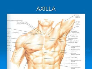
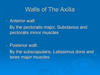
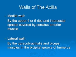
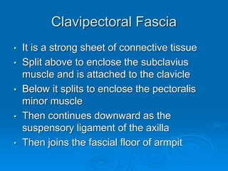
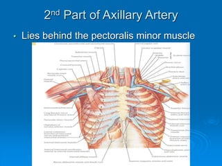
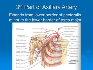
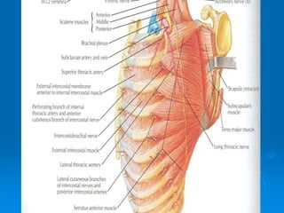
AXILLA
Boundaries
Contents
Axillary Vessels
By: Dr. Mujahid Khan
AXILLA
Definition
• It is a pyramid shaped space between the
upper part of the arm and the side of the
chest
• Important Nerves, Blood and Lymph
vessels travel through it from root of the
neck to the upper limb
Apex
• Upper end of axilla or APEX is directed
into the root of neck
• Bounded in front by the clavicle
• Behind by upper border of scapula
• Medially by outer border of the 1st rib
Base
• Lower end or Base is bounded in front by
the anterior axillary fold formed by
pectoralis major muscle
• Behind by posterior axillary fold formed by
the tendons of latissimus dorsi and teres
major muscles
• Medially by the chest wall
Walls of The Axilla
• Anterior wall:
By the pectoralis major, Subclavius and
pectoralis minor muscles
• Posterior wall:
By the subscapularis, Latissimus dorsi and
teres major muscles
Walls of The Axilla
• Medial wall:
By the upper 4 or 5 ribs and intercostal
spaces covered by serratus anterior
muscle
• Lateral wall:
By the coracobrachialis and biceps
muscles in the bicipital groove of humerus
Base
The Base of axilla is formed by the skin
stretching between the anterior and
posterior walls
Clavipectoral Fascia
• It is a strong sheet of connective tissue
• Split above to enclose the subclavius
muscle and is attached to the clavicle
• Below it splits to enclose the pectoralis
minor muscle
• Then continues downward as the
suspensory ligament of the axilla
• Then joins the fascial floor of armpit
Contents of Axilla
• Axillary artery and its branches
• Axillary vein and its tributaries
• Lymph vessels and lymph nodes
• Important nerve plexus the “Brachial
Plexus” which innervates the upper limb
Axillary Artery
• Is a continuation of subclavian artery
• Begins at the lateral border of the 1st rib
• Ends at the lower border of teres major
• It continues as the brachial artery
• Closely related to brachial plexus cords
• Enclosed with them in the axillary sheath
• Axillary sheath is continuous with the
prevertebral fascia
• Pectoralis minor divides it into 3 parts
1st Part of Axillary Artery
• Extends from the lateral border of the 1st
rib to the upper border of pectoralis minor
Relation
• Anterior: Pectoralis major, covering fascia,
skin, cephalic vein
• Posterior: Long thoracic nerve
• Lateral: Three cords of brachial plexus
• Medial: Axillary vein
2nd Part of Axillary Artery
• Lies behind the pectoralis minor muscle
Relation
• Anterior: Pectoralis minor and major,
covering fascia and skin
• Posterior: Posterior cord of brachial plexus
• Lateral: Lateral cord of brachial plexus
• Medial: medial cord of brachial plexus and
axillary vein
3rd Part of Axillary Artery
• Extends from lower border of pectoralis
minor to the lower border of teres major
Relation
• Anterior: Pectoralis major, medial root of
the median nerve
• Posterior: subscapularis, latissimus dorsi
and teres major
• Lateral: Coracobrachialis, biceps, humerus
• Medial: Ulnar nerve, axillary vein, medial
cutaneous nerve of the arm
Branches
• Branches of axillary artery supply the
thoracic wall and the shoulder region
• 1st Part: Highest thoracic artery
• 2nd Part: Thoracoacromial and lateral
thoracic arteries
• 3rd Part: Subscapular artery, anterior and
posterior circumflex humeral arteries
Pectoralis Major
• Origin: Medial half of clavicle, sternum,
upper 6 costal cartilages
• Insertion: Lateral lip of bicipital groove of
the humerus
• NS: Medial and Lateral pectoral Nerve
from medial and lateral pectoral cords of
brachial plexus
• Action: Adducts the arm and rotates it
medially, some fibers also cause flexion of
arm
Subclavius
• Origin: From the first costal cartilage
• Insertion: Fibers move upward and
laterally into the inferior surface of clavicle
• NS: Nerve to the subclavius from upper
trunk of brachial plexus
• Action: Depresses the clavicle and
steadies the bone
Pectoralis Minor
• Origin: From 3rd, 4th and 5th ribs
• Insertion: Coracoid Process
• NS: Medial pectoral nerve, a branch of the
medial cord of brachial plexus
• Action: Pulls the shoulder downward and
forward. Elevates the ribs of origin
Subscapularis
• Origin: Subscapular fossa on the anterior
surface of scapula
• Insertion: On the lesser tuberosity of the
humerus
• NS: Upper and Lower subscapular nerves
• Action: Medially rotates the arm and
stabilizes the shoulder joint
Latissimus Dorsi
• Origin: Posterior part of the iliac crest, lumbar
fascia and spines of lower 6 thoracic vertebrae,
lower 3 ribs
• Insertion: Floor of the bicipital groove of
humerus with teres major
• NS: Thoracodorsal nerve from posterior cord of
brachial plexus
• Action: Extends, adducts and medially rotates
the arm
Teres Major
• Origin: lower third of the lateral border of
scapula
• Insertion: Medial lip of bicipital groove of
humerus
• NS: Lower subscapular nerve from
posterior cord of brachial plexus
• Action: Adducts and medially rotates the
arm
Serratus Anterior
• Origin: From Outer surface of upper 8 ribs
• Insertion: Medial border of scapula in the
region of inferior angle
• NS: Long thoracic nerve
• Action: Draws the scapula forward, rotates
it
4-AXILLA.ppt

4-AXILLA.ppt

  • 1.
  • 2.
  • 3.
    Definition • It isa pyramid shaped space between the upper part of the arm and the side of the chest • Important Nerves, Blood and Lymph vessels travel through it from root of the neck to the upper limb
  • 4.
    Apex • Upper endof axilla or APEX is directed into the root of neck • Bounded in front by the clavicle • Behind by upper border of scapula • Medially by outer border of the 1st rib
  • 5.
    Base • Lower endor Base is bounded in front by the anterior axillary fold formed by pectoralis major muscle • Behind by posterior axillary fold formed by the tendons of latissimus dorsi and teres major muscles • Medially by the chest wall
  • 6.
    Walls of TheAxilla • Anterior wall: By the pectoralis major, Subclavius and pectoralis minor muscles • Posterior wall: By the subscapularis, Latissimus dorsi and teres major muscles
  • 7.
    Walls of TheAxilla • Medial wall: By the upper 4 or 5 ribs and intercostal spaces covered by serratus anterior muscle • Lateral wall: By the coracobrachialis and biceps muscles in the bicipital groove of humerus
  • 8.
    Base The Base ofaxilla is formed by the skin stretching between the anterior and posterior walls
  • 9.
    Clavipectoral Fascia • Itis a strong sheet of connective tissue • Split above to enclose the subclavius muscle and is attached to the clavicle • Below it splits to enclose the pectoralis minor muscle • Then continues downward as the suspensory ligament of the axilla • Then joins the fascial floor of armpit
  • 10.
    Contents of Axilla •Axillary artery and its branches • Axillary vein and its tributaries • Lymph vessels and lymph nodes • Important nerve plexus the “Brachial Plexus” which innervates the upper limb
  • 11.
    Axillary Artery • Isa continuation of subclavian artery • Begins at the lateral border of the 1st rib • Ends at the lower border of teres major • It continues as the brachial artery • Closely related to brachial plexus cords • Enclosed with them in the axillary sheath • Axillary sheath is continuous with the prevertebral fascia • Pectoralis minor divides it into 3 parts
  • 12.
    1st Part ofAxillary Artery • Extends from the lateral border of the 1st rib to the upper border of pectoralis minor
  • 14.
    Relation • Anterior: Pectoralismajor, covering fascia, skin, cephalic vein • Posterior: Long thoracic nerve • Lateral: Three cords of brachial plexus • Medial: Axillary vein
  • 15.
    2nd Part ofAxillary Artery • Lies behind the pectoralis minor muscle
  • 16.
    Relation • Anterior: Pectoralisminor and major, covering fascia and skin • Posterior: Posterior cord of brachial plexus • Lateral: Lateral cord of brachial plexus • Medial: medial cord of brachial plexus and axillary vein
  • 17.
    3rd Part ofAxillary Artery • Extends from lower border of pectoralis minor to the lower border of teres major
  • 18.
    Relation • Anterior: Pectoralismajor, medial root of the median nerve • Posterior: subscapularis, latissimus dorsi and teres major • Lateral: Coracobrachialis, biceps, humerus • Medial: Ulnar nerve, axillary vein, medial cutaneous nerve of the arm
  • 19.
    Branches • Branches ofaxillary artery supply the thoracic wall and the shoulder region • 1st Part: Highest thoracic artery • 2nd Part: Thoracoacromial and lateral thoracic arteries • 3rd Part: Subscapular artery, anterior and posterior circumflex humeral arteries
  • 20.
    Pectoralis Major • Origin:Medial half of clavicle, sternum, upper 6 costal cartilages • Insertion: Lateral lip of bicipital groove of the humerus • NS: Medial and Lateral pectoral Nerve from medial and lateral pectoral cords of brachial plexus • Action: Adducts the arm and rotates it medially, some fibers also cause flexion of arm
  • 22.
    Subclavius • Origin: Fromthe first costal cartilage • Insertion: Fibers move upward and laterally into the inferior surface of clavicle • NS: Nerve to the subclavius from upper trunk of brachial plexus • Action: Depresses the clavicle and steadies the bone
  • 23.
    Pectoralis Minor • Origin:From 3rd, 4th and 5th ribs • Insertion: Coracoid Process • NS: Medial pectoral nerve, a branch of the medial cord of brachial plexus • Action: Pulls the shoulder downward and forward. Elevates the ribs of origin
  • 25.
    Subscapularis • Origin: Subscapularfossa on the anterior surface of scapula • Insertion: On the lesser tuberosity of the humerus • NS: Upper and Lower subscapular nerves • Action: Medially rotates the arm and stabilizes the shoulder joint
  • 27.
    Latissimus Dorsi • Origin:Posterior part of the iliac crest, lumbar fascia and spines of lower 6 thoracic vertebrae, lower 3 ribs • Insertion: Floor of the bicipital groove of humerus with teres major • NS: Thoracodorsal nerve from posterior cord of brachial plexus • Action: Extends, adducts and medially rotates the arm
  • 29.
    Teres Major • Origin:lower third of the lateral border of scapula • Insertion: Medial lip of bicipital groove of humerus • NS: Lower subscapular nerve from posterior cord of brachial plexus • Action: Adducts and medially rotates the arm
  • 30.
    Serratus Anterior • Origin:From Outer surface of upper 8 ribs • Insertion: Medial border of scapula in the region of inferior angle • NS: Long thoracic nerve • Action: Draws the scapula forward, rotates it