Downloaded 416 times


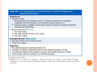
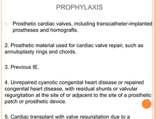
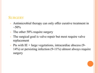
![MODIFIED DUKE CRITERIA
Major Diagnostic Criteria
Blood culture positive for IE
Typical microorganisms consistent with IE from 2 separate blood cultures:
Viridans streptococci, Streptococcus bovis, HACEK group, Staphylococcus
aureus; or community-acquired enterococci in the absence of a primary
focus, or microorganisms consistent with IE from persistently positive blood
cultures defined as follows: at least 2 positive cultures of blood samples
drawn >12 h apart or all 3 or a majority of ≥4 separate cultures of blood (with
first and last sample drawn at least 1 h apart)
Single positive blood culture for Coxiella burnetii or anti–phase 1 IgG
antibody titer ≥1:800
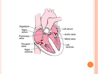
Evidence of endocardial involvement by echo.
Echocardiogram positive for IE (TEE recommended for patients with
prosthetic valves, rated at least possible IE by clinical criteria, or
complicated IE [paravalvular abscess]; TTE as first test in other
patients) defined as follows: oscillating intracardiac mass on valve or
supporting structures, in the path of regurgitant jets, or on implanted
material in the absence of an alternative anatomic explanation; abscess;
or new partial dehiscence of prosthetic valve or new valvular regurgitation
(worsening or changing or pre-existing murmur not sufficient)](https://image.slidesharecdn.com/infectiveendocarditis1-210619102705/85/Infective-endocarditis-31-320.jpg)
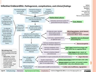

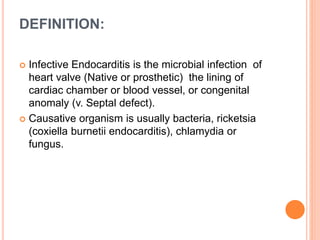
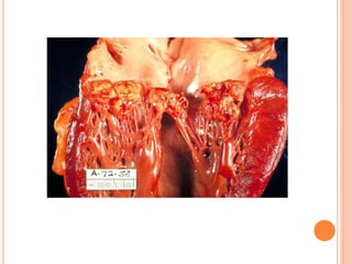
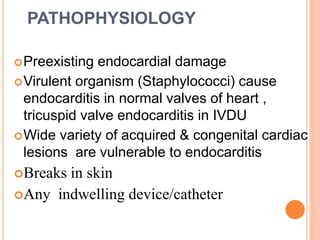
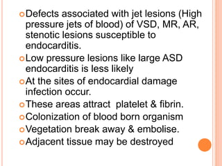
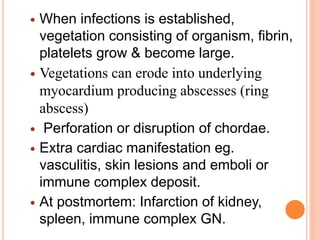
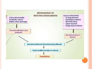
This document provides information about infective endocarditis: - Infective endocarditis is a microbial infection of the heart valves, heart lining, or blood vessels that is usually caused by bacteria. - It can affect both native and prosthetic heart valves. Staphylococcus aureus is now the most common cause. - Diagnosis is based on modified Duke criteria using clinical findings, blood cultures, and echocardiography findings. Treatment involves prolonged antibiotic therapy and may require surgery to remove infected tissues. - Complications can include heart valve damage, embolic events, heart failure, and extension of the infection. Proper antibiotic prophylaxis is important for those at high risk


