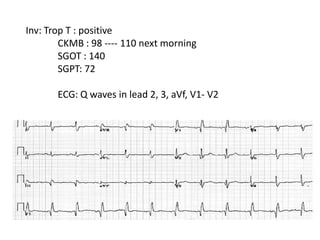
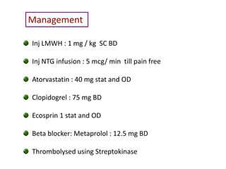
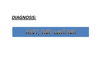
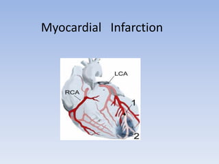
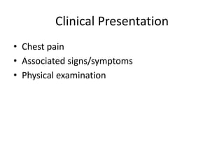
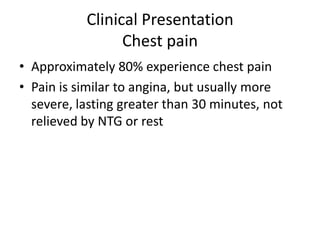
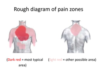
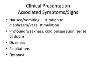
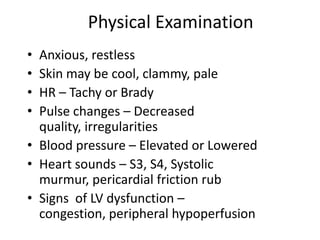
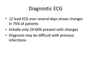
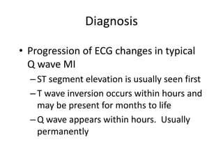
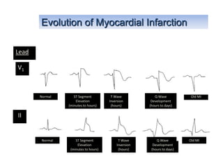
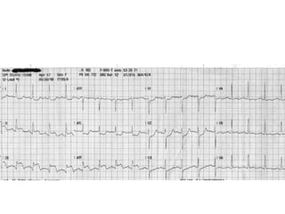
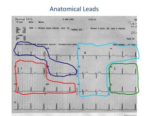
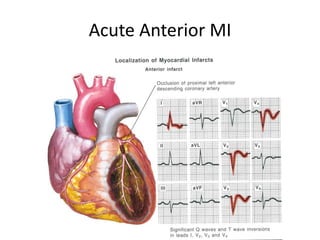
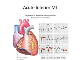
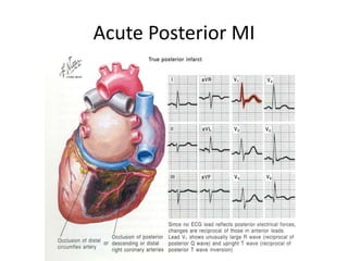
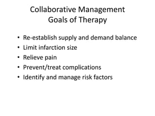
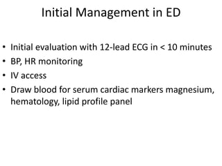
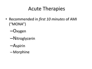
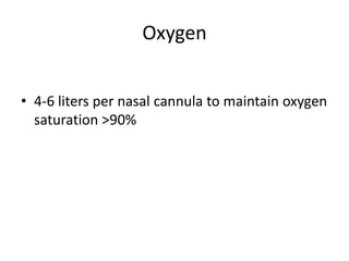
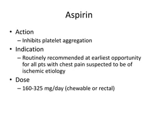
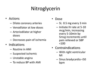
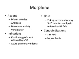
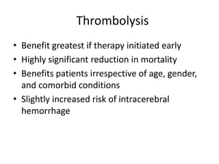
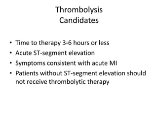
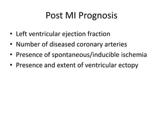
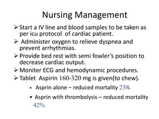
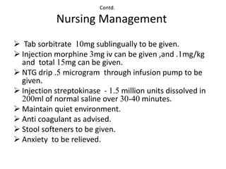
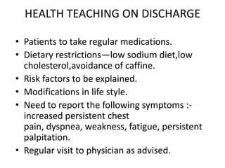
•CASE PRESENTATION

•ACUTE MYOCARDIAL INFARCTION

     •Definition
     •Etiology
     •Pathophysiology
     •Clinical features
     •Diagnosis
     •Management
     •Complication
     •Nursing care
Case :
  55 yr/M
  alcoholic and smoker


Presented with: pain in Left side of chest since 5 am
                radiating to lt. shoulder and back
O/E     GCS fair
        P-76
        BP 130/84

S/E :    WNL

Inv:    Hb- 12.9                        TLC-6700
        BLOOD SUGAR Random 146
        Urine normal
        Urea-25
        Creatinine -0.9
         CKMB 28
        Trop T - negative
        ECG:
        Chest x ray:      WNL

Pt was prescribed tab. Pantocid and syp Digene
And was send for TMT from ECHS empanelled hospital
At around 5 pm the same evening he came back with

       severe chest pain following TMT
       2 episodes of vomiting
       excessive sweating
       and TMT positive for provocation

Pt was admitted to ICU and was investigated:
       pulse : 97
       BP: 110/72mm hg
       resp:26

Chest : B/L basal crepts +
CVS
CNS                          WNL
P/A
Inv: Trop T : positive
        CKMB : 98 ---- 110 next morning
        SGOT : 140
        SGPT: 72

       ECG: Q waves in lead 2, 3, aVf, V1- V2
Management

Inj LMWH : 1 mg / kg SC BD

Inj NTG infusion : 5 mcg/ min till pain free

Atorvastatin : 40 mg stat and OD

Clopidogrel : 75 mg BD

Ecosprin 1 stat and OD

Beta blocker: Metaprolol : 12.5 mg BD

Thrombolysed using Streptokinase
DIAGNOSIS:
Myocardial Infarction
DEFINITION:-
 Myocardial Infarction ,

 Commonly known as HEART ATTACK

 is the rapid development of myocardial necrosis by a
 critical imbalance between oxygen supply and demand
 to the myocardium
Classification

• Acute coronary syndromes include
    ST-elevation MI (STEMI)
    Non ST-elevation MI ( NSTEMI)
    Unstable Angina
Classification
• Anatomic or morphologic
  Transmural= Full thickness
  Non-transmural= Partial thickness


• ECG
  Q wave MI
   Non Q wave MI
   Does not distinguish transmural from a non-
  transmural MI as determined by pathology
Risk Factors
•   Smoking
•   Family history
•   Hypertension
•   Diabetes mellitus
•   Elevated triglycerides and cholesterol levels
•   Obesity
•   Sedentary lifestyle
•   Aging
•   Stress
•   Men more than women (but women are increasing)
Causes
•   Athrosclerosis (90%)
•   Constriction or spasm of the coronary artery
•   Coronary artery embolus
•   Coronary artery thrombus
Pathophysiology
• 90% - coronary artery thrombosis
• 10% - vasospasm
• Irreversible cell death occurs within 20-40
  minutes of cessation of blood flow
• Wavefront of cellular death proceeds from
  endocardium to epicardium
FIGURE 25-1 Determinants of myocardial oxygen supply and
demand.
Pathophysiology
• Wavefront produces zones
  – Zone of necrosis
  – Zone of injury
  – Zone of ischemia
• Amount of damage/necrosis depends on
  duration of occlusion, which artery is blocked,
  degree of collateral blood flow
Zones of Ischemia Injury and Infarction with
Transmural and Subendocardial Infarction
Clinical Presentation
• Chest pain
• Associated signs/symptoms
• Physical examination
Clinical Presentation
                 Chest pain
• Approximately 80% experience chest pain
• Pain is similar to angina, but usually more
  severe, lasting greater than 30 minutes, not
  relieved by NTG or rest
Rough diagram of pain zones


(Dark red = most typical   (light red = other possible area)
        area)
Clinical Presentation
      Associated Symptoms/Signs
• Nausea/Vomiting – irritation to
  diaphragm/vagal stimulation
• Profound weakness, cold perspiration, sense
  of doom
• Dizziness
• Palpitations
• Dyspnea
Physical Examination
• Anxious, restless
• Skin may be cool, clammy, pale
• HR – Tachy or Brady
• Pulse changes – Decreased
  quality, irregularities
• Blood pressure – Elevated or Lowered
• Heart sounds – S3, S4, Systolic
  murmur, pericardial friction rub
• Signs of LV dysfunction –
  congestion, peripheral hypoperfusion
Diagnostic ECG
• 12 lead ECG over several days shows changes
  in 75% of patients
• Initially only 24-60% present with changes
• Diagnosis may be difficult with previous
  infarctions
Diagnosis: ECG
• Zone of Ischemia
  – T wave inversion
  – T wave inverts in leads facing ischemic zone and
    are upright in leads on opposite side of the heart
  – Usually symmetrical shape
Diagnosis: ECG
• Zone of Injury
  – ST segment elevation
  – ST segment elevation seen in leads facing
    injury zone
  – ST segment depression in opposite leads
  – Elevation is 1mm or greater in two contiguous
    leads
  – ST elevation in multiple leads is suggestive of
    pericarditis
Diagnosis: ECG

• Zone of Infarction – Q waves
  – Dead tissue does not depolarize
  – Q wave criteria
     • >0.03 seconds wide
     • Depth is at least 25% height of total QRS
Acute MI: ECG Changes
Diagnosis

• Progression of ECG changes in typical
  Q wave MI
  – ST segment elevation is usually seen first
  – T wave inversion occurs within hours and
    may be present for months to life
  – Q wave appears within hours. Usually
    permanently
Evolution of Myocardial Infarction

Lead

V1


       Normal      ST Segment          T Wave            Q Wave           Old MI
                    Elevation         Inversion        Development
                (minutes to hours)     (hours)        (hours to days)

II


       Normal       ST Segment             T Wave         Q Wave         Old MI
                     Elevation            Inversion     Development
                 (minutes to hours)        (hours)     (hours to days)
Region              Leads

Inferior wall     II , III , aVF
Anterior wall     V2 – V5
Lateral wall      I , aVL , V6
Posterior wall     V1
Rt. Ventricular   V1R – V6R
Anatomical Leads


L
               A


I                      L
Acute Anterior MI
Acute Inferior MI
Acute Posterior MI
Cardiac Biomarkers
• AMI damages cell membranes, releasing
  chemicals into plasma within 30-60 minutes

• The height and duration of these changes
  usually correlates with the size of AMI if
  allowed to run full course
Diagnosis:
           Cardiac Biomarkers

• Traditional biomarkers

• CK (CPK) – creatinine phosphokinase
  – Composed of 3 isoenzymes: MB, MM, BB
  – CK-MB is very sensitive to myocardial injury
Diagnosis:
           Cardiac Biomarkers

• Traditional biomarkers

• LDH – lactic dehydrogenase
  – Consists of 5 isoenzymes; LDH 1 being most
    reliable for myocardial damage
  – Increase in LDH 1 occurs after CK elevation
Diagnosis:
                Cardiac Biomarkers
• Newer biochemical markers
• Myoglobin
  – Found in both heart and skeletal muscles; rises
    within 2 hours, not specific to exclude skeletal
    damage
• Troponin
  – Structural protein only found in cardiac muscle
  – Negative if 0 - .6mg/ml; positive if 1.5ng/ml or
    greater
Acute MI: Serum Markers

             First        Peak        Duration
             Detectable
Troponin I   2-4 hrs      10-24 hrs   5-10 days

Troponin T   2-4 hrs      10-24 hrs   5-14 days

CK-MB        3-4 hrs      10-24 hrs   2-4 days
Acute MI: Serum Markers
Chest X-Ray

• Chest radiography may provide clues to an
  alternative diagnosis ( aortic dissection or
  pneumothorax)

• Chest radiography also reveals complications
  of myocardial infarction such as heart failure
MANAGEMENT:-
Collaborative Management
           Goals of Therapy

•   Re-establish supply and demand balance
•   Limit infarction size
•   Relieve pain
•   Prevent/treat complications
•   Identify and manage risk factors
Initial Management in ED

•   Initial evaluation with 12-lead ECG in < 10 minutes
•   BP, HR monitoring
•   IV access
•   Draw blood for serum cardiac markers magnesium,
    hematology, lipid profile panel
Acute Therapies
• Recommended in first 10 minutes of AMI
  (“MONA”)
  –Oxygen
  –Nitroglycerin
  –Aspirin
  – Morphine
Oxygen


• 4-6 liters per nasal cannula to maintain oxygen
  saturation >90%
Aspirin
• Action
  – Inhibits platelet aggregation
• Indication
  – Routinely recommended at earliest opportunity
    for all pts with chest pain suspected to be of
    ischemic etiology
• Dose
  – 160-325 mg/day (chewable or rectal)
Nitroglycerin
• Actions                         • Dose
   – Dilate coronary arteries        – SL 0.5 mg every 3 min
   – Venodilator at low doses        – Initiate IV rate at 5-10
   – Arteriodilator at higher          mcg/min, increasing
                                       every 5-10min by
     doses                             5mcg increments until
   – Decreases pain of ischemia        pain relieved or SBP
• Indications                          <100
   –   Routine in AMI             • Contraindications
                                     – With right ventricular
   –   Suspected ischemic
                                       MI
   –   Unstable angina               – Sinus bradycardia <50
   –   To reduce BP with AMI           bpm
Morphine
• Actions                    • Dose
   –   Dilates arteries         – 2-4mg increments every
   –   Analgesic                  5-10 minutes until pain
   –   Decreases anxiety          relieved or BP falls
   –   Venodilator           • Contraindications
• Indications                   – SBP <90
   – Continuing pain, not       – hypovolemia
     relieved by NTG
   – Acute pulmonary edema
ACE Inhibitors
• Actions               • Guidelines for initiation
  – Reduces preload &      – Give within 12-24 hours
    afterload              – Monitor closely for
  – Reduces infarct          hypotension
    expansion              – Monitor increase in
                             creatinine level
Beta Blockers
• Reduces mortality rates      • Indications
  by 20-25% when initiated        – All patients without
  days to weeks after AMI           contraindication to B
• Reduces myocardial                Blockers should be
                                    treated within 12
  demand                            hours of onset of AMI
   – Decreases automaticity.   • Contraindications
     arrhythmias
                                  – Bradycardia or
   – Decreases HTN                  presence of AV block
   – Decreases contractility      – Hypotension
                                  – Pulmonary edema
                                  – Hx of bronchospastic
                                    disease
Calcium Channel Blockers
• Not recommended as standard first-line
  therapy
• May be used for significant hypertension or
  refractory ischemia
Thrombolysis
• Benefit greatest if therapy initiated early
• Highly significant reduction in mortality
• Benefits patients irrespective of age, gender,
  and comorbid conditions
• Slightly increased risk of intracerebral
  hemorrhage
Thrombolysis
                  Candidates

•   Time to therapy 3-6 hours or less
•   Acute ST-segment elevation
•   Symptoms consistent with acute MI
•   Patients without ST-segment elevation should
    not receive thrombolytic therapy
Thrombolytic Therapy
              Contraindications
•   Active bleeding
•   Recent major surgery
•   Stroke within 2 months
•   Markedly elevated blood pressure
•   Significant bleeding diathesis
Thrombolytic Agents
• Nonspecific agents deplete coagulation factors
  A. Streptokinase
  B. Urokinase
  C.Anistreplase
• Specific agents do not deplete coagulation factors
  A. Alteplase (tPA)
  B. Reteplase
Primary PTCA
• Alternative to thrombolytic therapy if
  performed in a timely fashion by skilled
  individuals in high-volume centers
• Reperfusion strategy in patients with risk of
  bleeding contraindications to thrombolytic
  therapy
PTCA/Stent

• Clinical Indications
  – Stable & unstable angina
  – Evidence of myocardial ischemia upon treadmill
    testing
  – Acute MI
CABG
• Indications
  – Critical coronary artery obstruction or multiple
    ischemic sites
  – Emergent when hemodynamically unstable as
    result of dissecting artery during PTCA
CABG
Complications of MI
  Mechanical           Electrical

• Acute MR                 • Brady arrythmias
• RV infarction            • Tachy arrythmias
• Ventricular septal
rupture
• LV thrombus
• Pericarditis
Post MI Prognosis
•   Left ventricular ejection fraction
•   Number of diseased coronary arteries
•   Presence of spontaneous/inducible ischemia
•   Presence and extent of ventricular ectopy
Nursing Management
 Start a IV line and blood samples to be taken as
  per icu protocol of cardiac patient.
 Administer oxygen to relieve dyspnea and
  prevent arrhythmias.
 Provide bed rest with semi fowler’s position to
  decrease cardiac output.
 Moniter ECG and hemodynamic procedures.
 Tablet Aspirin 160-320 mg is given(to chew).
     •   Asprin alone – reduced mortality 23%
     • Asprin with thrombolysis – reduced mortality
      42%
Contd.

            Nursing Management
 Tab sorbitrate 10mg sublingually to be given.
 Injection morphine 3mg iv can be given ,and .1mg/kg
  and total 15mg can be given.
 NTG drip .5 microgram through infusion pump to be
  given.
 Injection streptokinase - 1.5 million units dissolved in
  200ml of normal saline over 30-40 minutes.
 Maintain quiet environment.
 Anti coagulant as advised.
 Stool softeners to be given.
 Anxiety to be relieved.
HEALTH TEACHING ON DISCHARGE

• Patients to take regular medications.
• Dietary restrictions—low sodium diet,low
  cholesterol,avoidance of caffine.
• Risk factors to be explained.
• Modifications in life style.
• Need to report the following symptoms :-
  increased persistent chest
  pain, dyspnea, weakness, fatigue, persistent
  palpitation.
• Regular visit to physician as advised.
Presentation on mi

Presentation on mi

  • 2.
    •CASE PRESENTATION •ACUTE MYOCARDIALINFARCTION •Definition •Etiology •Pathophysiology •Clinical features •Diagnosis •Management •Complication •Nursing care
  • 3.
    Case : 55 yr/M alcoholic and smoker Presented with: pain in Left side of chest since 5 am radiating to lt. shoulder and back
  • 4.
    O/E GCS fair P-76 BP 130/84 S/E : WNL Inv: Hb- 12.9 TLC-6700 BLOOD SUGAR Random 146 Urine normal Urea-25 Creatinine -0.9 CKMB 28 Trop T - negative ECG: Chest x ray: WNL Pt was prescribed tab. Pantocid and syp Digene And was send for TMT from ECHS empanelled hospital
  • 5.
    At around 5pm the same evening he came back with severe chest pain following TMT 2 episodes of vomiting excessive sweating and TMT positive for provocation Pt was admitted to ICU and was investigated: pulse : 97 BP: 110/72mm hg resp:26 Chest : B/L basal crepts + CVS CNS WNL P/A
  • 6.
    Inv: Trop T: positive CKMB : 98 ---- 110 next morning SGOT : 140 SGPT: 72 ECG: Q waves in lead 2, 3, aVf, V1- V2
  • 7.
    Management Inj LMWH :1 mg / kg SC BD Inj NTG infusion : 5 mcg/ min till pain free Atorvastatin : 40 mg stat and OD Clopidogrel : 75 mg BD Ecosprin 1 stat and OD Beta blocker: Metaprolol : 12.5 mg BD Thrombolysed using Streptokinase
  • 8.
  • 9.
  • 10.
    DEFINITION:- Myocardial Infarction, Commonly known as HEART ATTACK is the rapid development of myocardial necrosis by a critical imbalance between oxygen supply and demand to the myocardium
  • 11.
    Classification • Acute coronarysyndromes include ST-elevation MI (STEMI) Non ST-elevation MI ( NSTEMI) Unstable Angina
  • 12.
    Classification • Anatomic ormorphologic Transmural= Full thickness Non-transmural= Partial thickness • ECG Q wave MI Non Q wave MI Does not distinguish transmural from a non- transmural MI as determined by pathology
  • 13.
    Risk Factors • Smoking • Family history • Hypertension • Diabetes mellitus • Elevated triglycerides and cholesterol levels • Obesity • Sedentary lifestyle • Aging • Stress • Men more than women (but women are increasing)
  • 14.
    Causes • Athrosclerosis (90%) • Constriction or spasm of the coronary artery • Coronary artery embolus • Coronary artery thrombus
  • 15.
    Pathophysiology • 90% -coronary artery thrombosis • 10% - vasospasm • Irreversible cell death occurs within 20-40 minutes of cessation of blood flow • Wavefront of cellular death proceeds from endocardium to epicardium
  • 16.
    FIGURE 25-1 Determinantsof myocardial oxygen supply and demand.
  • 17.
    Pathophysiology • Wavefront produceszones – Zone of necrosis – Zone of injury – Zone of ischemia • Amount of damage/necrosis depends on duration of occlusion, which artery is blocked, degree of collateral blood flow
  • 18.
    Zones of IschemiaInjury and Infarction with Transmural and Subendocardial Infarction
  • 19.
    Clinical Presentation • Chestpain • Associated signs/symptoms • Physical examination
  • 20.
    Clinical Presentation Chest pain • Approximately 80% experience chest pain • Pain is similar to angina, but usually more severe, lasting greater than 30 minutes, not relieved by NTG or rest
  • 21.
    Rough diagram ofpain zones (Dark red = most typical (light red = other possible area) area)
  • 22.
    Clinical Presentation Associated Symptoms/Signs • Nausea/Vomiting – irritation to diaphragm/vagal stimulation • Profound weakness, cold perspiration, sense of doom • Dizziness • Palpitations • Dyspnea
  • 23.
    Physical Examination • Anxious,restless • Skin may be cool, clammy, pale • HR – Tachy or Brady • Pulse changes – Decreased quality, irregularities • Blood pressure – Elevated or Lowered • Heart sounds – S3, S4, Systolic murmur, pericardial friction rub • Signs of LV dysfunction – congestion, peripheral hypoperfusion
  • 24.
    Diagnostic ECG • 12lead ECG over several days shows changes in 75% of patients • Initially only 24-60% present with changes • Diagnosis may be difficult with previous infarctions
  • 26.
    Diagnosis: ECG • Zoneof Ischemia – T wave inversion – T wave inverts in leads facing ischemic zone and are upright in leads on opposite side of the heart – Usually symmetrical shape
  • 27.
    Diagnosis: ECG • Zoneof Injury – ST segment elevation – ST segment elevation seen in leads facing injury zone – ST segment depression in opposite leads – Elevation is 1mm or greater in two contiguous leads – ST elevation in multiple leads is suggestive of pericarditis
  • 28.
    Diagnosis: ECG • Zoneof Infarction – Q waves – Dead tissue does not depolarize – Q wave criteria • >0.03 seconds wide • Depth is at least 25% height of total QRS
  • 29.
  • 30.
    Diagnosis • Progression ofECG changes in typical Q wave MI – ST segment elevation is usually seen first – T wave inversion occurs within hours and may be present for months to life – Q wave appears within hours. Usually permanently
  • 31.
    Evolution of MyocardialInfarction Lead V1 Normal ST Segment T Wave Q Wave Old MI Elevation Inversion Development (minutes to hours) (hours) (hours to days) II Normal ST Segment T Wave Q Wave Old MI Elevation Inversion Development (minutes to hours) (hours) (hours to days)
  • 33.
    Region Leads Inferior wall II , III , aVF Anterior wall V2 – V5 Lateral wall I , aVL , V6 Posterior wall V1 Rt. Ventricular V1R – V6R
  • 34.
  • 35.
  • 36.
  • 37.
  • 38.
    Cardiac Biomarkers • AMIdamages cell membranes, releasing chemicals into plasma within 30-60 minutes • The height and duration of these changes usually correlates with the size of AMI if allowed to run full course
  • 39.
    Diagnosis: Cardiac Biomarkers • Traditional biomarkers • CK (CPK) – creatinine phosphokinase – Composed of 3 isoenzymes: MB, MM, BB – CK-MB is very sensitive to myocardial injury
  • 40.
    Diagnosis: Cardiac Biomarkers • Traditional biomarkers • LDH – lactic dehydrogenase – Consists of 5 isoenzymes; LDH 1 being most reliable for myocardial damage – Increase in LDH 1 occurs after CK elevation
  • 41.
    Diagnosis: Cardiac Biomarkers • Newer biochemical markers • Myoglobin – Found in both heart and skeletal muscles; rises within 2 hours, not specific to exclude skeletal damage • Troponin – Structural protein only found in cardiac muscle – Negative if 0 - .6mg/ml; positive if 1.5ng/ml or greater
  • 42.
    Acute MI: SerumMarkers First Peak Duration Detectable Troponin I 2-4 hrs 10-24 hrs 5-10 days Troponin T 2-4 hrs 10-24 hrs 5-14 days CK-MB 3-4 hrs 10-24 hrs 2-4 days
  • 43.
  • 44.
    Chest X-Ray • Chestradiography may provide clues to an alternative diagnosis ( aortic dissection or pneumothorax) • Chest radiography also reveals complications of myocardial infarction such as heart failure
  • 45.
  • 46.
    Collaborative Management Goals of Therapy • Re-establish supply and demand balance • Limit infarction size • Relieve pain • Prevent/treat complications • Identify and manage risk factors
  • 47.
    Initial Management inED • Initial evaluation with 12-lead ECG in < 10 minutes • BP, HR monitoring • IV access • Draw blood for serum cardiac markers magnesium, hematology, lipid profile panel
  • 48.
    Acute Therapies • Recommendedin first 10 minutes of AMI (“MONA”) –Oxygen –Nitroglycerin –Aspirin – Morphine
  • 49.
    Oxygen • 4-6 litersper nasal cannula to maintain oxygen saturation >90%
  • 50.
    Aspirin • Action – Inhibits platelet aggregation • Indication – Routinely recommended at earliest opportunity for all pts with chest pain suspected to be of ischemic etiology • Dose – 160-325 mg/day (chewable or rectal)
  • 51.
    Nitroglycerin • Actions • Dose – Dilate coronary arteries – SL 0.5 mg every 3 min – Venodilator at low doses – Initiate IV rate at 5-10 – Arteriodilator at higher mcg/min, increasing every 5-10min by doses 5mcg increments until – Decreases pain of ischemia pain relieved or SBP • Indications <100 – Routine in AMI • Contraindications – With right ventricular – Suspected ischemic MI – Unstable angina – Sinus bradycardia <50 – To reduce BP with AMI bpm
  • 52.
    Morphine • Actions • Dose – Dilates arteries – 2-4mg increments every – Analgesic 5-10 minutes until pain – Decreases anxiety relieved or BP falls – Venodilator • Contraindications • Indications – SBP <90 – Continuing pain, not – hypovolemia relieved by NTG – Acute pulmonary edema
  • 53.
    ACE Inhibitors • Actions • Guidelines for initiation – Reduces preload & – Give within 12-24 hours afterload – Monitor closely for – Reduces infarct hypotension expansion – Monitor increase in creatinine level
  • 54.
    Beta Blockers • Reducesmortality rates • Indications by 20-25% when initiated – All patients without days to weeks after AMI contraindication to B • Reduces myocardial Blockers should be treated within 12 demand hours of onset of AMI – Decreases automaticity. • Contraindications arrhythmias – Bradycardia or – Decreases HTN presence of AV block – Decreases contractility – Hypotension – Pulmonary edema – Hx of bronchospastic disease
  • 55.
    Calcium Channel Blockers •Not recommended as standard first-line therapy • May be used for significant hypertension or refractory ischemia
  • 56.
    Thrombolysis • Benefit greatestif therapy initiated early • Highly significant reduction in mortality • Benefits patients irrespective of age, gender, and comorbid conditions • Slightly increased risk of intracerebral hemorrhage
  • 57.
    Thrombolysis Candidates • Time to therapy 3-6 hours or less • Acute ST-segment elevation • Symptoms consistent with acute MI • Patients without ST-segment elevation should not receive thrombolytic therapy
  • 58.
    Thrombolytic Therapy Contraindications • Active bleeding • Recent major surgery • Stroke within 2 months • Markedly elevated blood pressure • Significant bleeding diathesis
  • 59.
    Thrombolytic Agents • Nonspecificagents deplete coagulation factors A. Streptokinase B. Urokinase C.Anistreplase • Specific agents do not deplete coagulation factors A. Alteplase (tPA) B. Reteplase
  • 60.
    Primary PTCA • Alternativeto thrombolytic therapy if performed in a timely fashion by skilled individuals in high-volume centers • Reperfusion strategy in patients with risk of bleeding contraindications to thrombolytic therapy
  • 61.
    PTCA/Stent • Clinical Indications – Stable & unstable angina – Evidence of myocardial ischemia upon treadmill testing – Acute MI
  • 63.
    CABG • Indications – Critical coronary artery obstruction or multiple ischemic sites – Emergent when hemodynamically unstable as result of dissecting artery during PTCA
  • 64.
  • 65.
    Complications of MI Mechanical Electrical • Acute MR • Brady arrythmias • RV infarction • Tachy arrythmias • Ventricular septal rupture • LV thrombus • Pericarditis
  • 66.
    Post MI Prognosis • Left ventricular ejection fraction • Number of diseased coronary arteries • Presence of spontaneous/inducible ischemia • Presence and extent of ventricular ectopy
  • 67.
    Nursing Management  Starta IV line and blood samples to be taken as per icu protocol of cardiac patient.  Administer oxygen to relieve dyspnea and prevent arrhythmias.  Provide bed rest with semi fowler’s position to decrease cardiac output.  Moniter ECG and hemodynamic procedures.  Tablet Aspirin 160-320 mg is given(to chew). • Asprin alone – reduced mortality 23% • Asprin with thrombolysis – reduced mortality 42%
  • 68.
    Contd. Nursing Management  Tab sorbitrate 10mg sublingually to be given.  Injection morphine 3mg iv can be given ,and .1mg/kg and total 15mg can be given.  NTG drip .5 microgram through infusion pump to be given.  Injection streptokinase - 1.5 million units dissolved in 200ml of normal saline over 30-40 minutes.  Maintain quiet environment.  Anti coagulant as advised.  Stool softeners to be given.  Anxiety to be relieved.
  • 69.
    HEALTH TEACHING ONDISCHARGE • Patients to take regular medications. • Dietary restrictions—low sodium diet,low cholesterol,avoidance of caffine. • Risk factors to be explained. • Modifications in life style. • Need to report the following symptoms :- increased persistent chest pain, dyspnea, weakness, fatigue, persistent palpitation. • Regular visit to physician as advised.