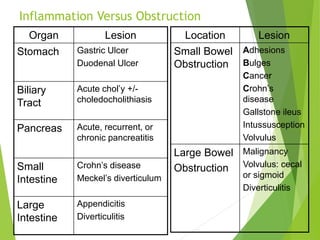
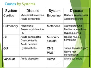
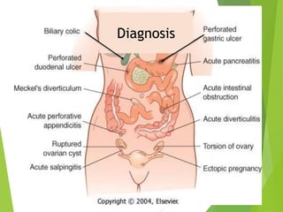
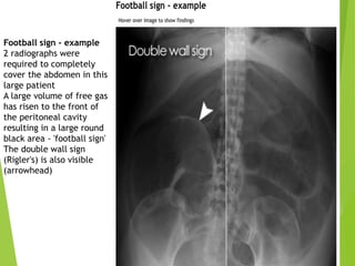
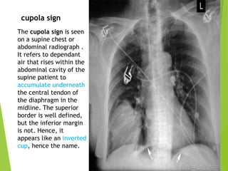
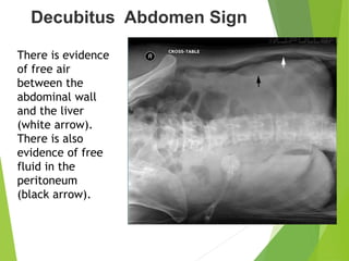
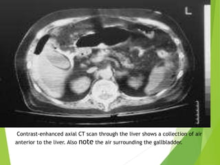
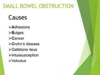
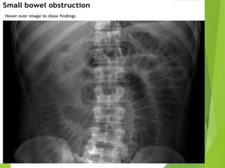
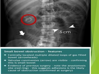
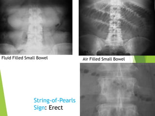
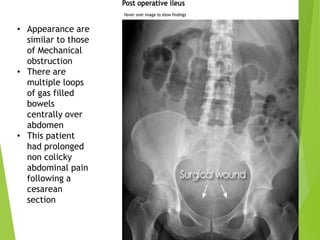
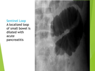
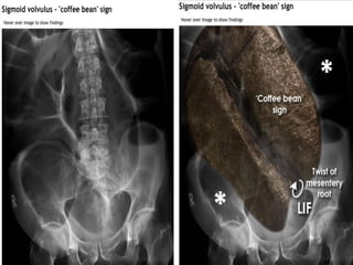
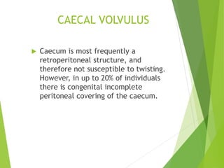
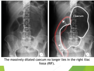
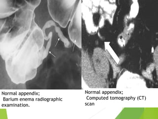
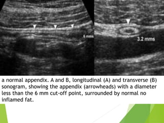
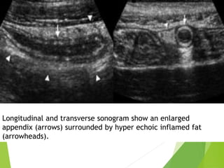
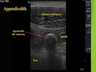
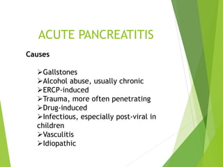
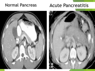
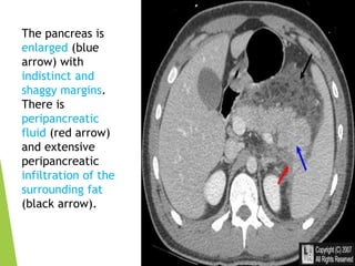
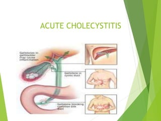
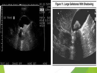
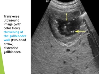
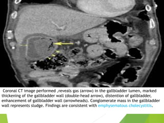
ACUTE ABDOMEN
Inflammation Versus Obstruction
Organ Lesion
Stomach Gastric Ulcer
Duodenal Ulcer
Biliary
Tract
Acute chol’y +/-
choledocholithiasis
Pancreas Acute, recurrent, or
chronic pancreatitis
Small
Intestine
Crohn’s disease
Meckel’s diverticulum
Large
Intestine
Appendicitis
Diverticulitis
Location Lesion
Small Bowel
Obstruction
Adhesions
Bulges
Cancer
Crohn’s
disease
Gallstone ileus
Intussusception
Volvulus
Large Bowel
Obstruction
Malignancy
Volvulus: cecal
or sigmoid
Diverticulitis
Causes by Systems
System Disease System Disease
Cardiac Myocardial infarction
Acute pericarditis
Endocrine Diabetic ketoacidosis
Addisonian crisis
Pulmonary Pneumonia
Pulmonary infarction
PE
Metabolic Acute porphyria
Mediterranean fever
Hyperlipidemia
GI Acute pancreatitis
Gastroenteritis
Acute hepatitis
Musculo-
skeletal
Rectus muscle
hematoma
GU Pyelonephritis CNS
PNS
Tabes dorsalis (syph)
Nerve root
compression
Vascular Aortic dissection Heme Sickle cell crisis
Diagnosis
PNEUMOPERITONEUM
CAUSES:
Bowel perforation
Insufflation of gas (CO2 or air)
during laparoscopy.
Abdominal X-ray
 Although the erect chest X-ray is a much more
sensitive investigation for pneumoperitoneum,
there are several signs that may be useful in
detecting free gas on an abdominal X-ray.
Rigler's/double wall sign
 Rigler's sign (also known as the double wall sign)
is the appearance of lucency (gas) on both sides
of the bowel wall.
Football sign - example
2 radiographs were
required to completely
cover the abdomen in this
large patient
A large volume of free gas
has risen to the front of
the peritoneal cavity
resulting in a large round
black area - 'football sign'
The double wall sign
(Rigler's) is also visible
(arrowhead)
The cupola sign is seen
on a supine chest or
abdominal radiograph .
It refers to dependant
air that rises within the
abdominal cavity of the
supine patient to
accumulate underneath
the central tendon of
the diaphragm in the
midline. The superior
border is well defined,
but the inferior margin
is not. Hence, it
appears like an inverted
cup, hence the name.
cupola sign
Decubitus Abdomen Sign
There is evidence
of free air
between the
abdominal wall
and the liver
(white arrow).
There is also
evidence of free
fluid in the
peritoneum
(black arrow).
Contrast-enhanced axial CT scan through the liver shows a collection of air
anterior to the liver. Also note the air surrounding the gallbladder.
SMALL BOWEL OBSTRUCTION
Causes
Adhesions
Bulges
Cancer
Crohn’s disease
Gallstone ileus
Intussusception
Volvulus
Fluid Filled Small Bowel Air Filled Small Bowel
String-of-Pearls
Sign: Erect
ILEUS
Hypomotility of the gastrointestinal tract in the
absence of mechanical bowel obstruction.
• Appearance are
similar to those
of Mechanical
obstruction
• There are
multiple loops
of gas filled
bowels
centrally over
abdomen
• This patient
had prolonged
non colicky
abdominal pain
following a
cesarean
section
Sentinel Loop
A localized loop
of small bowel is
dilated with
acute
pancreatitis
LARGE BOWEL OBSTRUCTION
CAUSES
 Colo-rectal carcinoma
 Diverticular strictures
 Hernias
 Volvulus
Adhesions do not commonly
cause large bowel obstruction.
Large bowel
obstruction
Here the colon is
dilated down to the
level of the distal
descending colon.
There is the
impression of soft
tissue density at the
level of obstruction
(X). No gas is seen
within the sigmoid
colon.
Obstruction is not
absolute in this
patient as a small
volume of gas has
reached the rectum
(arrow).
An obstructing colon
carcinoma was
confirmed on CT and
at surgery.
VOLVULUS
 Twisting of the bowel
 The two commonest types of bowel
twisting are sigmoid volvulus and
caecal volvulus.
SIGMOID Volvulus
 The sigmoid colon is more
prone to twisting than other
segments of the large bowel
because it is 'mobile' on its own
mesentery, which arises from a
fixed point in the left iliac
fossa (LIF).
CAECAL VOLVULUS
 Caecum is most frequently a
retroperitoneal structure, and
therefore not susceptible to twisting.
However, in up to 20% of individuals
there is congenital incomplete
peritoneal covering of the caecum.
The massively dilated caecum no longer lies in the right iliac
fossa (RIF).
ACUTE APPENDICITIS
Causes
 Stones, food,
mucus
 adhesions
 Tumors
 lymphoid
hyperplasia
Findings on plain film
 Normal
 Focal ileus
 Appendicolith
 Mass
 Free air is very rare
Normal appendix;
Barium enema radiographic
examination.
Normal appendix;
Computed tomography (CT)
scan
a normal appendix. A and B, longitudinal (A) and transverse (B)
sonogram, showing the appendix (arrowheads) with a diameter
less than the 6 mm cut-off point, surrounded by normal no
inflamed fat.
Longitudinal and transverse sonogram show an enlarged
appendix (arrows) surrounded by hyper echoic inflamed fat
(arrowheads).
ACUTE PANCREATITIS
Causes
Gallstones
Alcohol abuse, usually chronic
ERCP-induced
Trauma, more often penetrating
Drug-induced
Infectious, especially post-viral in
children
Vasculitis
Idiopathic
Normal Pancreas Acute Pancreatitis
The pancreas is
enlarged (blue
arrow) with
indistinct and
shaggy margins.
There is
peripancreatic
fluid (red arrow)
and extensive
peripancreatic
infiltration of the
surrounding fat
(black arrow).
ACUTE CHOLECYSTITIS
Transverse
ultrasound
image (with
color flow)
thickening of
the gallbladder
wall (two-head
arrow),
distended
gallbladder.
Coronal CT image performed ,reveals gas (arrow) in the gallbladder lumen, marked
thickening of the gallbladder wall (double-head arrow), distention of gallbladder,
enhancement of gallbladder wall (arrowheads). Conglomerate mass in the gallbladder
wall represents sludge. Findings are consistent with emphysematous cholecystitis.
Acute abdomen

Acute abdomen

  • 1.
  • 2.
    Inflammation Versus Obstruction OrganLesion Stomach Gastric Ulcer Duodenal Ulcer Biliary Tract Acute chol’y +/- choledocholithiasis Pancreas Acute, recurrent, or chronic pancreatitis Small Intestine Crohn’s disease Meckel’s diverticulum Large Intestine Appendicitis Diverticulitis Location Lesion Small Bowel Obstruction Adhesions Bulges Cancer Crohn’s disease Gallstone ileus Intussusception Volvulus Large Bowel Obstruction Malignancy Volvulus: cecal or sigmoid Diverticulitis
  • 3.
    Causes by Systems SystemDisease System Disease Cardiac Myocardial infarction Acute pericarditis Endocrine Diabetic ketoacidosis Addisonian crisis Pulmonary Pneumonia Pulmonary infarction PE Metabolic Acute porphyria Mediterranean fever Hyperlipidemia GI Acute pancreatitis Gastroenteritis Acute hepatitis Musculo- skeletal Rectus muscle hematoma GU Pyelonephritis CNS PNS Tabes dorsalis (syph) Nerve root compression Vascular Aortic dissection Heme Sickle cell crisis
  • 4.
  • 5.
  • 6.
    CAUSES: Bowel perforation Insufflation ofgas (CO2 or air) during laparoscopy.
  • 9.
    Abdominal X-ray  Althoughthe erect chest X-ray is a much more sensitive investigation for pneumoperitoneum, there are several signs that may be useful in detecting free gas on an abdominal X-ray. Rigler's/double wall sign  Rigler's sign (also known as the double wall sign) is the appearance of lucency (gas) on both sides of the bowel wall.
  • 11.
    Football sign -example 2 radiographs were required to completely cover the abdomen in this large patient A large volume of free gas has risen to the front of the peritoneal cavity resulting in a large round black area - 'football sign' The double wall sign (Rigler's) is also visible (arrowhead)
  • 13.
    The cupola signis seen on a supine chest or abdominal radiograph . It refers to dependant air that rises within the abdominal cavity of the supine patient to accumulate underneath the central tendon of the diaphragm in the midline. The superior border is well defined, but the inferior margin is not. Hence, it appears like an inverted cup, hence the name. cupola sign
  • 14.
    Decubitus Abdomen Sign Thereis evidence of free air between the abdominal wall and the liver (white arrow). There is also evidence of free fluid in the peritoneum (black arrow).
  • 15.
    Contrast-enhanced axial CTscan through the liver shows a collection of air anterior to the liver. Also note the air surrounding the gallbladder.
  • 16.
    SMALL BOWEL OBSTRUCTION Causes Adhesions Bulges Cancer Crohn’sdisease Gallstone ileus Intussusception Volvulus
  • 19.
    Fluid Filled SmallBowel Air Filled Small Bowel String-of-Pearls Sign: Erect
  • 20.
    ILEUS Hypomotility of thegastrointestinal tract in the absence of mechanical bowel obstruction.
  • 21.
    • Appearance are similarto those of Mechanical obstruction • There are multiple loops of gas filled bowels centrally over abdomen • This patient had prolonged non colicky abdominal pain following a cesarean section
  • 22.
    Sentinel Loop A localizedloop of small bowel is dilated with acute pancreatitis
  • 23.
    LARGE BOWEL OBSTRUCTION CAUSES Colo-rectal carcinoma  Diverticular strictures  Hernias  Volvulus Adhesions do not commonly cause large bowel obstruction.
  • 24.
    Large bowel obstruction Here thecolon is dilated down to the level of the distal descending colon. There is the impression of soft tissue density at the level of obstruction (X). No gas is seen within the sigmoid colon. Obstruction is not absolute in this patient as a small volume of gas has reached the rectum (arrow). An obstructing colon carcinoma was confirmed on CT and at surgery.
  • 25.
    VOLVULUS  Twisting ofthe bowel  The two commonest types of bowel twisting are sigmoid volvulus and caecal volvulus.
  • 26.
    SIGMOID Volvulus  Thesigmoid colon is more prone to twisting than other segments of the large bowel because it is 'mobile' on its own mesentery, which arises from a fixed point in the left iliac fossa (LIF).
  • 28.
    CAECAL VOLVULUS  Caecumis most frequently a retroperitoneal structure, and therefore not susceptible to twisting. However, in up to 20% of individuals there is congenital incomplete peritoneal covering of the caecum.
  • 29.
    The massively dilatedcaecum no longer lies in the right iliac fossa (RIF).
  • 30.
    ACUTE APPENDICITIS Causes  Stones,food, mucus  adhesions  Tumors  lymphoid hyperplasia Findings on plain film  Normal  Focal ileus  Appendicolith  Mass  Free air is very rare
  • 31.
    Normal appendix; Barium enemaradiographic examination. Normal appendix; Computed tomography (CT) scan
  • 32.
    a normal appendix.A and B, longitudinal (A) and transverse (B) sonogram, showing the appendix (arrowheads) with a diameter less than the 6 mm cut-off point, surrounded by normal no inflamed fat.
  • 33.
    Longitudinal and transversesonogram show an enlarged appendix (arrows) surrounded by hyper echoic inflamed fat (arrowheads).
  • 35.
    ACUTE PANCREATITIS Causes Gallstones Alcohol abuse,usually chronic ERCP-induced Trauma, more often penetrating Drug-induced Infectious, especially post-viral in children Vasculitis Idiopathic
  • 36.
  • 37.
    The pancreas is enlarged(blue arrow) with indistinct and shaggy margins. There is peripancreatic fluid (red arrow) and extensive peripancreatic infiltration of the surrounding fat (black arrow).
  • 38.
  • 40.
    Transverse ultrasound image (with color flow) thickeningof the gallbladder wall (two-head arrow), distended gallbladder.
  • 41.
    Coronal CT imageperformed ,reveals gas (arrow) in the gallbladder lumen, marked thickening of the gallbladder wall (double-head arrow), distention of gallbladder, enhancement of gallbladder wall (arrowheads). Conglomerate mass in the gallbladder wall represents sludge. Findings are consistent with emphysematous cholecystitis.