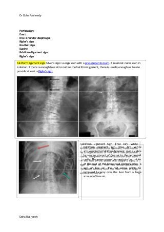
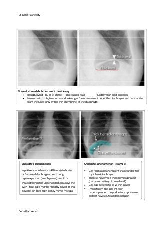
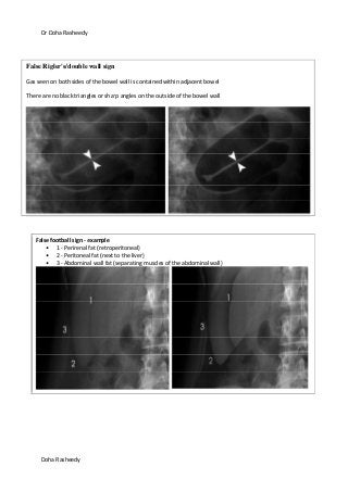
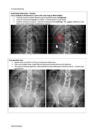
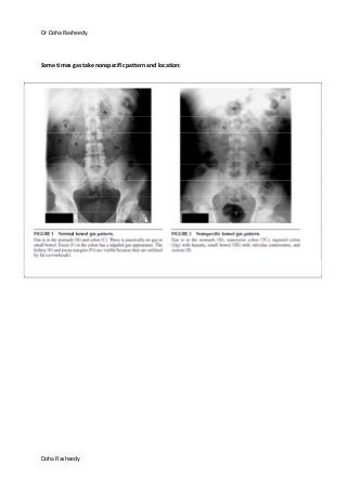
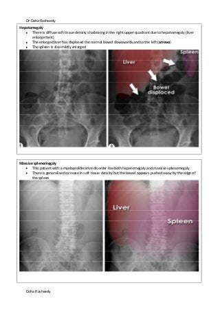
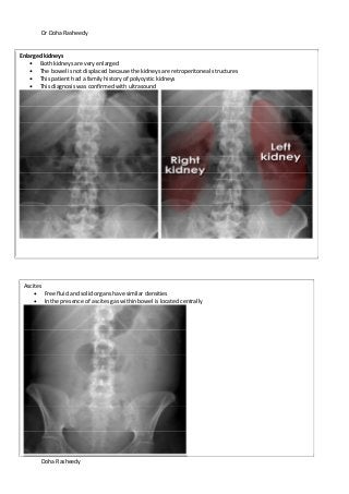
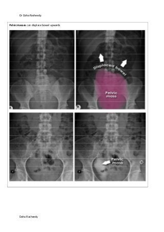
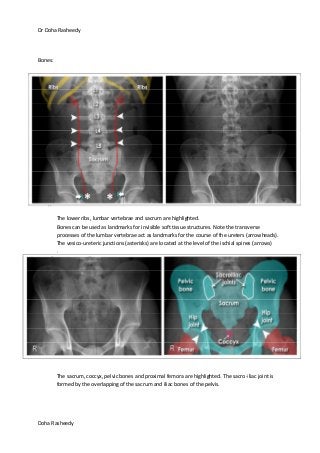
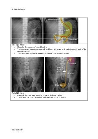
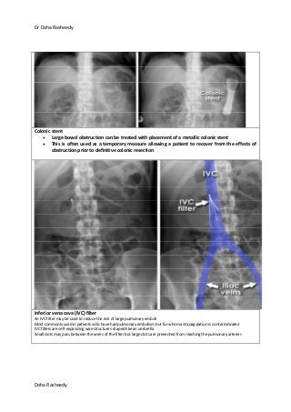
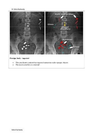
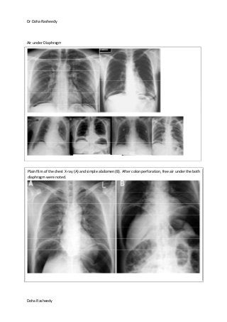
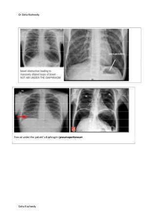
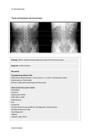
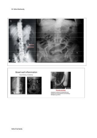
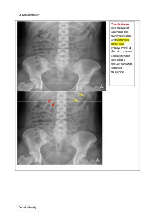
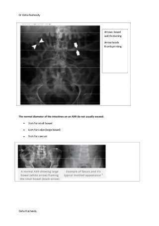
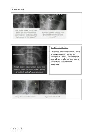
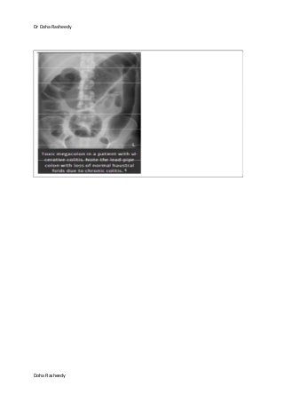
SlideShare a Scribd company logo
Dr Doha Rasheedy
Doha Rasheedy
X ray abdomen
Systematically review:
1. Bowel gas
2. Soft tissues
3. Bones and abnormal calcification
https://www.youtube.com/watch?v=SWd7onzmAPo
1. Bowel gas:
Normal stomach
If the stomach contains air it may be
visible in the left upper quadrant of
the abdomen. The lowest part of
the stomach crosses the midline.
Small bowel (duodenum to terminal
ileum)
Generally the small bowel lies centrally
within the abdomen. The valvulae
conniventes (also called plicae
circulares) are thin, circular, folds of
mucosa, some of which are
circumferential and are seen on an X-
ray to pass across the full width of the
lumen.
Normal <3cm
Dr Doha Rasheedy
Doha Rasheedy
If perforation of the bowel is suspected then an ERECT chest X-ray must be requested. This is the most
sensitive plain radiographic study to detect the presence of free gas in the abdomen.
Normal large bowel
Peripheral position in the
abdomen (the transverse and
sigmoid colon occupy very
variable positions)
Haustra (arrowheads)
Contains faeces
Dr Doha Rasheedy
Doha Rasheedy
air/gas under the diaphragm - erect chest X-ray
This patient has a large volume of free gas under the diaphragm. Dark crescents have formed
separating the thin diaphragm from the liver on the right, and bowel on the left.
This patient had a perforated duodenal ulcer
Rigler's/double wall sign - diagram
Normally only the inner wall of the bowel is visible
If there is pneumoperitoneum both sides of the bowel wall may be visible
Dr Doha Rasheedy
Doha Rasheedy
The double wall sign (Rigler's sign) is visible
Gas separates bowel segments and forms sharp angles and triangles (asterisks)
football sign - example
2 radiographs were required to completely cover the abdomen in this large patient
A large volume of free gas has risen to the front of the peritoneal cavity resulting in a large round black
area - 'football sign'
The double wall sign (Rigler's) is also visible (arrowhead)
Dr Doha Rasheedy
Doha Rasheedy
Perforation:
Erect:
Free air under diaphragm
Rigler’s sign
Football sign
Supine
Falciform ligament sign
Rigler’s sign
Falciform ligament sign: Silver's sign is a sign seen with a pneumoperitoneum. It is almost never seen in
isolation. If there is enough free air to outline the falciform ligament, there is usually enough air to also
provide at least a Rigler's sign.
Falciform Ligament Sign (Free Air). White
arrows point to falciform ligament, made visible
by a large amount of free air in the peritoneal
cavity. The green arrow demonstrate both sides
of the wall of the bowel wall (Rigler's sign), a
sign of free air. The red arrow points to
increased lucency over the liver from a large
amount of free air.
Dr Doha Rasheedy
Doha Rasheedy
Normal stomach bubble - erect chest X-ray
Round/ovoid - 'bubble' shape Thick upper wall Fluid level or food contents
In contrast to this, free intra-abdominal gas forms a crescent under the diaphragm, and is separated
from the lungs only by the thin membrane of the diaphragm
Chilaiditi's phenomenon
In patients who have small livers (cirrhosis),
or flattened diaphragms due to lung
hyperexpansion (emphysema), a void is
created within the upper abdomen above the
liver. This space may be filled by bowel. If this
bowel is air filled then it may mimic free gas
Chilaiditi's phenomenon - example
Gas forms a near crescent shape under the
right hemidiaphragm
There is however a thick hemidiaphragm
(partly consisting of bowel wall)
Gas can be seen to lie within bowel
Importantly, this patient with
hyperexpanded lungs, due to emphysema,
did not have acute abdominal pain
Dr Doha Rasheedy
Doha Rasheedy
False Rigler's/double wall sign
Gas seen on both sides of the bowel wall is contained within adjacent bowel
There are no black triangles or sharp angles on the outside of the bowel wall
False football sign - example
1 - Perirenal fat (retroperitoneal)
2 - Peritoneal fat (next to the liver)
3 - Abdominal wall fat (separating muscles of the abdominal wall)
Dr Doha Rasheedy
Doha Rasheedy
Small bowel obstruction – features
>3cm, multiple air fluid levels>2, some in the same loop at differe thights
Centrally located multiple dilated loops of gas filled bowel (arrowheads)
Valvulae conniventes (arrow) are visible - confirming this is small bowel
Evidence of previous surgery - note the anastomosis site (red ring) - this suggests adhesions is the
likely cause of obstruction (confirmed at surgery)
Post operative ileus
Appearances are similar to those of mechanical obstruction
There are multiple loops of gas filled bowel projected centrally over the abdomen
This patient had prolonged non-colicky abdominal pain following a Caesarian section - recovery was
spontaneous
Dr Doha Rasheedy
Doha Rasheedy
Small bowel obstruction – features
>3cm, multiple air fluid levels>2, some in the same loop at different hights
Features of small bowel obstruction include the central position of gas-filled and distended loops of
bowel.
The white lines passing across the full width of the bowel are 'valvulae conniventes' - these are only
found in the small bowel.
Dr Doha Rasheedy
Doha Rasheedy
Air–fluid levels on erect AXR—
associated with obstruction, ileus, ischaemia and gastroenteritis.
erect supine
Dr Doha Rasheedy
Doha Rasheedy
sentinel loop
Intra-abdominal inflammation, such as with pancreatitis, can lead to a localized ileus. This may appear as a
single loop of dilated bowel known as a 'sentinel loop
Large bowel obstruction: Dilatation of the caecum >9cm, and >6cm for the rest of the colon is considered
abnormal.
Large bowel obstruction
Here the colon is dilated down to the level of the distal descending colon. There is the impression of
soft tissue density at the level of obstruction (X). No gas is seen within the sigmoid colon.
Obstruction is not absolute in this patient as a small volume of gas has reached the rectum (arrow).
An obstructing colon carcinoma was confirmed on CT and at surgery.
Dr Doha Rasheedy
Doha Rasheedy
Volvulus is a specific cause of obstruction with characteristic X-ray appearances
The two commonest types of bowel twisting are sigmoid volvulus and caecal volvulus.
Sigmoid volvulus - 'coffee bean' sign
The sigmoid colon is very dilated because it is twisted at the root of its mesentery in the left iliac
fossa (LIF). The proximal large bowel is also dilated (asterisks).
The twisted loop of sigmoid colon is said to resemble a coffee bean. As in this case the loop of
dilated sigmoid colon - or 'coffee bean' - usually points upwards towards the diaphragm.
This patient is at high risk of perforation and/or bowel ischaemia
Caecal volvulus: The massively dilated caecum no longer lies in the right iliac fossa (RIF). Rather this is occupied by small bowel (red
outline). The small bowel is identified by the valvulae conniventes - mucosal folds that cross the full width of the bowel
(arrowheads). Caecal volvulus was confirmed at laparotomy
Dr Doha Rasheedy
Doha Rasheedy
Sigmoid volvulus: Coffee bean sign in RLQ
Dr Doha Rasheedy
Doha Rasheedy
Bowel wall inflammation
Abdominal X-rays sometimes demonstrate signs of bowel inflammation such as mucosal thickening
'thumb-printing' or a featureless colon 'lead pipe' colon.
Dr Doha Rasheedy
Doha Rasheedy
Toxic megacolon
The colon is very dilated in this patient with acute abdominal pain, sepsis, and a known history of ulcerative
colitis. The clinical features and X-ray appearances are consistent with toxic megacolon.
There is evidence of bowel wall oedema with 'thumbprinting', and pseudopolyps or 'mucosal islands' (red-
patches).
Dr Doha Rasheedy
Doha Rasheedy
The transverse colon is dilated and shows evidence of thumbprinting (black arrow)
The descending colon has a thickened featureless wall and possible pockets of intra-mural gas (white arrow). It is not
clear whether this appearance is due to intra-mural gas, or properitoneal fat interposed between the descending colon
and abdominal wall.
Patient has known Crohn's disease
Appearances are consistent with toxic megacolon
Intra-mural gas refers to abnormal gas in the wall of hollow abdominal viscus. Intra-mural gas is one of the most
serious findings on abdominal plain film requiring timely surgical intervention in adults. Linear streaks of intra-mural
gas indicate infarction of the bowel wall
Dr Doha Rasheedy
Doha Rasheedy
Other aberrant air:
Pneumobilia: Pneumobilia is typically seen as linear branching gas within the liver most prominent in
central large calibre ducts as the flow of bile pushes gas toward the hilum. This is in contrast to portal
venous gas where peripheral small calibre branching gas is usually seen due to the hepatopetal flow of
blood away from the hilum.
Causes
recent biliary instrumentation
o ERCP
o percutaneous or intraoperative cholangiography (small amount of gas only)
incompetent sphincter of Oddi
o sphincterotomy (~50% pneumobilia at 1 year)
o following passage of a gallstone
o scarring e.g. chronic pancreatitis
o drugs e.g. atropine
o congenital
biliary-enteric surgical anastomosis
o cholecystoenterostomy
o choledochoduodenostomy (with or without bile sump syndrome 2)
o Whipple procedure
spontaneous biliary-enteric fistula (cholecystoduodenal accounts for ~70% 3
)
o gallstone ileus
o peptic ulcer disease
o traumatic
o neoplasm, eg. cholangiocarcinoma, ampullary cancer
infection (rare)
o cholangitis
Dr Doha Rasheedy
Doha Rasheedy
o emphysematous cholecystitis (usually gallbladder gas only, ~20% will have gas in the
biliary tree also)
o liver abscess (if contains gas and communicates with the biliary tree)
biliary-bronchopleural fistula (rare)
Supine radiographs often
demonstrate a sword-shaped
lucency in the right paraspinal
region representing gas from the
common duct and the left hepatic
duct. This has been termed the
sabre sign and is present in ~50% of
patients with pneumobilia
Dr Doha Rasheedy
Doha Rasheedy
Portal venous gas: is the accumulation of gas in the portal vein and its branches. It needs to be
distinguished from pneumobilia, although this is usually not too problematic, when associated findings
are taken into account along with the pattern of gas (i.e. peripheral in portal venous gas, central in
pneumobilia).
Causes:
alterations of bowel wall
o ischaemic bowel (usually mural gas as well as mesenteric gas: mortality of 75-90%, but
gas is not an independent predictor)
o necrotic/ulcerated colorectal carcinoma (CRC)
o inflammatory bowel disease (IBD)
o perforated peptic ulcer
bowel luminal distention
o iatrogenic gastric and bowel dilatation (e.g. upper and lower endoscopic procedures,
enemas)
o paralytic ileus / mechanical bowel obstruction
o acute gastric dilatation
o barotrauma
intra-abdominal sepsis
o diverticulitis
o pelvic abscess
o cholecystitis/cholangitis
o appendicitis
unknown mechanism
o pneumatosis intestinalis
o chronic obstructive pulmonary disease (COPD)
o corticosteroid usage
Dr Doha Rasheedy
Doha Rasheedy
Gas forming infection in soft tissue
Air filled urinary bladder Wall: emphysematous cystitis!
An abdominal x-ray revealed gas surrounding the urine
bladder, shows curvilinear or mottled areas of increased
radiolucency in the region of the urinary bladder, separate
from more posterior rectal gas. Intraluminal gas will be seen as
an air-fluid level that changes with patient position, and, when
adjacent to the nondependent mucosal surface, may have a
cobblestone or “beaded necklace” appearance. This is thought
to reflect the irregular thickening produced by submucosal
blebs as seen at direct cystoscopy.
Dr Doha Rasheedy
Doha Rasheedy
Some times gas take nonspecific pattern and location:
Dr Doha Rasheedy
Doha Rasheedy
Soft tissues:
Abdominal X-rays provide a limited means of assessment of soft tissue structures,
Soft tissue organs visible on abdominal X-rays include the liver, spleen, kidneys, psoas muscles,
bladder (within pelvis), and lung bases (within thorax)
Liver on abdominal X-ray
The liver lies in the right upper quadrant
(RUQ) and is seen as a bland area of grey on
an abdominal X-ray.
The superior edge of the liver forms the right
hemi-diaphragm contour (arrowhead).
In this patient the breast shadow (red line)
overlies the liver, and markings of the right
lung are visible behind the liver.
The gallbladder is only rarely visible on an
abdominal X-ray. Its position is very variable.
This patient has had a cholecystectomy. The
clips mark the previous location of the
gallbladder.
Lung bases on abdominal X-ray
The lung bases, which pass behind the liver
and diaphragm in the posterior sulcus of
the thorax, may be visible on some
abdominal X-rays.
It is worth checking the lung bases as some
patients with lung pathology present with
abdominal symptoms.
If there is consolidation suspected from the
abdominal X-ray then a review of the
patient's respiratory system is necessary.
Dr Doha Rasheedy
Doha Rasheedy
Psoas edges on abdominal X-ray
The psoas muscles (red) arise from the transverse processes of the lumbar vertebrae (arrowheads) and
combine with the iliacus muscles. Together these powerful muscles form the iliopsoas tendon, which
attaches to the lesser trochanter of the femur (asterisk). The iliopsoas muscles are the flexors of the hip.
An abdominal X-ray often demonstrates the lateral edge of the psoas muscles as a near straight line. The
iliacus muscles are not visible, as they lie over the iliac bones of the pelvis.
Kidneys on abdominal X-ray
Natural contrast between the kidneys and the low density retroperitoneal fat that surrounds them means they are often visible on an X-
ray of the abdomen.They lie at the level of T12-L3 and lateral to the psoas muscles. The right kidney is usually slightly lower than the left
due to the position of the liver.
Dr Doha Rasheedy
Doha Rasheedy
Abnormal soft tissues
Spleen on abdominal X-ray: The spleen lies in the left upper quadrant immediately superior to the left
kidney
Bladder abdominal X-ray
The bladder has variable appearance
depending on how full it is. It has the
same density as other soft tissue
structures, due to its water content.
Dr Doha Rasheedy
Doha Rasheedy
Hepatomegaly
There is diffuse soft tissue density shadowing in the right upper quadrant due to hepatomegaly (liver
enlargement)
The enlarged liver has displaced the normal bowel downwards and to the left (arrows)
The spleen is also mildly enlarged
Massive splenomegaly
This patient with a myeloproliferative disorder has both hepatomegaly and massive splenomegaly
There is generalised increase in soft tissue density but the bowel appears pushed away by the edge of
the spleen
Dr Doha Rasheedy
Doha Rasheedy
Enlarged kidneys
Both kidneys are very enlarged
The bowel is not displaced because the kidneys are retroperitoneal structures
This patient had a family history of polycystic kidneys
This diagnosis was confirmed with ultrasound
Ascites
Free fluid and solid organs have similar densities
In the presence of ascites gas within bowel is located centrally
Dr Doha Rasheedy
Doha Rasheedy
Pelvic masses can displace bowel upwards
Dr Doha Rasheedy
Doha Rasheedy
Bones:
1.
2.
3.
4.
The lower ribs, lumbar vertebrae and sacrum are highlighted.
Bones can be used as landmarks for invisible soft tissue structures. Note the transverse
processes of the lumbar vertebrae act as landmarks for the course of the ureters (arrowheads).
The vesico-ureteric junctions (asterisks) are located at the level of the ischial spines (arrows)
.
The sacrum, coccyx, pelvic bones and proximal femora are highlighted. The sacro-iliac joint is
formed by the overlapping of the sacrum and iliac bones of the pelvis.
Dr Doha Rasheedy
Doha Rasheedy
4. Calcification and artifact
Ideally all jewellery that overlies anatomically important structures should be removed prior to acquiring
an X-ray
Vascular calcification and ring pessary
If seen, vascular (aorto-iliac) calcification implies a more generalised atherosclerosis.
Note the ring pessary in this elderly patient.
Dr Doha Rasheedy
Doha Rasheedy
Calcified structures
There are multiple incidental and asymptomatic calcified structures seen on this X-ray. The
patient is recovering from an appendicectomy (note surgical clips).
Gallstones are seen only if calcified (20% are calcified). Although they may cause symptoms they
are usually asymptomatic. If gallstone disease is suspected ultrasound examination is a more
appropriate investigation.
Costochondral calcification, calcified mesenteric lymph nodes, and phleboliths (calcified pelvic
veins) are rarely clinically significant. Occasionally additional investigations are required to
differentiate them from pathological calcium. For example phleboliths may be mistaken for
ureteric calculi. Other investigations such as intravenous urogram (IVU) or CT-KUB (CT Kidneys-
Ureters_bladder) should only be performed if there are typical clinical features of ureteric
calculi.
Dr Doha Rasheedy
Doha Rasheedy
Residual contrast
The large areas of very high density seen in the descending colon and rectum are caused by
residual contrast material in this patient who had a Barium enema 10 days previously.
Also note costochondral calcification, and phleboliths.
Do not mistake the tips of the transverse processes for ureteric calculi.
Dr Doha Rasheedy
Doha Rasheedy
Pelvic fracture and osteoarthritis
This elderly patient presented with abdominal pain with no clear history of trauma
Tenderness in the suprapubic regions was thought to be due to intra-abdominal pathology
The pubic ramus fractures was the cause of symptoms
Note the osteoarthritic appearances of the hips and lumbar spine
Bone metastases
There are numerous sclerotic
densities (white) of the vertebrae,
sacrum, pelvis and proximal
femora
This patient had a known history of
breast cancer
Dr Doha Rasheedy
Doha Rasheedy
Paget's disease
This patient has Paget's disease
which affects his lumbar spine
and right hemipelvis
This was an incidental finding
when looking for a cause of
abdominal pain
The typical features of Paget's are
bone expansion and coarsening of
the trabecular pattern involving
the whole of the bone(s) affected
Dr Doha Rasheedy
Doha Rasheedy
Ureteric stone/calculus
Look carefully for ureteric stones
which can be very subtle
Don't mistake a transverse
process for a stone
Bladder stones
Multiple well defined calcific densities are
seen within the bladder
Gallstones and mesenteric lymph node
Gallstones have a variable position
depending on the position of the
gallbladder and may be mistaken for renal
stones
Unlike renal stones they are often rounded
and cluster together
This X-ray also shows an incidental
calcified mesenteric node which may also
mimic renal stones
Dr Doha Rasheedy
Doha Rasheedy
Abdominal aortic aneurysm -
AAA
There is calcification of
the dilated aortic wall
Frequently only one
side of the aneurysm
is visible - as in this
image - the other
being projected over
the spine
Vascular
calcification
There is striking
calcification of the
aorta and iliac
vessels
This is a sign of
generalised
atherosclerosis
elsewhere in the
body
Appendicoliths are highly predictive of
appendicitis in patients presenting with
right iliac fossa pain
Dr Doha Rasheedy
Doha Rasheedy
Retroperitoneal calcification (pancreatic- adrenal):
Dr Doha Rasheedy
Doha Rasheedy
Naso-jejunal tube
Placed for the purpose of enteral feeding
The tube passes through the stomach and forms a C-shape as it navigates the 4 parts of the
duodenum (D1-4)
The tube tip lies beyond the duodenojejunal flexure which lies on the left
Pig-tail (JJ) stent
A ureteric stent has been placed to relieve ureteric obstruction
The catheter has loops (pig-tails) at both ends which hold it in place
Dr Doha Rasheedy
Doha Rasheedy
Colonic stent
Large bowel obstruction can be treated with placement of a metallic colonic stent
This is often used as a temporary measure allowing a patient to recover from the effects of
obstruction prior to definitive colonic resection
Inferior vena cava (IVC) filter
An IVC filter may be used to reduce the risk of large pulmonary emboli
Most commonly used in patients who have had pulmonary embolism but for whom anticoagulation is contraindicated
IVC filters are self-expanding wire structures shaped like an umbrella
Small clots may pass between the wires of the filter but large clots are prevented from reaching the pulmonary arteries
Dr Doha Rasheedy
Doha Rasheedy
Foreign body - ingested
This psychiatric patient has ingested numerous radio-opaque objects
The navel jewellery is external!
Dr Doha Rasheedy
Doha Rasheedy
Air under Diaphragm
Plain film of the chest X-ray (A) and simple abdomen (B). After colon perforation, free air under the both
diaphragm were noted.
Dr Doha Rasheedy
Doha Rasheedy
free air under the patient’s diaphragm (pneumoperitoneum
Dr Doha Rasheedy
Doha Rasheedy
Thumb printing (bowel wall inflammation)
Findings: Mild to moderate bowel edematous walls of the tranverse colon.
Diagnosis: Crohn's Disease
Discussion:
Thumbprinting of Bowel DDx:
Inflammatory Bowel Disease - most common. i.e. Crohn's and Ulcerative Colitis
Diverticulosis or Diverticulitis
Ischemic Colitis with hemorrhage into bowel wall
Other Uncommon causes include:
Amyloidosis
Carcinoid
Angineurotic Edema
CMV colitis in AIDS
Endometriosis
HUS
Lymphoma
Parasitic Infections esp Amebiasis, Strongyloides, Schistosomiasis
Pseudomembranous Colitis
Toxic Megacolon
Typhlitis
Urticaria "colon hives"
Dr Doha Rasheedy
Doha Rasheedy
Dr Doha Rasheedy
Doha Rasheedy
Thumbprinting
(red arrows) of
ascending and
transverse colon
and featureless
bowel wall
(yellow arrow) at
the left transverse
colon extending
into splenic
flexure, consistent
with wall
thickening.
Dr Doha Rasheedy
Doha Rasheedy
The normal diameter of the intestines on an AXR do not usually exceed:
3 cm for small bowel
6 cm for colon (large bowel)
9 cm for caecum
Arrows: bowel
wall thickening
Arrow heads:
thumb printing
Dr Doha Rasheedy
Doha Rasheedy
Small bowel obstruction
Small bowel obstruction can be visualised
on an AXR as dilatation of the small
bowel (>3cm). The valvulae conniventes
are much more visible and have what is
referred to as a “coiled spring
appearance”.
Dr Doha Rasheedy
Doha Rasheedy

More Related Content

What's hot

Ultrasound imaging of Bowel pathology
Ultrasound imaging of Bowel pathologyUltrasound imaging of Bowel pathology
Ultrasound imaging of Bowel pathology
airwave12
 
Ascites and Pleural Effusion
 Ascites and Pleural Effusion Ascites and Pleural Effusion
Ascites and Pleural Effusion
Media Genie
 
Signs in pneumoperitoneum
Signs in pneumoperitoneumSigns in pneumoperitoneum
Signs in pneumoperitoneum
Vikram Patil
 
Radiology Spotters
Radiology Spotters Radiology Spotters
Radiology Spotters
Anish Choudhary
 
Abdominal X ray
Abdominal X rayAbdominal X ray
Abdominal X ray
Richin Koshy
 
Radiology ----Classical Signs in GIT Dr. Muhammad Bin Zulfiqar
Radiology ----Classical Signs in GIT Dr. Muhammad Bin ZulfiqarRadiology ----Classical Signs in GIT Dr. Muhammad Bin Zulfiqar
Radiology ----Classical Signs in GIT Dr. Muhammad Bin Zulfiqar
Dr. Muhammad Bin Zulfiqar
 
Abdomen xray signs
Abdomen xray signsAbdomen xray signs
Abdomen xray signs
Badheeb
 
Patttern of lung disease on chest x ray
Patttern of lung disease on   chest x rayPatttern of lung disease on   chest x ray
Patttern of lung disease on chest x ray
Vrishit Saraswat
 
Diagnositc Imaging of the Esophagus
Diagnositc Imaging of the EsophagusDiagnositc Imaging of the Esophagus
Diagnositc Imaging of the Esophagus
Mohamed M.A. Zaitoun
 
Acute Abdomen Imaging
Acute Abdomen ImagingAcute Abdomen Imaging
Acute Abdomen Imaging
Wan Najwa Zaini
 
Gastric carcinoma radiology ppt
Gastric carcinoma radiology  ppt Gastric carcinoma radiology  ppt
Gastric carcinoma radiology ppt
Sidharthan Selvaraju
 
Ultrasound of acute & chronic cholecystitis
Ultrasound of acute & chronic cholecystitisUltrasound of acute & chronic cholecystitis
Ultrasound of acute & chronic cholecystitisSamir Haffar
 
Collapse and consolidation Lung Radiology
Collapse and consolidation Lung RadiologyCollapse and consolidation Lung Radiology
Collapse and consolidation Lung RadiologyNeelam Ashar
 
6 esophageal filling defects
6 esophageal filling defects6 esophageal filling defects
6 esophageal filling defects
Dr. Muhammad Bin Zulfiqar
 
Presentation1, radiological imaging of hypertrophic pyloric stenosis.
Presentation1, radiological imaging of hypertrophic pyloric stenosis.Presentation1, radiological imaging of hypertrophic pyloric stenosis.
Presentation1, radiological imaging of hypertrophic pyloric stenosis.
Abdellah Nazeer
 
Presentation1.pptx, radiological imaging of intusussception.
Presentation1.pptx, radiological imaging of intusussception.Presentation1.pptx, radiological imaging of intusussception.
Presentation1.pptx, radiological imaging of intusussception.Abdellah Nazeer
 
Signs in Chest Xray
Signs in Chest Xray Signs in Chest Xray
Signs in Chest Xray
Archana Koshy
 
The Radiology of Malrotation
The Radiology of MalrotationThe Radiology of Malrotation
The Radiology of Malrotationtboulden
 
Presentation1, abdominal ultrasound anatomy.
Presentation1, abdominal ultrasound anatomy.Presentation1, abdominal ultrasound anatomy.
Presentation1, abdominal ultrasound anatomy.
Abdellah Nazeer
 

What's hot (20)

Ultrasound imaging of Bowel pathology
Ultrasound imaging of Bowel pathologyUltrasound imaging of Bowel pathology
Ultrasound imaging of Bowel pathology
 
Ascites and Pleural Effusion
 Ascites and Pleural Effusion Ascites and Pleural Effusion
Ascites and Pleural Effusion
 
Signs in pneumoperitoneum
Signs in pneumoperitoneumSigns in pneumoperitoneum
Signs in pneumoperitoneum
 
Radiology Spotters
Radiology Spotters Radiology Spotters
Radiology Spotters
 
Abdominal X ray
Abdominal X rayAbdominal X ray
Abdominal X ray
 
Radiology ----Classical Signs in GIT Dr. Muhammad Bin Zulfiqar
Radiology ----Classical Signs in GIT Dr. Muhammad Bin ZulfiqarRadiology ----Classical Signs in GIT Dr. Muhammad Bin Zulfiqar
Radiology ----Classical Signs in GIT Dr. Muhammad Bin Zulfiqar
 
Abdomen xray signs
Abdomen xray signsAbdomen xray signs
Abdomen xray signs
 
Patttern of lung disease on chest x ray
Patttern of lung disease on   chest x rayPatttern of lung disease on   chest x ray
Patttern of lung disease on chest x ray
 
Diagnositc Imaging of the Esophagus
Diagnositc Imaging of the EsophagusDiagnositc Imaging of the Esophagus
Diagnositc Imaging of the Esophagus
 
Acute Abdomen Imaging
Acute Abdomen ImagingAcute Abdomen Imaging
Acute Abdomen Imaging
 
Gastric carcinoma radiology ppt
Gastric carcinoma radiology  ppt Gastric carcinoma radiology  ppt
Gastric carcinoma radiology ppt
 
Ultrasound of acute & chronic cholecystitis
Ultrasound of acute & chronic cholecystitisUltrasound of acute & chronic cholecystitis
Ultrasound of acute & chronic cholecystitis
 
Collapse and consolidation Lung Radiology
Collapse and consolidation Lung RadiologyCollapse and consolidation Lung Radiology
Collapse and consolidation Lung Radiology
 
6 esophageal filling defects
6 esophageal filling defects6 esophageal filling defects
6 esophageal filling defects
 
Presentation1, radiological imaging of hypertrophic pyloric stenosis.
Presentation1, radiological imaging of hypertrophic pyloric stenosis.Presentation1, radiological imaging of hypertrophic pyloric stenosis.
Presentation1, radiological imaging of hypertrophic pyloric stenosis.
 
Presentation1.pptx, radiological imaging of intusussception.
Presentation1.pptx, radiological imaging of intusussception.Presentation1.pptx, radiological imaging of intusussception.
Presentation1.pptx, radiological imaging of intusussception.
 
Signs in Chest Xray
Signs in Chest Xray Signs in Chest Xray
Signs in Chest Xray
 
The Radiology of Malrotation
The Radiology of MalrotationThe Radiology of Malrotation
The Radiology of Malrotation
 
Liver ultrasound
Liver ultrasoundLiver ultrasound
Liver ultrasound
 
Presentation1, abdominal ultrasound anatomy.
Presentation1, abdominal ultrasound anatomy.Presentation1, abdominal ultrasound anatomy.
Presentation1, abdominal ultrasound anatomy.
 

Similar to X ray abdomen

quick abdomen radiology review .pptx
quick abdomen radiology review .pptxquick abdomen radiology review .pptx
quick abdomen radiology review .pptx
Mohammed Ali
 
Plain abdomen
Plain abdomenPlain abdomen
Plain abdomen
khashayar cyrus
 
Abdominal Plain.pptx
Abdominal Plain.pptxAbdominal Plain.pptx
Abdominal Plain.pptx
hussainAltaher
 
Abdominal Plain.pptx
Abdominal Plain.pptxAbdominal Plain.pptx
Abdominal Plain.pptx
hussainAltaher
 
Approach to X ray Abdomen and KUB.pptx
Approach to X ray Abdomen and KUB.pptxApproach to X ray Abdomen and KUB.pptx
Approach to X ray Abdomen and KUB.pptx
ssuser2d8a16
 
inteestinal gas.pptx
inteestinal gas.pptxinteestinal gas.pptx
inteestinal gas.pptx
mohamedabosdira1
 
ACUTE ABDOMEN
ACUTE ABDOMENACUTE ABDOMEN
ACUTE ABDOMEN
radiologyoffice
 
abdominal x ray radiology
abdominal x ray radiologyabdominal x ray radiology
abdominal x ray radiology
sarfraj Ahmad
 
Presentation1aboutabdominalradiology.pptx
Presentation1aboutabdominalradiology.pptxPresentation1aboutabdominalradiology.pptx
Presentation1aboutabdominalradiology.pptx
ErumKhatri1
 
role of radiology in pediatric.pptx
role of radiology in pediatric.pptxrole of radiology in pediatric.pptx
role of radiology in pediatric.pptx
dypradio
 
Radiographic anatomy of gastrointestinal tract
Radiographic anatomy of gastrointestinal tractRadiographic anatomy of gastrointestinal tract
Radiographic anatomy of gastrointestinal tract
airwave12
 
Acute abdomen
Acute abdomenAcute abdomen
Acute abdomen
arvapally koushik
 
acute abdomen.pdf
acute abdomen.pdfacute abdomen.pdf
acute abdomen.pdf
DrMuhammadOmairChaud
 
Xray cases radiology residents-must know
Xray cases radiology residents-must knowXray cases radiology residents-must know
Xray cases radiology residents-must know
Lê Huỳnh Ngọc
 
Spontaneous Esophageal Rupture 修改后
Spontaneous Esophageal Rupture  修改后Spontaneous Esophageal Rupture  修改后
Spontaneous Esophageal Rupture 修改后
Deep Deep
 
VISCUS INJURY .ppt
VISCUS INJURY .pptVISCUS INJURY .ppt
VISCUS INJURY .ppt
farhanashafie3
 
Dr. kawa bilateral
Dr. kawa bilateralDr. kawa bilateral
Dr. kawa bilateral
Rzgar Tayeb
 
7-170509160536-171107152009 (1).pptx
7-170509160536-171107152009 (1).pptx7-170509160536-171107152009 (1).pptx
7-170509160536-171107152009 (1).pptx
SheikhTuhin1
 
Pictures of acute abdomen.pdf
Pictures of acute abdomen.pdfPictures of acute abdomen.pdf
Pictures of acute abdomen.pdf
DrMuhammadOmairChaud
 
Abdominal xray images
Abdominal xray imagesAbdominal xray images
Abdominal xray imagesLe Jang
 

Similar to X ray abdomen (20)

quick abdomen radiology review .pptx
quick abdomen radiology review .pptxquick abdomen radiology review .pptx
quick abdomen radiology review .pptx
 
Plain abdomen
Plain abdomenPlain abdomen
Plain abdomen
 
Abdominal Plain.pptx
Abdominal Plain.pptxAbdominal Plain.pptx
Abdominal Plain.pptx
 
Abdominal Plain.pptx
Abdominal Plain.pptxAbdominal Plain.pptx
Abdominal Plain.pptx
 
Approach to X ray Abdomen and KUB.pptx
Approach to X ray Abdomen and KUB.pptxApproach to X ray Abdomen and KUB.pptx
Approach to X ray Abdomen and KUB.pptx
 
inteestinal gas.pptx
inteestinal gas.pptxinteestinal gas.pptx
inteestinal gas.pptx
 
ACUTE ABDOMEN
ACUTE ABDOMENACUTE ABDOMEN
ACUTE ABDOMEN
 
abdominal x ray radiology
abdominal x ray radiologyabdominal x ray radiology
abdominal x ray radiology
 
Presentation1aboutabdominalradiology.pptx
Presentation1aboutabdominalradiology.pptxPresentation1aboutabdominalradiology.pptx
Presentation1aboutabdominalradiology.pptx
 
role of radiology in pediatric.pptx
role of radiology in pediatric.pptxrole of radiology in pediatric.pptx
role of radiology in pediatric.pptx
 
Radiographic anatomy of gastrointestinal tract
Radiographic anatomy of gastrointestinal tractRadiographic anatomy of gastrointestinal tract
Radiographic anatomy of gastrointestinal tract
 
Acute abdomen
Acute abdomenAcute abdomen
Acute abdomen
 
acute abdomen.pdf
acute abdomen.pdfacute abdomen.pdf
acute abdomen.pdf
 
Xray cases radiology residents-must know
Xray cases radiology residents-must knowXray cases radiology residents-must know
Xray cases radiology residents-must know
 
Spontaneous Esophageal Rupture 修改后
Spontaneous Esophageal Rupture  修改后Spontaneous Esophageal Rupture  修改后
Spontaneous Esophageal Rupture 修改后
 
VISCUS INJURY .ppt
VISCUS INJURY .pptVISCUS INJURY .ppt
VISCUS INJURY .ppt
 
Dr. kawa bilateral
Dr. kawa bilateralDr. kawa bilateral
Dr. kawa bilateral
 
7-170509160536-171107152009 (1).pptx
7-170509160536-171107152009 (1).pptx7-170509160536-171107152009 (1).pptx
7-170509160536-171107152009 (1).pptx
 
Pictures of acute abdomen.pdf
Pictures of acute abdomen.pdfPictures of acute abdomen.pdf
Pictures of acute abdomen.pdf
 
Abdominal xray images
Abdominal xray imagesAbdominal xray images
Abdominal xray images
 

More from Doha Rasheedy

social cognition domains and impairment.pptx
social cognition domains and impairment.pptxsocial cognition domains and impairment.pptx
social cognition domains and impairment.pptx
Doha Rasheedy
 
The Value of Collateral History in Screening for Mild Cognitive Impairment in...
The Value of Collateral History in Screening for Mild Cognitive Impairment in...The Value of Collateral History in Screening for Mild Cognitive Impairment in...
The Value of Collateral History in Screening for Mild Cognitive Impairment in...
Doha Rasheedy
 
geriatric nutritional tips.pptx
geriatric nutritional tips.pptxgeriatric nutritional tips.pptx
geriatric nutritional tips.pptx
Doha Rasheedy
 
Pulmonology 2023.pptx
Pulmonology 2023.pptxPulmonology 2023.pptx
Pulmonology 2023.pptx
Doha Rasheedy
 
NEW paradigm of CGA.pdf
NEW paradigm of CGA.pdfNEW paradigm of CGA.pdf
NEW paradigm of CGA.pdf
Doha Rasheedy
 
nutritional frailty.pdf
nutritional frailty.pdfnutritional frailty.pdf
nutritional frailty.pdf
Doha Rasheedy
 
Frailty in older adults: Myths and Facts
Frailty in older adults: Myths and FactsFrailty in older adults: Myths and Facts
Frailty in older adults: Myths and Facts
Doha Rasheedy
 
EASL Clinical Practice Guidelines for the management of patients with decompe...
EASL Clinical Practice Guidelines for the management of patients withdecompe...EASL Clinical Practice Guidelines for the management of patients withdecompe...
EASL Clinical Practice Guidelines for the management of patients with decompe...
Doha Rasheedy
 
non atherosclerotic angina final Doha Rasheedy.docx
non atherosclerotic angina  final  Doha Rasheedy.docxnon atherosclerotic angina  final  Doha Rasheedy.docx
non atherosclerotic angina final Doha Rasheedy.docx
Doha Rasheedy
 
Non Atherosclerotic angina Final Doha Rasheedy.pptx
Non Atherosclerotic angina  Final Doha Rasheedy.pptxNon Atherosclerotic angina  Final Doha Rasheedy.pptx
Non Atherosclerotic angina Final Doha Rasheedy.pptx
Doha Rasheedy
 
Thiazide diuretics.pptx
Thiazide diuretics.pptxThiazide diuretics.pptx
Thiazide diuretics.pptx
Doha Rasheedy
 
Adverse Effects Associated with Proton Pump Inhibitor Use.pptx
Adverse Effects Associated with Proton Pump Inhibitor Use.pptxAdverse Effects Associated with Proton Pump Inhibitor Use.pptx
Adverse Effects Associated with Proton Pump Inhibitor Use.pptx
Doha Rasheedy
 
Adrenal insufficiency.pptx
Adrenal insufficiency.pptxAdrenal insufficiency.pptx
Adrenal insufficiency.pptx
Doha Rasheedy
 
Respiratory part 2
Respiratory part 2Respiratory part 2
Respiratory part 2
Doha Rasheedy
 
Basic of geriatrics and internal medicine for physiotherapist
Basic of geriatrics and internal medicine for physiotherapistBasic of geriatrics and internal medicine for physiotherapist
Basic of geriatrics and internal medicine for physiotherapist
Doha Rasheedy
 
perioperative care of elderly patients
perioperative care of elderly patientsperioperative care of elderly patients
perioperative care of elderly patients
Doha Rasheedy
 
inflammatory bowel disease in elderly
inflammatory  bowel disease in elderlyinflammatory  bowel disease in elderly
inflammatory bowel disease in elderly
Doha Rasheedy
 
Cognition and cognitive syndromes cme
Cognition and cognitive syndromes cmeCognition and cognitive syndromes cme
Cognition and cognitive syndromes cme
Doha Rasheedy
 
Sarcopenia
SarcopeniaSarcopenia
Sarcopenia
Doha Rasheedy
 
Orthostatic hypotension
Orthostatic hypotensionOrthostatic hypotension
Orthostatic hypotension
Doha Rasheedy
 

More from Doha Rasheedy (20)

social cognition domains and impairment.pptx
social cognition domains and impairment.pptxsocial cognition domains and impairment.pptx
social cognition domains and impairment.pptx
 
The Value of Collateral History in Screening for Mild Cognitive Impairment in...
The Value of Collateral History in Screening for Mild Cognitive Impairment in...The Value of Collateral History in Screening for Mild Cognitive Impairment in...
The Value of Collateral History in Screening for Mild Cognitive Impairment in...
 
geriatric nutritional tips.pptx
geriatric nutritional tips.pptxgeriatric nutritional tips.pptx
geriatric nutritional tips.pptx
 
Pulmonology 2023.pptx
Pulmonology 2023.pptxPulmonology 2023.pptx
Pulmonology 2023.pptx
 
NEW paradigm of CGA.pdf
NEW paradigm of CGA.pdfNEW paradigm of CGA.pdf
NEW paradigm of CGA.pdf
 
nutritional frailty.pdf
nutritional frailty.pdfnutritional frailty.pdf
nutritional frailty.pdf
 
Frailty in older adults: Myths and Facts
Frailty in older adults: Myths and FactsFrailty in older adults: Myths and Facts
Frailty in older adults: Myths and Facts
 
EASL Clinical Practice Guidelines for the management of patients with decompe...
EASL Clinical Practice Guidelines for the management of patients withdecompe...EASL Clinical Practice Guidelines for the management of patients withdecompe...
EASL Clinical Practice Guidelines for the management of patients with decompe...
 
non atherosclerotic angina final Doha Rasheedy.docx
non atherosclerotic angina  final  Doha Rasheedy.docxnon atherosclerotic angina  final  Doha Rasheedy.docx
non atherosclerotic angina final Doha Rasheedy.docx
 
Non Atherosclerotic angina Final Doha Rasheedy.pptx
Non Atherosclerotic angina  Final Doha Rasheedy.pptxNon Atherosclerotic angina  Final Doha Rasheedy.pptx
Non Atherosclerotic angina Final Doha Rasheedy.pptx
 
Thiazide diuretics.pptx
Thiazide diuretics.pptxThiazide diuretics.pptx
Thiazide diuretics.pptx
 
Adverse Effects Associated with Proton Pump Inhibitor Use.pptx
Adverse Effects Associated with Proton Pump Inhibitor Use.pptxAdverse Effects Associated with Proton Pump Inhibitor Use.pptx
Adverse Effects Associated with Proton Pump Inhibitor Use.pptx
 
Adrenal insufficiency.pptx
Adrenal insufficiency.pptxAdrenal insufficiency.pptx
Adrenal insufficiency.pptx
 
Respiratory part 2
Respiratory part 2Respiratory part 2
Respiratory part 2
 
Basic of geriatrics and internal medicine for physiotherapist
Basic of geriatrics and internal medicine for physiotherapistBasic of geriatrics and internal medicine for physiotherapist
Basic of geriatrics and internal medicine for physiotherapist
 
perioperative care of elderly patients
perioperative care of elderly patientsperioperative care of elderly patients
perioperative care of elderly patients
 
inflammatory bowel disease in elderly
inflammatory  bowel disease in elderlyinflammatory  bowel disease in elderly
inflammatory bowel disease in elderly
 
Cognition and cognitive syndromes cme
Cognition and cognitive syndromes cmeCognition and cognitive syndromes cme
Cognition and cognitive syndromes cme
 
Sarcopenia
SarcopeniaSarcopenia
Sarcopenia
 
Orthostatic hypotension
Orthostatic hypotensionOrthostatic hypotension
Orthostatic hypotension
 

Recently uploaded

Ocular injury ppt Upendra pal optometrist upums saifai etawah
Ocular injury  ppt  Upendra pal  optometrist upums saifai etawahOcular injury  ppt  Upendra pal  optometrist upums saifai etawah
Ocular injury ppt Upendra pal optometrist upums saifai etawah
pal078100
 
The hemodynamic and autonomic determinants of elevated blood pressure in obes...
The hemodynamic and autonomic determinants of elevated blood pressure in obes...The hemodynamic and autonomic determinants of elevated blood pressure in obes...
The hemodynamic and autonomic determinants of elevated blood pressure in obes...
Catherine Liao
 
New Directions in Targeted Therapeutic Approaches for Older Adults With Mantl...
New Directions in Targeted Therapeutic Approaches for Older Adults With Mantl...New Directions in Targeted Therapeutic Approaches for Older Adults With Mantl...
New Directions in Targeted Therapeutic Approaches for Older Adults With Mantl...
i3 Health
 
How to Give Better Lectures: Some Tips for Doctors
How to Give Better Lectures: Some Tips for DoctorsHow to Give Better Lectures: Some Tips for Doctors
How to Give Better Lectures: Some Tips for Doctors
LanceCatedral
 
Phone Us ❤85270-49040❤ #ℂall #gIRLS In Surat By Surat @ℂall @Girls Hotel With...
Phone Us ❤85270-49040❤ #ℂall #gIRLS In Surat By Surat @ℂall @Girls Hotel With...Phone Us ❤85270-49040❤ #ℂall #gIRLS In Surat By Surat @ℂall @Girls Hotel With...
Phone Us ❤85270-49040❤ #ℂall #gIRLS In Surat By Surat @ℂall @Girls Hotel With...
Savita Shen $i11
 
Ophthalmology Clinical Tests for OSCE exam
Ophthalmology Clinical Tests for OSCE examOphthalmology Clinical Tests for OSCE exam
Ophthalmology Clinical Tests for OSCE exam
KafrELShiekh University
 
Charaka Samhita Sutra Sthana 9 Chapter khuddakachatuspadadhyaya
Charaka Samhita Sutra Sthana 9 Chapter khuddakachatuspadadhyayaCharaka Samhita Sutra Sthana 9 Chapter khuddakachatuspadadhyaya
Charaka Samhita Sutra Sthana 9 Chapter khuddakachatuspadadhyaya
Dr KHALID B.M
 
heat stroke and heat exhaustion in children
heat stroke and heat exhaustion in childrenheat stroke and heat exhaustion in children
heat stroke and heat exhaustion in children
SumeraAhmad5
 
Factory Supply Best Quality Pmk Oil CAS 28578–16–7 PMK Powder in Stock
Factory Supply Best Quality Pmk Oil CAS 28578–16–7 PMK Powder in StockFactory Supply Best Quality Pmk Oil CAS 28578–16–7 PMK Powder in Stock
Factory Supply Best Quality Pmk Oil CAS 28578–16–7 PMK Powder in Stock
rebeccabio
 
BRACHYTHERAPY OVERVIEW AND APPLICATORS
BRACHYTHERAPY OVERVIEW  AND  APPLICATORSBRACHYTHERAPY OVERVIEW  AND  APPLICATORS
BRACHYTHERAPY OVERVIEW AND APPLICATORS
Krishan Murari
 
For Better Surat #ℂall #Girl Service ❤85270-49040❤ Surat #ℂall #Girls
For Better Surat #ℂall #Girl Service ❤85270-49040❤ Surat #ℂall #GirlsFor Better Surat #ℂall #Girl Service ❤85270-49040❤ Surat #ℂall #Girls
For Better Surat #ℂall #Girl Service ❤85270-49040❤ Surat #ℂall #Girls
Savita Shen $i11
 
Evaluation of antidepressant activity of clitoris ternatea in animals
Evaluation of antidepressant activity of clitoris ternatea in animalsEvaluation of antidepressant activity of clitoris ternatea in animals
Evaluation of antidepressant activity of clitoris ternatea in animals
Shweta
 
Physiology of Special Chemical Sensation of Taste
Physiology of Special Chemical Sensation of TastePhysiology of Special Chemical Sensation of Taste
Physiology of Special Chemical Sensation of Taste
MedicoseAcademics
 
HOT NEW PRODUCT! BIG SALES FAST SHIPPING NOW FROM CHINA!! EU KU DB BK substit...
HOT NEW PRODUCT! BIG SALES FAST SHIPPING NOW FROM CHINA!! EU KU DB BK substit...HOT NEW PRODUCT! BIG SALES FAST SHIPPING NOW FROM CHINA!! EU KU DB BK substit...
HOT NEW PRODUCT! BIG SALES FAST SHIPPING NOW FROM CHINA!! EU KU DB BK substit...
GL Anaacs
 
ARTHROLOGY PPT NCISM SYLLABUS AYURVEDA STUDENTS
ARTHROLOGY PPT NCISM SYLLABUS AYURVEDA STUDENTSARTHROLOGY PPT NCISM SYLLABUS AYURVEDA STUDENTS
ARTHROLOGY PPT NCISM SYLLABUS AYURVEDA STUDENTS
Dr. Vinay Pareek
 
The Normal Electrocardiogram - Part I of II
The Normal Electrocardiogram - Part I of IIThe Normal Electrocardiogram - Part I of II
The Normal Electrocardiogram - Part I of II
MedicoseAcademics
 
Report Back from SGO 2024: What’s the Latest in Cervical Cancer?
Report Back from SGO 2024: What’s the Latest in Cervical Cancer?Report Back from SGO 2024: What’s the Latest in Cervical Cancer?
Report Back from SGO 2024: What’s the Latest in Cervical Cancer?
bkling
 
Surat @ℂall @Girls ꧁❤8527049040❤꧂@ℂall @Girls Service Vip Top Model Safe
Surat @ℂall @Girls ꧁❤8527049040❤꧂@ℂall @Girls Service Vip Top Model SafeSurat @ℂall @Girls ꧁❤8527049040❤꧂@ℂall @Girls Service Vip Top Model Safe
Surat @ℂall @Girls ꧁❤8527049040❤꧂@ℂall @Girls Service Vip Top Model Safe
Savita Shen $i11
 
Ozempic: Preoperative Management of Patients on GLP-1 Receptor Agonists
Ozempic: Preoperative Management of Patients on GLP-1 Receptor Agonists  Ozempic: Preoperative Management of Patients on GLP-1 Receptor Agonists
Ozempic: Preoperative Management of Patients on GLP-1 Receptor Agonists
Saeid Safari
 
micro teaching on communication m.sc nursing.pdf
micro teaching on communication m.sc nursing.pdfmicro teaching on communication m.sc nursing.pdf
micro teaching on communication m.sc nursing.pdf
Anurag Sharma
 

Recently uploaded (20)

Ocular injury ppt Upendra pal optometrist upums saifai etawah
Ocular injury  ppt  Upendra pal  optometrist upums saifai etawahOcular injury  ppt  Upendra pal  optometrist upums saifai etawah
Ocular injury ppt Upendra pal optometrist upums saifai etawah
 
The hemodynamic and autonomic determinants of elevated blood pressure in obes...
The hemodynamic and autonomic determinants of elevated blood pressure in obes...The hemodynamic and autonomic determinants of elevated blood pressure in obes...
The hemodynamic and autonomic determinants of elevated blood pressure in obes...
 
New Directions in Targeted Therapeutic Approaches for Older Adults With Mantl...
New Directions in Targeted Therapeutic Approaches for Older Adults With Mantl...New Directions in Targeted Therapeutic Approaches for Older Adults With Mantl...
New Directions in Targeted Therapeutic Approaches for Older Adults With Mantl...
 
How to Give Better Lectures: Some Tips for Doctors
How to Give Better Lectures: Some Tips for DoctorsHow to Give Better Lectures: Some Tips for Doctors
How to Give Better Lectures: Some Tips for Doctors
 
Phone Us ❤85270-49040❤ #ℂall #gIRLS In Surat By Surat @ℂall @Girls Hotel With...
Phone Us ❤85270-49040❤ #ℂall #gIRLS In Surat By Surat @ℂall @Girls Hotel With...Phone Us ❤85270-49040❤ #ℂall #gIRLS In Surat By Surat @ℂall @Girls Hotel With...
Phone Us ❤85270-49040❤ #ℂall #gIRLS In Surat By Surat @ℂall @Girls Hotel With...
 
Ophthalmology Clinical Tests for OSCE exam
Ophthalmology Clinical Tests for OSCE examOphthalmology Clinical Tests for OSCE exam
Ophthalmology Clinical Tests for OSCE exam
 
Charaka Samhita Sutra Sthana 9 Chapter khuddakachatuspadadhyaya
Charaka Samhita Sutra Sthana 9 Chapter khuddakachatuspadadhyayaCharaka Samhita Sutra Sthana 9 Chapter khuddakachatuspadadhyaya
Charaka Samhita Sutra Sthana 9 Chapter khuddakachatuspadadhyaya
 
heat stroke and heat exhaustion in children
heat stroke and heat exhaustion in childrenheat stroke and heat exhaustion in children
heat stroke and heat exhaustion in children
 
Factory Supply Best Quality Pmk Oil CAS 28578–16–7 PMK Powder in Stock
Factory Supply Best Quality Pmk Oil CAS 28578–16–7 PMK Powder in StockFactory Supply Best Quality Pmk Oil CAS 28578–16–7 PMK Powder in Stock
Factory Supply Best Quality Pmk Oil CAS 28578–16–7 PMK Powder in Stock
 
BRACHYTHERAPY OVERVIEW AND APPLICATORS
BRACHYTHERAPY OVERVIEW  AND  APPLICATORSBRACHYTHERAPY OVERVIEW  AND  APPLICATORS
BRACHYTHERAPY OVERVIEW AND APPLICATORS
 
For Better Surat #ℂall #Girl Service ❤85270-49040❤ Surat #ℂall #Girls
For Better Surat #ℂall #Girl Service ❤85270-49040❤ Surat #ℂall #GirlsFor Better Surat #ℂall #Girl Service ❤85270-49040❤ Surat #ℂall #Girls
For Better Surat #ℂall #Girl Service ❤85270-49040❤ Surat #ℂall #Girls
 
Evaluation of antidepressant activity of clitoris ternatea in animals
Evaluation of antidepressant activity of clitoris ternatea in animalsEvaluation of antidepressant activity of clitoris ternatea in animals
Evaluation of antidepressant activity of clitoris ternatea in animals
 
Physiology of Special Chemical Sensation of Taste
Physiology of Special Chemical Sensation of TastePhysiology of Special Chemical Sensation of Taste
Physiology of Special Chemical Sensation of Taste
 
HOT NEW PRODUCT! BIG SALES FAST SHIPPING NOW FROM CHINA!! EU KU DB BK substit...
HOT NEW PRODUCT! BIG SALES FAST SHIPPING NOW FROM CHINA!! EU KU DB BK substit...HOT NEW PRODUCT! BIG SALES FAST SHIPPING NOW FROM CHINA!! EU KU DB BK substit...
HOT NEW PRODUCT! BIG SALES FAST SHIPPING NOW FROM CHINA!! EU KU DB BK substit...
 
ARTHROLOGY PPT NCISM SYLLABUS AYURVEDA STUDENTS
ARTHROLOGY PPT NCISM SYLLABUS AYURVEDA STUDENTSARTHROLOGY PPT NCISM SYLLABUS AYURVEDA STUDENTS
ARTHROLOGY PPT NCISM SYLLABUS AYURVEDA STUDENTS
 
The Normal Electrocardiogram - Part I of II
The Normal Electrocardiogram - Part I of IIThe Normal Electrocardiogram - Part I of II
The Normal Electrocardiogram - Part I of II
 
Report Back from SGO 2024: What’s the Latest in Cervical Cancer?
Report Back from SGO 2024: What’s the Latest in Cervical Cancer?Report Back from SGO 2024: What’s the Latest in Cervical Cancer?
Report Back from SGO 2024: What’s the Latest in Cervical Cancer?
 
Surat @ℂall @Girls ꧁❤8527049040❤꧂@ℂall @Girls Service Vip Top Model Safe
Surat @ℂall @Girls ꧁❤8527049040❤꧂@ℂall @Girls Service Vip Top Model SafeSurat @ℂall @Girls ꧁❤8527049040❤꧂@ℂall @Girls Service Vip Top Model Safe
Surat @ℂall @Girls ꧁❤8527049040❤꧂@ℂall @Girls Service Vip Top Model Safe
 
Ozempic: Preoperative Management of Patients on GLP-1 Receptor Agonists
Ozempic: Preoperative Management of Patients on GLP-1 Receptor Agonists  Ozempic: Preoperative Management of Patients on GLP-1 Receptor Agonists
Ozempic: Preoperative Management of Patients on GLP-1 Receptor Agonists
 
micro teaching on communication m.sc nursing.pdf
micro teaching on communication m.sc nursing.pdfmicro teaching on communication m.sc nursing.pdf
micro teaching on communication m.sc nursing.pdf
 

X ray abdomen

  • 1. Dr Doha Rasheedy Doha Rasheedy X ray abdomen Systematically review: 1. Bowel gas 2. Soft tissues 3. Bones and abnormal calcification https://www.youtube.com/watch?v=SWd7onzmAPo 1. Bowel gas: Normal stomach If the stomach contains air it may be visible in the left upper quadrant of the abdomen. The lowest part of the stomach crosses the midline. Small bowel (duodenum to terminal ileum) Generally the small bowel lies centrally within the abdomen. The valvulae conniventes (also called plicae circulares) are thin, circular, folds of mucosa, some of which are circumferential and are seen on an X- ray to pass across the full width of the lumen. Normal <3cm
  • 2. Dr Doha Rasheedy Doha Rasheedy If perforation of the bowel is suspected then an ERECT chest X-ray must be requested. This is the most sensitive plain radiographic study to detect the presence of free gas in the abdomen. Normal large bowel Peripheral position in the abdomen (the transverse and sigmoid colon occupy very variable positions) Haustra (arrowheads) Contains faeces
  • 3. Dr Doha Rasheedy Doha Rasheedy air/gas under the diaphragm - erect chest X-ray This patient has a large volume of free gas under the diaphragm. Dark crescents have formed separating the thin diaphragm from the liver on the right, and bowel on the left. This patient had a perforated duodenal ulcer Rigler's/double wall sign - diagram Normally only the inner wall of the bowel is visible If there is pneumoperitoneum both sides of the bowel wall may be visible
  • 4. Dr Doha Rasheedy Doha Rasheedy The double wall sign (Rigler's sign) is visible Gas separates bowel segments and forms sharp angles and triangles (asterisks) football sign - example 2 radiographs were required to completely cover the abdomen in this large patient A large volume of free gas has risen to the front of the peritoneal cavity resulting in a large round black area - 'football sign' The double wall sign (Rigler's) is also visible (arrowhead)
  • 5. Dr Doha Rasheedy Doha Rasheedy Perforation: Erect: Free air under diaphragm Rigler’s sign Football sign Supine Falciform ligament sign Rigler’s sign Falciform ligament sign: Silver's sign is a sign seen with a pneumoperitoneum. It is almost never seen in isolation. If there is enough free air to outline the falciform ligament, there is usually enough air to also provide at least a Rigler's sign. Falciform Ligament Sign (Free Air). White arrows point to falciform ligament, made visible by a large amount of free air in the peritoneal cavity. The green arrow demonstrate both sides of the wall of the bowel wall (Rigler's sign), a sign of free air. The red arrow points to increased lucency over the liver from a large amount of free air. Falciform Ligament Sign (Free Air). White arrows point to falciform ligament, made visible by a large amount of free air in the peritoneal cavity. The green arrow demonstrate both sides of the wall of the bowel wall (Rigler's sign), a sign of free air. The red arrow points to increased lucency over the liver from a large amount of free air.
  • 6. Dr Doha Rasheedy Doha Rasheedy Normal stomach bubble - erect chest X-ray Round/ovoid - 'bubble' shape Thick upper wall Fluid level or food contents In contrast to this, free intra-abdominal gas forms a crescent under the diaphragm, and is separated from the lungs only by the thin membrane of the diaphragm Chilaiditi's phenomenon In patients who have small livers (cirrhosis), or flattened diaphragms due to lung hyperexpansion (emphysema), a void is created within the upper abdomen above the liver. This space may be filled by bowel. If this bowel is air filled then it may mimic free gas Chilaiditi's phenomenon - example Gas forms a near crescent shape under the right hemidiaphragm There is however a thick hemidiaphragm (partly consisting of bowel wall) Gas can be seen to lie within bowel Importantly, this patient with hyperexpanded lungs, due to emphysema, did not have acute abdominal pain
  • 7. Dr Doha Rasheedy Doha Rasheedy False Rigler's/double wall sign Gas seen on both sides of the bowel wall is contained within adjacent bowel There are no black triangles or sharp angles on the outside of the bowel wall False football sign - example 1 - Perirenal fat (retroperitoneal) 2 - Peritoneal fat (next to the liver) 3 - Abdominal wall fat (separating muscles of the abdominal wall)
  • 8. Dr Doha Rasheedy Doha Rasheedy Small bowel obstruction – features >3cm, multiple air fluid levels>2, some in the same loop at differe thights Centrally located multiple dilated loops of gas filled bowel (arrowheads) Valvulae conniventes (arrow) are visible - confirming this is small bowel Evidence of previous surgery - note the anastomosis site (red ring) - this suggests adhesions is the likely cause of obstruction (confirmed at surgery) Post operative ileus Appearances are similar to those of mechanical obstruction There are multiple loops of gas filled bowel projected centrally over the abdomen This patient had prolonged non-colicky abdominal pain following a Caesarian section - recovery was spontaneous
  • 9. Dr Doha Rasheedy Doha Rasheedy Small bowel obstruction – features >3cm, multiple air fluid levels>2, some in the same loop at different hights Features of small bowel obstruction include the central position of gas-filled and distended loops of bowel. The white lines passing across the full width of the bowel are 'valvulae conniventes' - these are only found in the small bowel.
  • 10. Dr Doha Rasheedy Doha Rasheedy Air–fluid levels on erect AXR— associated with obstruction, ileus, ischaemia and gastroenteritis. erect supine
  • 11. Dr Doha Rasheedy Doha Rasheedy sentinel loop Intra-abdominal inflammation, such as with pancreatitis, can lead to a localized ileus. This may appear as a single loop of dilated bowel known as a 'sentinel loop Large bowel obstruction: Dilatation of the caecum >9cm, and >6cm for the rest of the colon is considered abnormal. Large bowel obstruction Here the colon is dilated down to the level of the distal descending colon. There is the impression of soft tissue density at the level of obstruction (X). No gas is seen within the sigmoid colon. Obstruction is not absolute in this patient as a small volume of gas has reached the rectum (arrow). An obstructing colon carcinoma was confirmed on CT and at surgery.
  • 12. Dr Doha Rasheedy Doha Rasheedy Volvulus is a specific cause of obstruction with characteristic X-ray appearances The two commonest types of bowel twisting are sigmoid volvulus and caecal volvulus. Sigmoid volvulus - 'coffee bean' sign The sigmoid colon is very dilated because it is twisted at the root of its mesentery in the left iliac fossa (LIF). The proximal large bowel is also dilated (asterisks). The twisted loop of sigmoid colon is said to resemble a coffee bean. As in this case the loop of dilated sigmoid colon - or 'coffee bean' - usually points upwards towards the diaphragm. This patient is at high risk of perforation and/or bowel ischaemia Caecal volvulus: The massively dilated caecum no longer lies in the right iliac fossa (RIF). Rather this is occupied by small bowel (red outline). The small bowel is identified by the valvulae conniventes - mucosal folds that cross the full width of the bowel (arrowheads). Caecal volvulus was confirmed at laparotomy
  • 13. Dr Doha Rasheedy Doha Rasheedy Sigmoid volvulus: Coffee bean sign in RLQ
  • 14. Dr Doha Rasheedy Doha Rasheedy Bowel wall inflammation Abdominal X-rays sometimes demonstrate signs of bowel inflammation such as mucosal thickening 'thumb-printing' or a featureless colon 'lead pipe' colon.
  • 15. Dr Doha Rasheedy Doha Rasheedy Toxic megacolon The colon is very dilated in this patient with acute abdominal pain, sepsis, and a known history of ulcerative colitis. The clinical features and X-ray appearances are consistent with toxic megacolon. There is evidence of bowel wall oedema with 'thumbprinting', and pseudopolyps or 'mucosal islands' (red- patches).
  • 16. Dr Doha Rasheedy Doha Rasheedy The transverse colon is dilated and shows evidence of thumbprinting (black arrow) The descending colon has a thickened featureless wall and possible pockets of intra-mural gas (white arrow). It is not clear whether this appearance is due to intra-mural gas, or properitoneal fat interposed between the descending colon and abdominal wall. Patient has known Crohn's disease Appearances are consistent with toxic megacolon Intra-mural gas refers to abnormal gas in the wall of hollow abdominal viscus. Intra-mural gas is one of the most serious findings on abdominal plain film requiring timely surgical intervention in adults. Linear streaks of intra-mural gas indicate infarction of the bowel wall
  • 17. Dr Doha Rasheedy Doha Rasheedy Other aberrant air: Pneumobilia: Pneumobilia is typically seen as linear branching gas within the liver most prominent in central large calibre ducts as the flow of bile pushes gas toward the hilum. This is in contrast to portal venous gas where peripheral small calibre branching gas is usually seen due to the hepatopetal flow of blood away from the hilum. Causes recent biliary instrumentation o ERCP o percutaneous or intraoperative cholangiography (small amount of gas only) incompetent sphincter of Oddi o sphincterotomy (~50% pneumobilia at 1 year) o following passage of a gallstone o scarring e.g. chronic pancreatitis o drugs e.g. atropine o congenital biliary-enteric surgical anastomosis o cholecystoenterostomy o choledochoduodenostomy (with or without bile sump syndrome 2) o Whipple procedure spontaneous biliary-enteric fistula (cholecystoduodenal accounts for ~70% 3 ) o gallstone ileus o peptic ulcer disease o traumatic o neoplasm, eg. cholangiocarcinoma, ampullary cancer infection (rare) o cholangitis
  • 18. Dr Doha Rasheedy Doha Rasheedy o emphysematous cholecystitis (usually gallbladder gas only, ~20% will have gas in the biliary tree also) o liver abscess (if contains gas and communicates with the biliary tree) biliary-bronchopleural fistula (rare) Supine radiographs often demonstrate a sword-shaped lucency in the right paraspinal region representing gas from the common duct and the left hepatic duct. This has been termed the sabre sign and is present in ~50% of patients with pneumobilia
  • 19. Dr Doha Rasheedy Doha Rasheedy Portal venous gas: is the accumulation of gas in the portal vein and its branches. It needs to be distinguished from pneumobilia, although this is usually not too problematic, when associated findings are taken into account along with the pattern of gas (i.e. peripheral in portal venous gas, central in pneumobilia). Causes: alterations of bowel wall o ischaemic bowel (usually mural gas as well as mesenteric gas: mortality of 75-90%, but gas is not an independent predictor) o necrotic/ulcerated colorectal carcinoma (CRC) o inflammatory bowel disease (IBD) o perforated peptic ulcer bowel luminal distention o iatrogenic gastric and bowel dilatation (e.g. upper and lower endoscopic procedures, enemas) o paralytic ileus / mechanical bowel obstruction o acute gastric dilatation o barotrauma intra-abdominal sepsis o diverticulitis o pelvic abscess o cholecystitis/cholangitis o appendicitis unknown mechanism o pneumatosis intestinalis o chronic obstructive pulmonary disease (COPD) o corticosteroid usage
  • 20. Dr Doha Rasheedy Doha Rasheedy Gas forming infection in soft tissue Air filled urinary bladder Wall: emphysematous cystitis! An abdominal x-ray revealed gas surrounding the urine bladder, shows curvilinear or mottled areas of increased radiolucency in the region of the urinary bladder, separate from more posterior rectal gas. Intraluminal gas will be seen as an air-fluid level that changes with patient position, and, when adjacent to the nondependent mucosal surface, may have a cobblestone or “beaded necklace” appearance. This is thought to reflect the irregular thickening produced by submucosal blebs as seen at direct cystoscopy.
  • 21. Dr Doha Rasheedy Doha Rasheedy Some times gas take nonspecific pattern and location:
  • 22. Dr Doha Rasheedy Doha Rasheedy Soft tissues: Abdominal X-rays provide a limited means of assessment of soft tissue structures, Soft tissue organs visible on abdominal X-rays include the liver, spleen, kidneys, psoas muscles, bladder (within pelvis), and lung bases (within thorax) Liver on abdominal X-ray The liver lies in the right upper quadrant (RUQ) and is seen as a bland area of grey on an abdominal X-ray. The superior edge of the liver forms the right hemi-diaphragm contour (arrowhead). In this patient the breast shadow (red line) overlies the liver, and markings of the right lung are visible behind the liver. The gallbladder is only rarely visible on an abdominal X-ray. Its position is very variable. This patient has had a cholecystectomy. The clips mark the previous location of the gallbladder. Lung bases on abdominal X-ray The lung bases, which pass behind the liver and diaphragm in the posterior sulcus of the thorax, may be visible on some abdominal X-rays. It is worth checking the lung bases as some patients with lung pathology present with abdominal symptoms. If there is consolidation suspected from the abdominal X-ray then a review of the patient's respiratory system is necessary.
  • 23. Dr Doha Rasheedy Doha Rasheedy Psoas edges on abdominal X-ray The psoas muscles (red) arise from the transverse processes of the lumbar vertebrae (arrowheads) and combine with the iliacus muscles. Together these powerful muscles form the iliopsoas tendon, which attaches to the lesser trochanter of the femur (asterisk). The iliopsoas muscles are the flexors of the hip. An abdominal X-ray often demonstrates the lateral edge of the psoas muscles as a near straight line. The iliacus muscles are not visible, as they lie over the iliac bones of the pelvis. Kidneys on abdominal X-ray Natural contrast between the kidneys and the low density retroperitoneal fat that surrounds them means they are often visible on an X- ray of the abdomen.They lie at the level of T12-L3 and lateral to the psoas muscles. The right kidney is usually slightly lower than the left due to the position of the liver.
  • 24. Dr Doha Rasheedy Doha Rasheedy Abnormal soft tissues Spleen on abdominal X-ray: The spleen lies in the left upper quadrant immediately superior to the left kidney Bladder abdominal X-ray The bladder has variable appearance depending on how full it is. It has the same density as other soft tissue structures, due to its water content.
  • 25. Dr Doha Rasheedy Doha Rasheedy Hepatomegaly There is diffuse soft tissue density shadowing in the right upper quadrant due to hepatomegaly (liver enlargement) The enlarged liver has displaced the normal bowel downwards and to the left (arrows) The spleen is also mildly enlarged Massive splenomegaly This patient with a myeloproliferative disorder has both hepatomegaly and massive splenomegaly There is generalised increase in soft tissue density but the bowel appears pushed away by the edge of the spleen
  • 26. Dr Doha Rasheedy Doha Rasheedy Enlarged kidneys Both kidneys are very enlarged The bowel is not displaced because the kidneys are retroperitoneal structures This patient had a family history of polycystic kidneys This diagnosis was confirmed with ultrasound Ascites Free fluid and solid organs have similar densities In the presence of ascites gas within bowel is located centrally
  • 27. Dr Doha Rasheedy Doha Rasheedy Pelvic masses can displace bowel upwards
  • 28. Dr Doha Rasheedy Doha Rasheedy Bones: 1. 2. 3. 4. The lower ribs, lumbar vertebrae and sacrum are highlighted. Bones can be used as landmarks for invisible soft tissue structures. Note the transverse processes of the lumbar vertebrae act as landmarks for the course of the ureters (arrowheads). The vesico-ureteric junctions (asterisks) are located at the level of the ischial spines (arrows) . The sacrum, coccyx, pelvic bones and proximal femora are highlighted. The sacro-iliac joint is formed by the overlapping of the sacrum and iliac bones of the pelvis.
  • 29. Dr Doha Rasheedy Doha Rasheedy 4. Calcification and artifact Ideally all jewellery that overlies anatomically important structures should be removed prior to acquiring an X-ray Vascular calcification and ring pessary If seen, vascular (aorto-iliac) calcification implies a more generalised atherosclerosis. Note the ring pessary in this elderly patient.
  • 30. Dr Doha Rasheedy Doha Rasheedy Calcified structures There are multiple incidental and asymptomatic calcified structures seen on this X-ray. The patient is recovering from an appendicectomy (note surgical clips). Gallstones are seen only if calcified (20% are calcified). Although they may cause symptoms they are usually asymptomatic. If gallstone disease is suspected ultrasound examination is a more appropriate investigation. Costochondral calcification, calcified mesenteric lymph nodes, and phleboliths (calcified pelvic veins) are rarely clinically significant. Occasionally additional investigations are required to differentiate them from pathological calcium. For example phleboliths may be mistaken for ureteric calculi. Other investigations such as intravenous urogram (IVU) or CT-KUB (CT Kidneys- Ureters_bladder) should only be performed if there are typical clinical features of ureteric calculi.
  • 31. Dr Doha Rasheedy Doha Rasheedy Residual contrast The large areas of very high density seen in the descending colon and rectum are caused by residual contrast material in this patient who had a Barium enema 10 days previously. Also note costochondral calcification, and phleboliths. Do not mistake the tips of the transverse processes for ureteric calculi.
  • 32. Dr Doha Rasheedy Doha Rasheedy Pelvic fracture and osteoarthritis This elderly patient presented with abdominal pain with no clear history of trauma Tenderness in the suprapubic regions was thought to be due to intra-abdominal pathology The pubic ramus fractures was the cause of symptoms Note the osteoarthritic appearances of the hips and lumbar spine Bone metastases There are numerous sclerotic densities (white) of the vertebrae, sacrum, pelvis and proximal femora This patient had a known history of breast cancer
  • 33. Dr Doha Rasheedy Doha Rasheedy Paget's disease This patient has Paget's disease which affects his lumbar spine and right hemipelvis This was an incidental finding when looking for a cause of abdominal pain The typical features of Paget's are bone expansion and coarsening of the trabecular pattern involving the whole of the bone(s) affected
  • 34. Dr Doha Rasheedy Doha Rasheedy Ureteric stone/calculus Look carefully for ureteric stones which can be very subtle Don't mistake a transverse process for a stone Bladder stones Multiple well defined calcific densities are seen within the bladder Gallstones and mesenteric lymph node Gallstones have a variable position depending on the position of the gallbladder and may be mistaken for renal stones Unlike renal stones they are often rounded and cluster together This X-ray also shows an incidental calcified mesenteric node which may also mimic renal stones
  • 35. Dr Doha Rasheedy Doha Rasheedy Abdominal aortic aneurysm - AAA There is calcification of the dilated aortic wall Frequently only one side of the aneurysm is visible - as in this image - the other being projected over the spine Vascular calcification There is striking calcification of the aorta and iliac vessels This is a sign of generalised atherosclerosis elsewhere in the body Appendicoliths are highly predictive of appendicitis in patients presenting with right iliac fossa pain
  • 36. Dr Doha Rasheedy Doha Rasheedy Retroperitoneal calcification (pancreatic- adrenal):
  • 37. Dr Doha Rasheedy Doha Rasheedy Naso-jejunal tube Placed for the purpose of enteral feeding The tube passes through the stomach and forms a C-shape as it navigates the 4 parts of the duodenum (D1-4) The tube tip lies beyond the duodenojejunal flexure which lies on the left Pig-tail (JJ) stent A ureteric stent has been placed to relieve ureteric obstruction The catheter has loops (pig-tails) at both ends which hold it in place
  • 38. Dr Doha Rasheedy Doha Rasheedy Colonic stent Large bowel obstruction can be treated with placement of a metallic colonic stent This is often used as a temporary measure allowing a patient to recover from the effects of obstruction prior to definitive colonic resection Inferior vena cava (IVC) filter An IVC filter may be used to reduce the risk of large pulmonary emboli Most commonly used in patients who have had pulmonary embolism but for whom anticoagulation is contraindicated IVC filters are self-expanding wire structures shaped like an umbrella Small clots may pass between the wires of the filter but large clots are prevented from reaching the pulmonary arteries
  • 39. Dr Doha Rasheedy Doha Rasheedy Foreign body - ingested This psychiatric patient has ingested numerous radio-opaque objects The navel jewellery is external!
  • 40. Dr Doha Rasheedy Doha Rasheedy Air under Diaphragm Plain film of the chest X-ray (A) and simple abdomen (B). After colon perforation, free air under the both diaphragm were noted.
  • 41. Dr Doha Rasheedy Doha Rasheedy free air under the patient’s diaphragm (pneumoperitoneum
  • 42. Dr Doha Rasheedy Doha Rasheedy Thumb printing (bowel wall inflammation) Findings: Mild to moderate bowel edematous walls of the tranverse colon. Diagnosis: Crohn's Disease Discussion: Thumbprinting of Bowel DDx: Inflammatory Bowel Disease - most common. i.e. Crohn's and Ulcerative Colitis Diverticulosis or Diverticulitis Ischemic Colitis with hemorrhage into bowel wall Other Uncommon causes include: Amyloidosis Carcinoid Angineurotic Edema CMV colitis in AIDS Endometriosis HUS Lymphoma Parasitic Infections esp Amebiasis, Strongyloides, Schistosomiasis Pseudomembranous Colitis Toxic Megacolon Typhlitis Urticaria "colon hives"
  • 44. Dr Doha Rasheedy Doha Rasheedy Thumbprinting (red arrows) of ascending and transverse colon and featureless bowel wall (yellow arrow) at the left transverse colon extending into splenic flexure, consistent with wall thickening.
  • 45. Dr Doha Rasheedy Doha Rasheedy The normal diameter of the intestines on an AXR do not usually exceed: 3 cm for small bowel 6 cm for colon (large bowel) 9 cm for caecum Arrows: bowel wall thickening Arrow heads: thumb printing
  • 46. Dr Doha Rasheedy Doha Rasheedy Small bowel obstruction Small bowel obstruction can be visualised on an AXR as dilatation of the small bowel (>3cm). The valvulae conniventes are much more visible and have what is referred to as a “coiled spring appearance”.