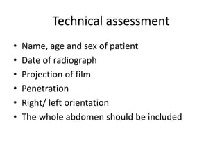
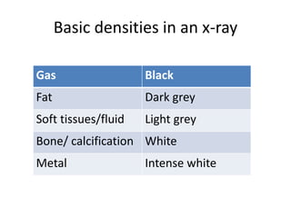
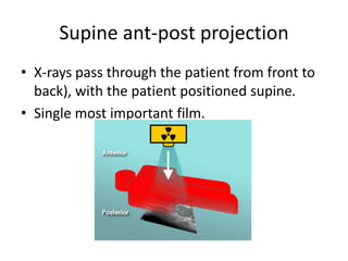
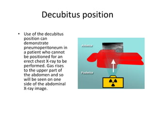
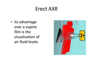
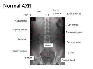
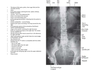
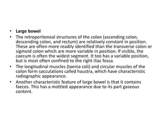
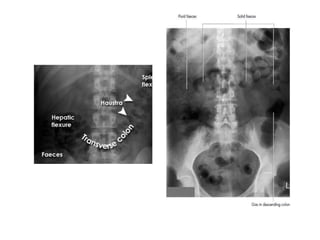
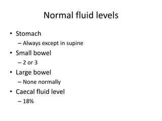
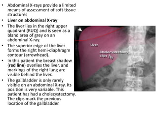
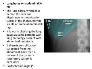
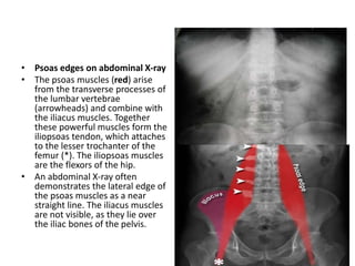
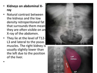
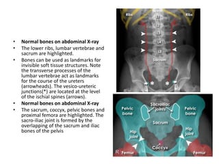
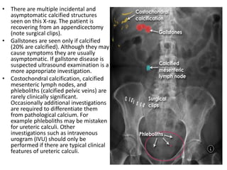
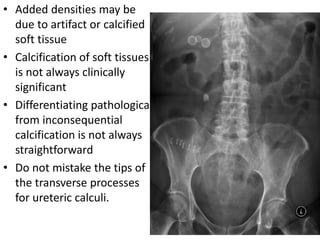
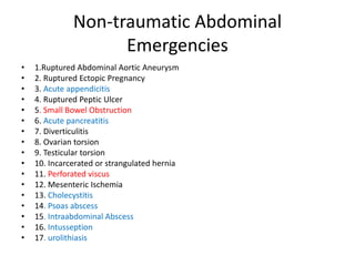
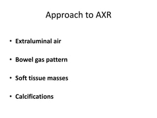
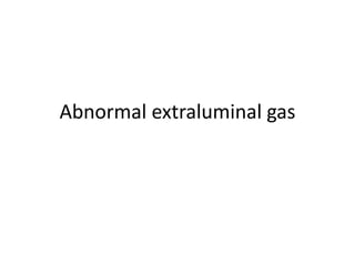
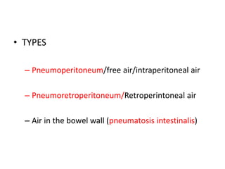
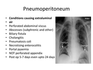
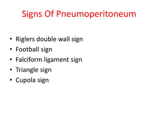
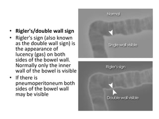
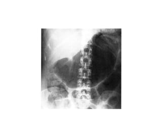
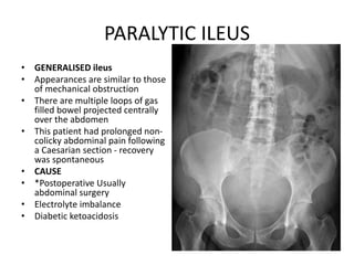
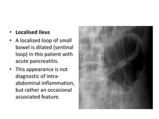
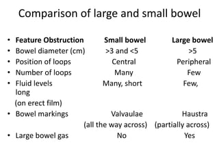
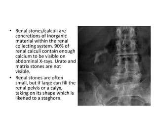
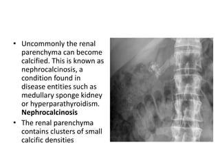
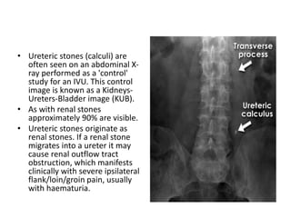
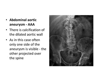
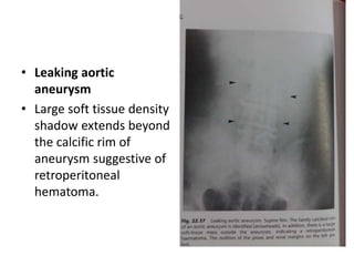
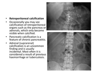
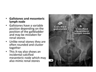
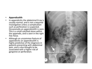
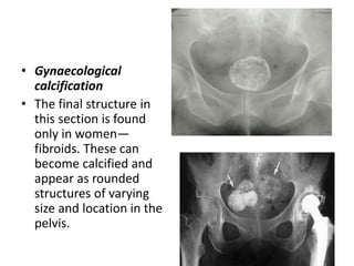
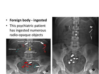
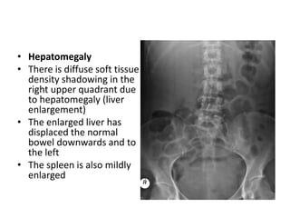
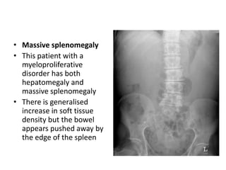
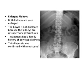
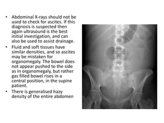
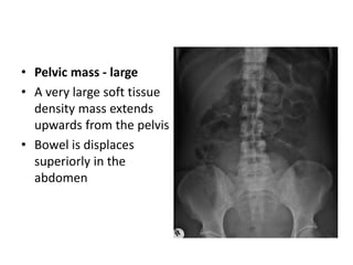
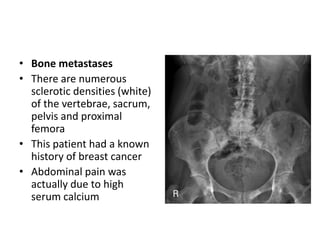
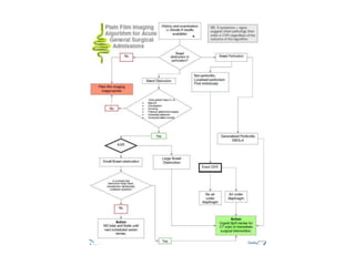
NORMAL
ABDOMINAL
RADIOGRAPH
&
PLAIN
RADIOGRAPHY IN
NON TRAUMATIC
EMERGENCY
• Postgraduate:
Dr Inayat Ellahi
Normal abdominal radiograph
• Plain abdominal radiographs are traditionally
the initial and most useful method in the
evaluation of acute abdomen
• Interpretation of plain films may lead to
– A specific diagnosis
– Non- specific findings
– Misleading conclusions
Technical assessment
• Name, age and sex of patient
• Date of radiograph
• Projection of film
• Penetration
• Right/ left orientation
• The whole abdomen should be included
Basic densities in an x-ray
Gas Black
Fat Dark grey
Soft tissues/fluid Light grey
Bone/ calcification White
Metal Intense white
Supine ant-post projection
• X-rays pass through the patient from front to
back), with the patient positioned supine.
• Single most important film.
Decubitus position
• Use of the decubitus
position can
demonstrate
pneumoperitoneum in
a patient who cannot
be positioned for an
erect chest X-ray to be
performed. Gas rises
to the upper part of
the abdomen and so
will be seen on one
side of the abdominal
X-ray image.
Erect AXR
• its advantage
over a supine
film is the
visualisation of
air-fluid levels
Viewing the Xray
• A conventional X-ray should only ever be seriously
inspected by uniform transmitted light coming through
it, i.e. a viewing box. There is no place for waving it
about in the wind when irregular illumination and
reflections will prevent 10–20% of the useful
information on it being visualized.
• Even when viewing digital images on a monitor, care
should be taken to minimize bright reflections off the
screen and keep the background illumination down.
Normal AXR
11th rib
Hepatic flexure
Gas in
stomach
T12
Gas in caecum
Iliac crest
Femoral head
SI joint
Gas in sigmoid
Transverse colon
Splenic flexure
Psoas margin
Sacrum
Left kidney
Liver
Bladder
• The bones of the spine, pelvis, chest cage (ribs) and the
sacroiliac and hip
• joints
• ● The dark margins outlining the liver, spleen, kidneys,
bladder and psoas
• muscles – this is intra-abdominal fat
• ● Gas in the body of the stomach
• ● Gas in the descending colon
• ● The wide gynaecoid pelvis, indicating that the patient is
female
• ● Pelvic phleboliths – normal finding
• ● Minor joint space narrowing in the hips (normal for this
age)
• ● The granular texture of the amorphous fluid faecal
matter containing pockets
• of gas in the caecum, overlying the right iliac bone
• ● The ‘R’ marked low down on the right side. The marker
can be anywhere on
• the film and you often have to search for it. All references
to ‘right’ and ‘left’
• refer to the patient’s right and left. Note the name badge
at the bottom, not the
• top, (blacked out)
• ● Check that the ‘R’ marker is compatible with the visible
anatomy, e.g.
• – liver on the right
• – left kidney higher than the right
• – stomach on the left
• – spleen on the left
• – heart on the left, when visible
• ● The dark skin fold going right across the upper abdomen
(normal).
• Psoas muscle
• Bowel gas pattern
• Any part of the bowel may be visible if it contains gas/air within the
lumen. Gas/air is of low density and forms a natural contrast against
surrounding denser soft tissues. It is often difficult to differentiate
between normal small and large bowel, but this often becomes
easier when the bowel is abnormally distended.
• The upper limit of normal diameter of the bowel is generally
accepted as 3cm for the small bowel, 6cm for the colon and 9cm for
the caecum (3/6/9 rule).
• Normal bowel sections are sometimes identified by the following
features.
• Stomach
• The stomach may be visible if it contains gas/air. It is not visible if it
is either completely empty, or fluid filled.
• Small bowel (duodenum to terminal ileum)
• Generally the small bowel lies centrally within the abdomen. The
valvulae conniventes (also called plicae circulares) are thin, circular,
folds of mucosa, some of which are circumferential and are seen on
an X-ray to pass across the full width of the lumen.
• Large bowel
• The retroperitoneal structures of the colon (ascending colon,
descending colon, and rectum) are relatively constant in position.
These are often more readily identified than the transverse colon or
sigmoid colon which are more variable in position. If visible, the
caecum is often the widest segment. It too has a variable position,
but is most often confined to the right iliac fossa.
• The longitudinal muscles (taenia coli) and circular muscles of the
colon form sacculations called haustra, which have characteristic
radiographic appearance.
• Another characteristic feature of large bowel is that it contains
faeces. This has a mottled appearance due to its part gaseous
content.
Normal fluid levels
• Stomach
– Always except in supine
• Small bowel
– 2 or 3
• Large bowel
– None normally
• Caecal fluid level
– 18%
• Abdominal X-rays provide a limited
means of assessment of soft tissue
structures
• Liver on abdominal X-ray
• The liver lies in the right upper
quadrant (RUQ) and is seen as a
bland area of grey on an
abdominal X-ray.
• The superior edge of the liver
forms the right hemi-diaphragm
contour (arrowhead).
• In this patient the breast shadow
(red line) overlies the liver, and
markings of the right lung are
visible behind the liver.
• The gallbladder is only rarely
visible on an abdominal X-ray. Its
position is very variable. This
patient has had a cholecystectomy.
The clips mark the previous
location of the gallbladder.
• Lung bases on abdominal X-
ray
• The lung bases, which pass
behind the liver and
diaphragm in the posterior
sulcus of the thorax, may be
visible on some abdominal X-
rays.
• It is worth checking the lung
bases as some patients with
lung pathology present with
abdominal symptoms.
• If there is consolidation
suspected from the
abdominal X-ray then a
review of the patient's
respiratory system is
necessary.
• Costophrenic angle (*)
• Psoas edges on abdominal X-ray
• The psoas muscles (red) arise
from the transverse processes of
the lumbar vertebrae
(arrowheads) and combine with
the iliacus muscles. Together
these powerful muscles form the
iliopsoas tendon, which attaches
to the lesser trochanter of the
femur (*). The iliopsoas muscles
are the flexors of the hip.
• An abdominal X-ray often
demonstrates the lateral edge of
the psoas muscles as a near
straight line. The iliacus muscles
are not visible, as they lie over
the iliac bones of the pelvis.
• Kidneys on abdominal X-
ray
• Natural contrast between
the kidneys and the low
density retroperitoneal fat
that surrounds them means
they are often visible on an
X-ray of the abdomen.
• They lie at the level of T12-
L3 and lateral to the psoas
muscles. The right kidney is
usually slightly lower than
the left due to the position
of the liver.
•
• Spleen on abdominal X-
ray
• The spleen lies in the left
upper quadrant
(LUQ)immediately
superior to the left
kidney.
• Bladder on abdominal
X-ray
• The bladder has
variable appearance
depending on how full
it is. It has the same
density as other soft
tissue structures, due to
its water content.
• Normal bones on abdominal X-ray
• The lower ribs, lumbar vertebrae and
sacrum are highlighted.
• Bones can be used as landmarks for
invisible soft tissue structures. Note
the transverse processes of the
lumbar vertebrae act as landmarks
for the course of the ureters
(arrowheads). The vesico-ureteric
junctions(*) are located at the level
of the ischial spines (arrows).
• Normal bones on abdominal X-ray
• The sacrum, coccyx, pelvic bones and
proximal femora are highlighted. The
sacro-iliac joint is formed by the
overlapping of the sacrum and iliac
bones of the pelvis
• There are multiple incidental and
asymptomatic calcified structures
seen on this X-ray. The patient is
recovering from an appendicectomy
(note surgical clips).
• Gallstones are seen only if calcified
(20% are calcified). Although they may
cause symptoms they are usually
asymptomatic. If gallstone disease is
suspected ultrasound examination is a
more appropriate investigation.
• Costochondral calcification, calcified
mesenteric lymph nodes, and
phleboliths (calcified pelvic veins) are
rarely clinically significant.
Occasionally additional investigations
are required to differentiate them
from pathological calcium. For
example phleboliths may be mistaken
for ureteric calculi. Other
investigations such as intravenous
urogram (IVU) should only be
performed if there are typical clinical
features of ureteric calculi.
• Added densities may be
due to artifact or calcified
soft tissue
• Calcification of soft tissues
is not always clinically
significant
• Differentiating pathological
from inconsequential
calcification is not always
straightforward
• Do not mistake the tips of
the transverse processes
for ureteric calculi.
Non-traumatic Abdominal
Emergencies
• 1.Ruptured Abdominal Aortic Aneurysm
• 2. Ruptured Ectopic Pregnancy
• 3. Acute appendicitis
• 4. Ruptured Peptic Ulcer
• 5. Small Bowel Obstruction
• 6. Acute pancreatitis
• 7. Diverticulitis
• 8. Ovarian torsion
• 9. Testicular torsion
• 10. Incarcerated or strangulated hernia
• 11. Perforated viscus
• 12. Mesenteric Ischemia
• 13. Cholecystitis
• 14. Psoas abscess
• 15. Intraabdominal Abscess
• 16. Intusseption
• 17. urolithiasis
Approach to AXR
• Extraluminal air
• Bowel gas pattern
• Soft tissue masses
• Calcifications
Abnormal extraluminal gas
Extraluminal air
• TYPES
– Pneumoperitoneum/free air/intraperitoneal air
– Pneumoretroperitoneum/Retroperintoneal air
– Air in the bowel wall (pneumatosis intestinalis)
Pneumoperitoneum
• Conditions causing extraluminal
• air
• Perforated abdominal viscus
• Abscesses (subphrenic and other)
• Biliary fistula
• Cholangitis
• Pneumatosis coli
• Necrotising enterocolitis
• Portal pyaemia
• NOT perforated appendix
• Post op 5-7 days even upto 24 days
Signs Of Pneumoperitoneum
• Riglers double wall sign
• Football sign
• Falciform ligament sign
• Triangle sign
• Cupola sign
• Rigler's/double wall sign
• Rigler's sign (also known
as the double wall sign) is
the appearance of
lucency (gas) on both
sides of the bowel wall.
Normally only the inner
wall of the bowel is visible
• If there is
pneumoperitoneum both
sides of the bowel wall
may be visible
• The falciform ligament sign (also
called the Silver's sign) is a sign
seen with pneumoperitoneum.
• It is almost never seen in
isolation. If there is enough free
air to outline the falciform
ligament, there is usually enough
air to also provide at least
a Rigler's sign.
• The falciform ligament connects
the anterior abdominal wall to
the liver. The ligament continues
to extend inferiorly beyond the
liver where it becomes the round
ligament (white arrow). Given
that the falciform ligament is
situated against the anterior
abdominal wall, it is not
surprising that it becomes
outlined with air in a supine
patient with free abdominal gas.
• Football sign - example
• 2 radiographs were
required to completely
cover the abdomen in
this large patient
• A large volume of free
gas has risen to the
front of the peritoneal
cavity resulting in a
large round black area -
'football sign'
• The cupola sign is seen at
supine radiography as an
arcuate lucency overlying the
lower thoracic spine and
projecting caudad to the heart.
The superior border is well
defined and the inferior margin
is poorly delineated. The term
cupola is used to indicate the
inverted cup shaped
configuration of the lucency.
Triangle Sign
• The triangle sign
refers to small
triangles of free gas
that can typically be
positioned between
the large bowel and
the flank
• Chilaiditi's phenomenon
• In patients who have
small livers (cirrhosis), or
flattened diaphragms due
to lung hyperexpansion
(emphysema), a void is
created within the upper
abdomen above the liver.
This space may be filled
by bowel. If this bowel is
air filled then it may
mimic free gas.
• Subphrenic abscess
• This is a localised
collection of free gas
and fluid, which usually
forms under the right
hemidiaphragm, above
the solid liver. This
gas collection usually
occurs above the 11th
rib
Retroperitoneal Air
• Recognised by:
– Streaky, linear appearance outlining retroperitoneal
structures
– Mottled, blotchy appearance
– Relatively fixed position
• May outline:
– Psoas muscles
– Kidneys, ureters, bladder
– Aorta or IVC
– Subphrenic spaces
Causes of retroperitoneal air
• Bowel perforation (appendix, ileum, colon)
• Trauma (blunt or penetrating)
• Iatrogenic
• Foreign body
• Gas producing infection
Pneumoretroperitoneum
• This patient has free air in
the retroperitoneal space.
The air is seen surrounding
the lateral border of the right
kidney (white arrow). There is
other evidence of free gas
including Rigler's sign.
• If you are not confident that
the appearance is
pneumoretroperitoneum,
you can try an erect and
decubitus view to see if the
gas moves. If the gas is seen
to move, it's not in the
retroperitoneum.
Pneumatosis intestinalis
• Intramural air, may be
in the form of cysts
(benign) or linear
streaks (bowel necrosis)
• Abnormal gas may be found
in abdominal organs. Gas in
the biliary tree and portal
veins is discussed earlier. Gas
may also be seen in the
kidneys (emphysematous
pyelonephritis),pancreas
(infected necrosis or abscess),
gallbladder wall and urinary
bladder
• Emphysematous
pyelonephritis. Patient with
diabetes mellitus and sepsis.
The left renal collecting
system and ureter are
distended and gas filled.
There are also multiple dense
gallstones in the gallbladder.
Emphysematous cholecystitis
Emphysematous cystitis
Abnormal intraluminal gas
• If a patient presents with clinical features of
obstruction then radiological assessment can
be very helpful in determining the level of
obstruction, and occasionally the cause.
GASTRIC DILATATION
• Causes
• Paralytic ileus (Acute Gastric Dilatation)
• Mechanical gastric outlet obstruction
• Gastric volvulus
• Air swallowing
• Intubation
PARALYTIC ILEUS
• GENERALISED ileus
• Appearances are similar to those
of mechanical obstruction
• There are multiple loops of gas
filled bowel projected centrally
over the abdomen
• This patient had prolonged non-
colicky abdominal pain following
a Caesarian section - recovery
was spontaneous
• CAUSE
• *Postoperative Usually
abdominal surgery
• Electrolyte imbalance
• Diabetic ketoacidosis
• Localised Ileus
• A localized loop of small
bowel is dilated (sentinal
loop) in this patient with
acute pancreatitis.
• This appearance is not
diagnostic of intra-
abdominal inflammation,
but rather an occasional
associated feature.
Causes of Localised Ileus
by location
SITE OF DILATED LOOPS CAUSE
Right upper quadrant Cholecystitis
Left upper quadrant Pancreatitis
Right lower quadrant Appendicitis
Left lower quadrant Diverticulitis
Mid-abdomen Ulcer or kidney/ureteric calculi
Colon cut off sign
Explanation:
Inflammatory exudate in acute
pancreatitis extends into the
phrenicocolic ligament via lateral
attachment of the transverse
mesocolon
Infiltration of the phrenicocolic
ligament results in functional spasm
and/or mechanical narrowing of the
splenic flexure at the level where the
colon returns to the
retroperitoneum.
Abrupt cutoff of colonic gas column at the splenic flexure (arrow). The colon is usually
decompressed beyond this point.
Large bowel obstruction
• Dilatation of the caecum >9cm is abnormal
• Dilatation of any other part of the colon >6cm
is abnormal
• Abdominal X-ray may demonstrate the level of
obstruction
• Abdominal X-ray cannot reliably differentiate
mechanical obstruction from pseudo-
obstruction
• Large bowel obstruction
• Here the colon is dilated
down to the level of the
distal descending colon.
There is the impression of
soft tissue density at the
level of obstruction (X). No
gas is seen within the
sigmoid colon.
• Obstruction is not absolute in
this patient as a small
volume of gas has reached
the rectum (arrow).
• An obstructing colon
carcinoma was confirmed on
CT and at surgery.
• Pseudo-obstruction
• Pseudo-obstruction is a poorly understood
functional abnormality of bowel, most often
occurring in the elderly population, in those with
underlying systemic medical conditions, or due to
certain drugs. The clinical features can be similar
to true obstruction, but no mechanical cause is
found. An abdominal X-ray cannot reliably
differentiate true mechanical obstruction from
pseudo-obstruction.
Small bowel obstruction
• In small bowel obstruction, dilated small bowel loops are seen
centrally on the radiograph.
• The valvulae conniventes should be visible across the whole width
of this dilated bowel.
• The dilated bowel diameter is greater than 3 cm but usually less
than 5 cm. There are likely to be several dilated bowel loops.
• The number of small bowel loops gives an indication of the level at
which the obstruction within the small bowel has occurred: the
higher the obstruction, the fewer the number of loops seen.
• No gas should be seen within the large bowel.
• An erect film tends to show multiple small Fluid levels, a
“stepladder” appearance.
String of pearls sign
Considered diagnostic of obstruction (as opposed to ileus)
and is caused by small bubbles of air trapped in the
valvulae of the small bowel.
Coil spring sign
Comparison of large and small bowel
• Feature Obstruction Small bowel Large bowel
• Bowel diameter (cm) >3 and <5 >5
• Position of loops Central Peripheral
• Number of loops Many Few
• Fluid levels Many, short Few,
long
(on erect film)
• Bowel markings Valvaulae Haustra
(all the way across) (partially across)
• Large bowel gas No Yes
Mechanical SBO Adynamic SBO
Volvulus
• Twisting of the bowel, or volvulus, is a specific
cause of bowel obstruction which can have
characteristic appearances on an abdominal X-
ray.
• The two commonest types of bowel twisting
are sigmoid volvulus and caecal volvulus.
• Sigmoid volvulus - coffee
bean sign
• Sigmoid volvulus classically
results in the formation of a
loop of sigmoid colon,
which is twisted at the root
of the sigmoid mesentery,
which lies in the left iliac
fossa (LIF). The loop of
dilated bowel usually points
upwards towards the
diaphragm.
• This image demonstrates
dilatation of the twisted
sigmoid loop 'coffee bean'
and of the proximal large
bowel (*). This patient is at
high risk of perforation
and/or bowel ischaemia.
Coffee Bean Sign
Sigmoid volvulus
Massively
dilated
sigmoid loop
• Caecal volvulus
• The caecum is most
frequently a retroperitoneal
structure, and therefore not
susceptible to twisting.
However, in up to 11% of
individuals there is
congenital incomplete
peritoneal covering of the
caecum with formation of a
'mobile' caecum on a
mesentery, such that it no
longer lies in the right iliac
fossa. This is a normal
variant but is associated with
increased incidence of
folding or twisting of the
caecum (caecal volvulus),
which may be complicated
by obstruction, vascular
compromise, or perforation.
Gastric volvulus
• Spherical viscus
• Displaced upwards & to
the left
• Raised left
hemidiaphargm
• Contains both air and
fluid
• Usually no gas in bowel
beyond stomach
Gastric volvulus Caecal volvulus
Gallstone ileus
• 2% of SBO
• Specific radiological signs in 40%
• High operative mortality
• Features
– Branching pattern of gas n biliary tree
– Gas in portal vein (peripheral)
– Obstructing gallstone in ileum
• Obscured by sacrum/overlying dilated gut
Biliary tree air Portal venous air
Intussussception
• Plain films tray show evidence of
small-bowel obstruction, or the
intussusception itself may be
identified as a soft-tissue mass
someftimes surrounded by a crescent
of gas and most frequently i dentified
in the right hypochondrium.
• More recently the `target sign' has
been described, comprising two
concentric circles of fat density lying
to the right of the spine-often
superimposed on the kidney. It is
probably due to the layers of
peritoneal fat surrounding and within
the intussusceptum alternating with
the layers of mucosa and muscle but
seen `end on' as it passes forward
from the right paraspinal gutter in
the transverse colon.
Intraperitoneal fliud
• Abdominal X-rays should not be
used to check for ascites. If this
diagnosis is suspected then again
ultrasound is the best initial
investigation, and can also be
used to assist drainage.
• Fluid and soft tissues have similar
densities, and so ascites may be
mistaken for organomegaly. The
bowel does not appear pushed to
the side as in organomegaly, but
rather gas filled bowel rises in a
central position, in the supine
patient.
• There is generalized hazy
densityof entiree abdomen
• Bowel wall inflammation
• Occasionally, abdominal X-rays show signs of
inflammation in patients with inflammatory
bowel disease. Abnormalities may relate to
either acute or chronic stages of disease.
• Mucosal thickening -
'thumbprinting'
• This patient presented with
an exacerbation of
symptoms of ulcerative
colitis .
• The distance between loops
of bowel is increased
(arrows) due to thickening
of the bowel wall. The
haustral folds are very thick
(arrowheads), leading to a
sign known as
'thumbprinting.'
• Lead pipe colon
• This patient with ulcerative
colitis has a featureless
segment of transverse colon
with shows loss of the normal
haustral markings.
• This 'lead pipe' appearance is
associated with longstanding
ulcerative colitis.
• The distal bowel is always
involved in this disease but, as
there is no air in the
descending colon, this
segment of colon is not
evidently abnormal.
• Toxic megacolon is a
potentially life-threatening
condition characterized by
dilatation of the large bowel
without obstruction, in the
context of acute bowel
inflammation. This may be
due to inflammatory bowel
disease, especially
ulcerative colitis, or other
causes of colitis such as
infection.
• Meteorism (excessive
swallowed air) is
particularly common in
crying children and
hyperventilating adults.
Although there are
prominent bowel loops,
there is no cut off point:
the bowel has
beenlikened to crazy
paving.
Abnormal calcification
• Renal stones/calculi are
concretions of inorganic
material within the renal
collecting system. 90% of
renal calculi contain enough
calcium to be visible on
abdominal X-rays. Urate and
matrix stones are not
visible.
• Renal stones are often
small, but if large can fill the
renal pelvis or a calyx,
taking on its shape which is
likened to a staghorn.
• Uncommonly the renal
parenchyma can become
calcified. This is known as
nephrocalcinosis, a
condition found in
disease entities such as
medullary sponge kidney
or hyperparathyroidism.
Nephrocalcinosis
• The renal parenchyma
contains clusters of small
calcific densities
• Ureteric stones (calculi) are
often seen on an abdominal X-
ray performed as a 'control'
study for an IVU. This control
image is known as a Kidneys-
Ureters-Bladder image (KUB).
• As with renal stones
approximately 90% are visible.
• Ureteric stones originate as
renal stones. If a renal stone
migrates into a ureter it may
cause renal outflow tract
obstruction, which manifests
clinically with severe ipsilateral
flank/loin/groin pain, usually
with haematuria.
• Bladder stones generally form
in the bladder itself. They arise
as a result of urinary stasis
such as in bladder outflow
obstruction (enlarged
prostate) or in patients with a
neurogenic bladder (loss of
bladder function due to spinal
cord injury/disease). Those
with bladder wall
abnormalities (ureterocele,
diverticulum) or those with
recurrent urinary infections
are also at higher risk of
forming bladder stones.
• When seen on an
abdominal/pelvic X-ray they
are often multiple and
rounded.
• Calcification of arteries
seen on x-rays is a sign of
more generalised
atherosclerosis.
• Occasionally vascular
calcification seen on an
abdominal X-ray reveals
an unexpected aneurysm.
Remember that
abdominal pain is not
only caused by
gastrointestinal disease.
Chinese Dragon Sign
Calcified splenic artery
• Abdominal aortic
aneurysm - AAA
• There is calcification of
the dilated aortic wall
• As in this case often
only one side of the
aneurysm is visible - the
other projected over
the spine
• Leaking aortic
aneurysm
• Large soft tissue density
shadow extends beyond
the calcific rim of
aneurysm suggestive of
retroperitoneal
hematoma.
• Retroperitoneal calcification
• Occasionally you may see
calcification of retroperitoneal
organs such as the pancreas or
adrenals, which only become
visible when calcified.
• Pancreatic calcification is a
feature of chronic pancreatitis.
• Adrenal (suprarenal)
calcification is an uncommon
finding and is usually
incidental. Most often it is
considered a result of previous
haemorrhage or tuberculosis.
• Gallstones and mesenteric
lymph node
• Gallstones have a variable
position depending on the
position of the gallbladder
and may be mistaken for
renal stones
• Unlike renal stones they are
often rounded and cluster
together
• This X-ray also shows an
incidental calcified
mesenteric node which may
also mimic renal stones
Radiological signs in acute appendicitis
• Appendicolith
• Sentinal loop
• Dilated caecum
• Right lower quadrent haze
• RIF mass indenting caecum
• Blurring of right psoas outline
• Intestinal obstruction
• Appendicolith
• In appendicitis the abdominal X-ray is
usually normal, and is not a required
investigation unless a complication
such as perforation is suspected.
Occasionally an appendicolith is seen.
This is a small calcified stone within
the appendix, and is seen in the right
iliac fossa.
• Although an uncommon feature of
appendicitis an appendicolith is
highly predictive of the diagnosis in
patients presenting with abdominal
pain, and is also thought to be
associated with a higher risk of
gangrene or perforation.
• Gynaecological
calcification
• The final structure in
this section is found
only in women—
fibroids. These can
become calcified and
appear as rounded
structures of varying
size and location in the
pelvis.
• Foreign body - ingested
• This psychiatric patient
has ingested numerous
radio-opaque objects
Abnormal soft tissues
• Hepatomegaly
• There is diffuse soft tissue
density shadowing in the
right upper quadrant due
to hepatomegaly (liver
enlargement)
• The enlarged liver has
displaced the normal
bowel downwards and to
the left
• The spleen is also mildly
enlarged
• Massive splenomegaly
• This patient with a
myeloproliferative
disorder has both
hepatomegaly and
massive splenomegaly
• There is generalised
increase in soft tissue
density but the bowel
appears pushed away by
the edge of the spleen
• Enlarged kidneys
• Both kidneys are very
enlarged
• The bowel is not displaced
because the kidneys are
retroperitoneal structures
• This patient had a family
history of polycystic kidneys
• This diagnosis was
confirmed with ultrasound
• Abdominal X-rays should not be
used to check for ascites. If this
diagnosis is suspected then
again ultrasound is the best
initial investigation, and can
also be used to assist drainage.
• Fluid and soft tissues have
similar densities, and so ascites
may be mistaken for
organomegaly. The bowel does
not appear pushed to the side
as in organomegaly, but rather
gas filled bowel rises in a
central position, in the supine
patient.
• There is generalised hazy
density of the entire abdomen
• Pelvic mass - large
• A very large soft tissue
density mass extends
upwards from the pelvis
• Bowel is displaces
superiorly in the
abdomen
• Bone metastases
• There are numerous
sclerotic densities (white)
of the vertebrae, sacrum,
pelvis and proximal
femora
• This patient had a known
history of breast cancer
• Abdominal pain was
actually due to high
serum calcium
THANK YOU

Plain radiography in acute abdomen

  • 1.
  • 2.
  • 3.
    • Plain abdominalradiographs are traditionally the initial and most useful method in the evaluation of acute abdomen • Interpretation of plain films may lead to – A specific diagnosis – Non- specific findings – Misleading conclusions
  • 4.
    Technical assessment • Name,age and sex of patient • Date of radiograph • Projection of film • Penetration • Right/ left orientation • The whole abdomen should be included
  • 5.
    Basic densities inan x-ray Gas Black Fat Dark grey Soft tissues/fluid Light grey Bone/ calcification White Metal Intense white
  • 6.
    Supine ant-post projection •X-rays pass through the patient from front to back), with the patient positioned supine. • Single most important film.
  • 7.
    Decubitus position • Useof the decubitus position can demonstrate pneumoperitoneum in a patient who cannot be positioned for an erect chest X-ray to be performed. Gas rises to the upper part of the abdomen and so will be seen on one side of the abdominal X-ray image.
  • 8.
    Erect AXR • itsadvantage over a supine film is the visualisation of air-fluid levels
  • 9.
    Viewing the Xray •A conventional X-ray should only ever be seriously inspected by uniform transmitted light coming through it, i.e. a viewing box. There is no place for waving it about in the wind when irregular illumination and reflections will prevent 10–20% of the useful information on it being visualized. • Even when viewing digital images on a monitor, care should be taken to minimize bright reflections off the screen and keep the background illumination down.
  • 10.
    Normal AXR 11th rib Hepaticflexure Gas in stomach T12 Gas in caecum Iliac crest Femoral head SI joint Gas in sigmoid Transverse colon Splenic flexure Psoas margin Sacrum Left kidney Liver Bladder
  • 11.
    • The bonesof the spine, pelvis, chest cage (ribs) and the sacroiliac and hip • joints • ● The dark margins outlining the liver, spleen, kidneys, bladder and psoas • muscles – this is intra-abdominal fat • ● Gas in the body of the stomach • ● Gas in the descending colon • ● The wide gynaecoid pelvis, indicating that the patient is female • ● Pelvic phleboliths – normal finding • ● Minor joint space narrowing in the hips (normal for this age) • ● The granular texture of the amorphous fluid faecal matter containing pockets • of gas in the caecum, overlying the right iliac bone • ● The ‘R’ marked low down on the right side. The marker can be anywhere on • the film and you often have to search for it. All references to ‘right’ and ‘left’ • refer to the patient’s right and left. Note the name badge at the bottom, not the • top, (blacked out) • ● Check that the ‘R’ marker is compatible with the visible anatomy, e.g. • – liver on the right • – left kidney higher than the right • – stomach on the left • – spleen on the left • – heart on the left, when visible • ● The dark skin fold going right across the upper abdomen (normal). • Psoas muscle
  • 12.
    • Bowel gaspattern • Any part of the bowel may be visible if it contains gas/air within the lumen. Gas/air is of low density and forms a natural contrast against surrounding denser soft tissues. It is often difficult to differentiate between normal small and large bowel, but this often becomes easier when the bowel is abnormally distended. • The upper limit of normal diameter of the bowel is generally accepted as 3cm for the small bowel, 6cm for the colon and 9cm for the caecum (3/6/9 rule). • Normal bowel sections are sometimes identified by the following features. • Stomach • The stomach may be visible if it contains gas/air. It is not visible if it is either completely empty, or fluid filled. • Small bowel (duodenum to terminal ileum) • Generally the small bowel lies centrally within the abdomen. The valvulae conniventes (also called plicae circulares) are thin, circular, folds of mucosa, some of which are circumferential and are seen on an X-ray to pass across the full width of the lumen.
  • 14.
    • Large bowel •The retroperitoneal structures of the colon (ascending colon, descending colon, and rectum) are relatively constant in position. These are often more readily identified than the transverse colon or sigmoid colon which are more variable in position. If visible, the caecum is often the widest segment. It too has a variable position, but is most often confined to the right iliac fossa. • The longitudinal muscles (taenia coli) and circular muscles of the colon form sacculations called haustra, which have characteristic radiographic appearance. • Another characteristic feature of large bowel is that it contains faeces. This has a mottled appearance due to its part gaseous content.
  • 16.
    Normal fluid levels •Stomach – Always except in supine • Small bowel – 2 or 3 • Large bowel – None normally • Caecal fluid level – 18%
  • 17.
    • Abdominal X-raysprovide a limited means of assessment of soft tissue structures • Liver on abdominal X-ray • The liver lies in the right upper quadrant (RUQ) and is seen as a bland area of grey on an abdominal X-ray. • The superior edge of the liver forms the right hemi-diaphragm contour (arrowhead). • In this patient the breast shadow (red line) overlies the liver, and markings of the right lung are visible behind the liver. • The gallbladder is only rarely visible on an abdominal X-ray. Its position is very variable. This patient has had a cholecystectomy. The clips mark the previous location of the gallbladder.
  • 18.
    • Lung baseson abdominal X- ray • The lung bases, which pass behind the liver and diaphragm in the posterior sulcus of the thorax, may be visible on some abdominal X- rays. • It is worth checking the lung bases as some patients with lung pathology present with abdominal symptoms. • If there is consolidation suspected from the abdominal X-ray then a review of the patient's respiratory system is necessary. • Costophrenic angle (*)
  • 19.
    • Psoas edgeson abdominal X-ray • The psoas muscles (red) arise from the transverse processes of the lumbar vertebrae (arrowheads) and combine with the iliacus muscles. Together these powerful muscles form the iliopsoas tendon, which attaches to the lesser trochanter of the femur (*). The iliopsoas muscles are the flexors of the hip. • An abdominal X-ray often demonstrates the lateral edge of the psoas muscles as a near straight line. The iliacus muscles are not visible, as they lie over the iliac bones of the pelvis.
  • 20.
    • Kidneys onabdominal X- ray • Natural contrast between the kidneys and the low density retroperitoneal fat that surrounds them means they are often visible on an X-ray of the abdomen. • They lie at the level of T12- L3 and lateral to the psoas muscles. The right kidney is usually slightly lower than the left due to the position of the liver. •
  • 21.
    • Spleen onabdominal X- ray • The spleen lies in the left upper quadrant (LUQ)immediately superior to the left kidney.
  • 22.
    • Bladder onabdominal X-ray • The bladder has variable appearance depending on how full it is. It has the same density as other soft tissue structures, due to its water content.
  • 23.
    • Normal boneson abdominal X-ray • The lower ribs, lumbar vertebrae and sacrum are highlighted. • Bones can be used as landmarks for invisible soft tissue structures. Note the transverse processes of the lumbar vertebrae act as landmarks for the course of the ureters (arrowheads). The vesico-ureteric junctions(*) are located at the level of the ischial spines (arrows). • Normal bones on abdominal X-ray • The sacrum, coccyx, pelvic bones and proximal femora are highlighted. The sacro-iliac joint is formed by the overlapping of the sacrum and iliac bones of the pelvis
  • 24.
    • There aremultiple incidental and asymptomatic calcified structures seen on this X-ray. The patient is recovering from an appendicectomy (note surgical clips). • Gallstones are seen only if calcified (20% are calcified). Although they may cause symptoms they are usually asymptomatic. If gallstone disease is suspected ultrasound examination is a more appropriate investigation. • Costochondral calcification, calcified mesenteric lymph nodes, and phleboliths (calcified pelvic veins) are rarely clinically significant. Occasionally additional investigations are required to differentiate them from pathological calcium. For example phleboliths may be mistaken for ureteric calculi. Other investigations such as intravenous urogram (IVU) should only be performed if there are typical clinical features of ureteric calculi.
  • 25.
    • Added densitiesmay be due to artifact or calcified soft tissue • Calcification of soft tissues is not always clinically significant • Differentiating pathological from inconsequential calcification is not always straightforward • Do not mistake the tips of the transverse processes for ureteric calculi.
  • 26.
    Non-traumatic Abdominal Emergencies • 1.RupturedAbdominal Aortic Aneurysm • 2. Ruptured Ectopic Pregnancy • 3. Acute appendicitis • 4. Ruptured Peptic Ulcer • 5. Small Bowel Obstruction • 6. Acute pancreatitis • 7. Diverticulitis • 8. Ovarian torsion • 9. Testicular torsion • 10. Incarcerated or strangulated hernia • 11. Perforated viscus • 12. Mesenteric Ischemia • 13. Cholecystitis • 14. Psoas abscess • 15. Intraabdominal Abscess • 16. Intusseption • 17. urolithiasis
  • 27.
    Approach to AXR •Extraluminal air • Bowel gas pattern • Soft tissue masses • Calcifications
  • 28.
  • 29.
    Extraluminal air • TYPES –Pneumoperitoneum/free air/intraperitoneal air – Pneumoretroperitoneum/Retroperintoneal air – Air in the bowel wall (pneumatosis intestinalis)
  • 30.
    Pneumoperitoneum • Conditions causingextraluminal • air • Perforated abdominal viscus • Abscesses (subphrenic and other) • Biliary fistula • Cholangitis • Pneumatosis coli • Necrotising enterocolitis • Portal pyaemia • NOT perforated appendix • Post op 5-7 days even upto 24 days
  • 31.
    Signs Of Pneumoperitoneum •Riglers double wall sign • Football sign • Falciform ligament sign • Triangle sign • Cupola sign
  • 32.
    • Rigler's/double wallsign • Rigler's sign (also known as the double wall sign) is the appearance of lucency (gas) on both sides of the bowel wall. Normally only the inner wall of the bowel is visible • If there is pneumoperitoneum both sides of the bowel wall may be visible
  • 34.
    • The falciformligament sign (also called the Silver's sign) is a sign seen with pneumoperitoneum. • It is almost never seen in isolation. If there is enough free air to outline the falciform ligament, there is usually enough air to also provide at least a Rigler's sign. • The falciform ligament connects the anterior abdominal wall to the liver. The ligament continues to extend inferiorly beyond the liver where it becomes the round ligament (white arrow). Given that the falciform ligament is situated against the anterior abdominal wall, it is not surprising that it becomes outlined with air in a supine patient with free abdominal gas.
  • 35.
    • Football sign- example • 2 radiographs were required to completely cover the abdomen in this large patient • A large volume of free gas has risen to the front of the peritoneal cavity resulting in a large round black area - 'football sign'
  • 36.
    • The cupolasign is seen at supine radiography as an arcuate lucency overlying the lower thoracic spine and projecting caudad to the heart. The superior border is well defined and the inferior margin is poorly delineated. The term cupola is used to indicate the inverted cup shaped configuration of the lucency.
  • 37.
    Triangle Sign • Thetriangle sign refers to small triangles of free gas that can typically be positioned between the large bowel and the flank
  • 38.
    • Chilaiditi's phenomenon •In patients who have small livers (cirrhosis), or flattened diaphragms due to lung hyperexpansion (emphysema), a void is created within the upper abdomen above the liver. This space may be filled by bowel. If this bowel is air filled then it may mimic free gas.
  • 39.
    • Subphrenic abscess •This is a localised collection of free gas and fluid, which usually forms under the right hemidiaphragm, above the solid liver. This gas collection usually occurs above the 11th rib
  • 40.
    Retroperitoneal Air • Recognisedby: – Streaky, linear appearance outlining retroperitoneal structures – Mottled, blotchy appearance – Relatively fixed position • May outline: – Psoas muscles – Kidneys, ureters, bladder – Aorta or IVC – Subphrenic spaces
  • 41.
    Causes of retroperitonealair • Bowel perforation (appendix, ileum, colon) • Trauma (blunt or penetrating) • Iatrogenic • Foreign body • Gas producing infection
  • 42.
    Pneumoretroperitoneum • This patienthas free air in the retroperitoneal space. The air is seen surrounding the lateral border of the right kidney (white arrow). There is other evidence of free gas including Rigler's sign. • If you are not confident that the appearance is pneumoretroperitoneum, you can try an erect and decubitus view to see if the gas moves. If the gas is seen to move, it's not in the retroperitoneum.
  • 43.
    Pneumatosis intestinalis • Intramuralair, may be in the form of cysts (benign) or linear streaks (bowel necrosis)
  • 44.
    • Abnormal gasmay be found in abdominal organs. Gas in the biliary tree and portal veins is discussed earlier. Gas may also be seen in the kidneys (emphysematous pyelonephritis),pancreas (infected necrosis or abscess), gallbladder wall and urinary bladder • Emphysematous pyelonephritis. Patient with diabetes mellitus and sepsis. The left renal collecting system and ureter are distended and gas filled. There are also multiple dense gallstones in the gallbladder.
  • 45.
  • 46.
  • 47.
  • 48.
    • If apatient presents with clinical features of obstruction then radiological assessment can be very helpful in determining the level of obstruction, and occasionally the cause.
  • 49.
    GASTRIC DILATATION • Causes •Paralytic ileus (Acute Gastric Dilatation) • Mechanical gastric outlet obstruction • Gastric volvulus • Air swallowing • Intubation
  • 51.
    PARALYTIC ILEUS • GENERALISEDileus • Appearances are similar to those of mechanical obstruction • There are multiple loops of gas filled bowel projected centrally over the abdomen • This patient had prolonged non- colicky abdominal pain following a Caesarian section - recovery was spontaneous • CAUSE • *Postoperative Usually abdominal surgery • Electrolyte imbalance • Diabetic ketoacidosis
  • 52.
    • Localised Ileus •A localized loop of small bowel is dilated (sentinal loop) in this patient with acute pancreatitis. • This appearance is not diagnostic of intra- abdominal inflammation, but rather an occasional associated feature.
  • 53.
    Causes of LocalisedIleus by location SITE OF DILATED LOOPS CAUSE Right upper quadrant Cholecystitis Left upper quadrant Pancreatitis Right lower quadrant Appendicitis Left lower quadrant Diverticulitis Mid-abdomen Ulcer or kidney/ureteric calculi
  • 54.
    Colon cut offsign Explanation: Inflammatory exudate in acute pancreatitis extends into the phrenicocolic ligament via lateral attachment of the transverse mesocolon Infiltration of the phrenicocolic ligament results in functional spasm and/or mechanical narrowing of the splenic flexure at the level where the colon returns to the retroperitoneum. Abrupt cutoff of colonic gas column at the splenic flexure (arrow). The colon is usually decompressed beyond this point.
  • 55.
    Large bowel obstruction •Dilatation of the caecum >9cm is abnormal • Dilatation of any other part of the colon >6cm is abnormal • Abdominal X-ray may demonstrate the level of obstruction • Abdominal X-ray cannot reliably differentiate mechanical obstruction from pseudo- obstruction
  • 56.
    • Large bowelobstruction • Here the colon is dilated down to the level of the distal descending colon. There is the impression of soft tissue density at the level of obstruction (X). No gas is seen within the sigmoid colon. • Obstruction is not absolute in this patient as a small volume of gas has reached the rectum (arrow). • An obstructing colon carcinoma was confirmed on CT and at surgery.
  • 57.
    • Pseudo-obstruction • Pseudo-obstructionis a poorly understood functional abnormality of bowel, most often occurring in the elderly population, in those with underlying systemic medical conditions, or due to certain drugs. The clinical features can be similar to true obstruction, but no mechanical cause is found. An abdominal X-ray cannot reliably differentiate true mechanical obstruction from pseudo-obstruction.
  • 58.
    Small bowel obstruction •In small bowel obstruction, dilated small bowel loops are seen centrally on the radiograph. • The valvulae conniventes should be visible across the whole width of this dilated bowel. • The dilated bowel diameter is greater than 3 cm but usually less than 5 cm. There are likely to be several dilated bowel loops. • The number of small bowel loops gives an indication of the level at which the obstruction within the small bowel has occurred: the higher the obstruction, the fewer the number of loops seen. • No gas should be seen within the large bowel. • An erect film tends to show multiple small Fluid levels, a “stepladder” appearance.
  • 60.
    String of pearlssign Considered diagnostic of obstruction (as opposed to ileus) and is caused by small bubbles of air trapped in the valvulae of the small bowel.
  • 61.
  • 62.
    Comparison of largeand small bowel • Feature Obstruction Small bowel Large bowel • Bowel diameter (cm) >3 and <5 >5 • Position of loops Central Peripheral • Number of loops Many Few • Fluid levels Many, short Few, long (on erect film) • Bowel markings Valvaulae Haustra (all the way across) (partially across) • Large bowel gas No Yes
  • 63.
  • 64.
    Volvulus • Twisting ofthe bowel, or volvulus, is a specific cause of bowel obstruction which can have characteristic appearances on an abdominal X- ray. • The two commonest types of bowel twisting are sigmoid volvulus and caecal volvulus.
  • 65.
    • Sigmoid volvulus- coffee bean sign • Sigmoid volvulus classically results in the formation of a loop of sigmoid colon, which is twisted at the root of the sigmoid mesentery, which lies in the left iliac fossa (LIF). The loop of dilated bowel usually points upwards towards the diaphragm. • This image demonstrates dilatation of the twisted sigmoid loop 'coffee bean' and of the proximal large bowel (*). This patient is at high risk of perforation and/or bowel ischaemia.
  • 66.
    Coffee Bean Sign Sigmoidvolvulus Massively dilated sigmoid loop
  • 67.
    • Caecal volvulus •The caecum is most frequently a retroperitoneal structure, and therefore not susceptible to twisting. However, in up to 11% of individuals there is congenital incomplete peritoneal covering of the caecum with formation of a 'mobile' caecum on a mesentery, such that it no longer lies in the right iliac fossa. This is a normal variant but is associated with increased incidence of folding or twisting of the caecum (caecal volvulus), which may be complicated by obstruction, vascular compromise, or perforation.
  • 68.
    Gastric volvulus • Sphericalviscus • Displaced upwards & to the left • Raised left hemidiaphargm • Contains both air and fluid • Usually no gas in bowel beyond stomach
  • 69.
  • 70.
    Gallstone ileus • 2%of SBO • Specific radiological signs in 40% • High operative mortality • Features – Branching pattern of gas n biliary tree – Gas in portal vein (peripheral) – Obstructing gallstone in ileum • Obscured by sacrum/overlying dilated gut
  • 72.
    Biliary tree airPortal venous air
  • 73.
    Intussussception • Plain filmstray show evidence of small-bowel obstruction, or the intussusception itself may be identified as a soft-tissue mass someftimes surrounded by a crescent of gas and most frequently i dentified in the right hypochondrium. • More recently the `target sign' has been described, comprising two concentric circles of fat density lying to the right of the spine-often superimposed on the kidney. It is probably due to the layers of peritoneal fat surrounding and within the intussusceptum alternating with the layers of mucosa and muscle but seen `end on' as it passes forward from the right paraspinal gutter in the transverse colon.
  • 74.
    Intraperitoneal fliud • AbdominalX-rays should not be used to check for ascites. If this diagnosis is suspected then again ultrasound is the best initial investigation, and can also be used to assist drainage. • Fluid and soft tissues have similar densities, and so ascites may be mistaken for organomegaly. The bowel does not appear pushed to the side as in organomegaly, but rather gas filled bowel rises in a central position, in the supine patient. • There is generalized hazy densityof entiree abdomen
  • 75.
    • Bowel wallinflammation • Occasionally, abdominal X-rays show signs of inflammation in patients with inflammatory bowel disease. Abnormalities may relate to either acute or chronic stages of disease.
  • 76.
    • Mucosal thickening- 'thumbprinting' • This patient presented with an exacerbation of symptoms of ulcerative colitis . • The distance between loops of bowel is increased (arrows) due to thickening of the bowel wall. The haustral folds are very thick (arrowheads), leading to a sign known as 'thumbprinting.'
  • 78.
    • Lead pipecolon • This patient with ulcerative colitis has a featureless segment of transverse colon with shows loss of the normal haustral markings. • This 'lead pipe' appearance is associated with longstanding ulcerative colitis. • The distal bowel is always involved in this disease but, as there is no air in the descending colon, this segment of colon is not evidently abnormal.
  • 79.
    • Toxic megacolonis a potentially life-threatening condition characterized by dilatation of the large bowel without obstruction, in the context of acute bowel inflammation. This may be due to inflammatory bowel disease, especially ulcerative colitis, or other causes of colitis such as infection.
  • 80.
    • Meteorism (excessive swallowedair) is particularly common in crying children and hyperventilating adults. Although there are prominent bowel loops, there is no cut off point: the bowel has beenlikened to crazy paving.
  • 81.
  • 82.
    • Renal stones/calculiare concretions of inorganic material within the renal collecting system. 90% of renal calculi contain enough calcium to be visible on abdominal X-rays. Urate and matrix stones are not visible. • Renal stones are often small, but if large can fill the renal pelvis or a calyx, taking on its shape which is likened to a staghorn.
  • 83.
    • Uncommonly therenal parenchyma can become calcified. This is known as nephrocalcinosis, a condition found in disease entities such as medullary sponge kidney or hyperparathyroidism. Nephrocalcinosis • The renal parenchyma contains clusters of small calcific densities
  • 84.
    • Ureteric stones(calculi) are often seen on an abdominal X- ray performed as a 'control' study for an IVU. This control image is known as a Kidneys- Ureters-Bladder image (KUB). • As with renal stones approximately 90% are visible. • Ureteric stones originate as renal stones. If a renal stone migrates into a ureter it may cause renal outflow tract obstruction, which manifests clinically with severe ipsilateral flank/loin/groin pain, usually with haematuria.
  • 86.
    • Bladder stonesgenerally form in the bladder itself. They arise as a result of urinary stasis such as in bladder outflow obstruction (enlarged prostate) or in patients with a neurogenic bladder (loss of bladder function due to spinal cord injury/disease). Those with bladder wall abnormalities (ureterocele, diverticulum) or those with recurrent urinary infections are also at higher risk of forming bladder stones. • When seen on an abdominal/pelvic X-ray they are often multiple and rounded.
  • 87.
    • Calcification ofarteries seen on x-rays is a sign of more generalised atherosclerosis. • Occasionally vascular calcification seen on an abdominal X-ray reveals an unexpected aneurysm. Remember that abdominal pain is not only caused by gastrointestinal disease.
  • 88.
  • 89.
    • Abdominal aortic aneurysm- AAA • There is calcification of the dilated aortic wall • As in this case often only one side of the aneurysm is visible - the other projected over the spine
  • 90.
    • Leaking aortic aneurysm •Large soft tissue density shadow extends beyond the calcific rim of aneurysm suggestive of retroperitoneal hematoma.
  • 91.
    • Retroperitoneal calcification •Occasionally you may see calcification of retroperitoneal organs such as the pancreas or adrenals, which only become visible when calcified. • Pancreatic calcification is a feature of chronic pancreatitis. • Adrenal (suprarenal) calcification is an uncommon finding and is usually incidental. Most often it is considered a result of previous haemorrhage or tuberculosis.
  • 92.
    • Gallstones andmesenteric lymph node • Gallstones have a variable position depending on the position of the gallbladder and may be mistaken for renal stones • Unlike renal stones they are often rounded and cluster together • This X-ray also shows an incidental calcified mesenteric node which may also mimic renal stones
  • 93.
    Radiological signs inacute appendicitis • Appendicolith • Sentinal loop • Dilated caecum • Right lower quadrent haze • RIF mass indenting caecum • Blurring of right psoas outline • Intestinal obstruction
  • 94.
    • Appendicolith • Inappendicitis the abdominal X-ray is usually normal, and is not a required investigation unless a complication such as perforation is suspected. Occasionally an appendicolith is seen. This is a small calcified stone within the appendix, and is seen in the right iliac fossa. • Although an uncommon feature of appendicitis an appendicolith is highly predictive of the diagnosis in patients presenting with abdominal pain, and is also thought to be associated with a higher risk of gangrene or perforation.
  • 95.
    • Gynaecological calcification • Thefinal structure in this section is found only in women— fibroids. These can become calcified and appear as rounded structures of varying size and location in the pelvis.
  • 96.
    • Foreign body- ingested • This psychiatric patient has ingested numerous radio-opaque objects
  • 97.
  • 98.
    • Hepatomegaly • Thereis diffuse soft tissue density shadowing in the right upper quadrant due to hepatomegaly (liver enlargement) • The enlarged liver has displaced the normal bowel downwards and to the left • The spleen is also mildly enlarged
  • 99.
    • Massive splenomegaly •This patient with a myeloproliferative disorder has both hepatomegaly and massive splenomegaly • There is generalised increase in soft tissue density but the bowel appears pushed away by the edge of the spleen
  • 100.
    • Enlarged kidneys •Both kidneys are very enlarged • The bowel is not displaced because the kidneys are retroperitoneal structures • This patient had a family history of polycystic kidneys • This diagnosis was confirmed with ultrasound
  • 101.
    • Abdominal X-raysshould not be used to check for ascites. If this diagnosis is suspected then again ultrasound is the best initial investigation, and can also be used to assist drainage. • Fluid and soft tissues have similar densities, and so ascites may be mistaken for organomegaly. The bowel does not appear pushed to the side as in organomegaly, but rather gas filled bowel rises in a central position, in the supine patient. • There is generalised hazy density of the entire abdomen
  • 102.
    • Pelvic mass- large • A very large soft tissue density mass extends upwards from the pelvis • Bowel is displaces superiorly in the abdomen
  • 103.
    • Bone metastases •There are numerous sclerotic densities (white) of the vertebrae, sacrum, pelvis and proximal femora • This patient had a known history of breast cancer • Abdominal pain was actually due to high serum calcium
  • 105.

Editor's Notes

  • #7 A supine abdomen and an erect chest can be regarded as the basic standard radiographs in the acute abdomen. The clinical condition of the patient will determine whether he or she can stand or sit for the erect radiograph. In a patient who is too ill to be moved it may only be possible to obtain a lateral decubitus with a horizontal ray ( Fig. 29.1 ). The patient should ideally remain in a given position for 10 minutes before the horizontal-ray radiograph to allow time for any free gas to rise to the highest point, although this is rarely achieved in practice. The supine radiograph should ideally be taken with an empty bladder, and should include the area from the diaphragm to the hernial orifices.
  • #8 ● Decubitus films can be identified by fluid levels lying parallel to the long axis of the body, as opposed to at right-angles to it on conventional erect films A ‘right decubitus’ means the patient is lying with his right side down. A ‘left decubitus’ means the patient is lying with his left side down. For technical reasons decubitus films tend to come out very dark (i.e. over exposed) and frequently require bright lights behind them to allow them to be studied properly or thoroughly digitally interrogated.
  • #9 Under the effects of gravity much changes when an abdominal X-ray is taken in the erect position. The major events are: ● Air rises ● Fluid sinks ● Kidneys drop ● Transverse colon drops ● Small bowel drops ● Breasts drop (females: they lie laterally when supine) ● Lower abdomen bulges and increases in X-ray density ● Diaphragm descends, causing increased clarity of lung bases. In perforation of the bowel an erect film may confirm a pneumoperitoneum, when gas has risen to the classic subdiaphragmatic position.
  • #13 The calibre of the normal small bowel should not exceed 2.5–3 cm, increasing slightly distally
  • #14 In the supine position, depending on how much is present, the gas in the stomach will rise anteriorly to outline variable volumes of the body and antrum of this structure, to the left of and across the spine around the lowermost thoracic or upper lumbar levels. Simultaneously the resting gastric fluid will form a pool in the fundus beneath the diaphragm, posteriorly on the left-hand side, creating a circular outline – the ‘gastric pseudotumour’ – which should not be mistaken for an abnormal renal, adrenal or splenic mass, although occasionally it is and requests are received in X-ray to ‘investigate the left upper quadrant mass’. Try to avoid this mistake. The mass can be made to disappear by turning the patient prone or sitting him upright, when the familiar fundal gas bubble, commonly best seen on chest X-rays, will appear with a fluid level directly beneath the medial aspect of the left hemidiaphragm
  • #16 The calibre of the normal small bowel should not exceed 2.5–3 cm, increasing slightly distally
  • #17 Fluid levels are common in normal people, and they usually lie in the colon. Three to five fluid levels less than 2.5 cm in length may be seen, particularly in the right lower quadrant, without any evidence of intestinal obstruction or paralytic ileus. However, more than two fluid levels in dilated small bowel (calibre greater than 2.5 cm) are said to be abnormal, and usually indicate paralytic ileus or intestinal obstruction[4]. Fluid levels at different heights in the same loop of small bowel do not help differentiate obstruction from paralytic ileus and may occur in normal people. The significance of air–fluid levels is often overstated. Small-bowel fluid levels are by no means specific for obstruction, and reference to Table 29.1 illustrates the number of alternative causes of such an abnormality. The value of the erect abdominal radiograph in diagnosing intestinal obstruction is therefore highly questionable
  • #19 Gas in the right upper quadrant within the biliary tree is a “normal” finding after sphincterotomy or biliary surgery, but it can indicate the presence of a fistula between the biliary tree and the gut. Beware of gas in the portal vein, as this can look very similar to biliary air. Gas in the portal vein is always pathological and frequently fatal. It occurs in ischaemic states, such as toxic megacolon, and it may be accompanied by gas within the bowel wall (intramural gas).
  • #25 Costal cartilages may be mistaken for ........ Biliary and renal calculi Hepatic and splenic calcification Old TB in lung bases Aorta may be mistaken for .......................... Aortic aneurysm (if tortuous or bent) Iliac arteries may be mistaken for ............... Iliac aneurysms (if tortuous or bent) Splenic artery, ‘The Chinese dragon sign’, may be mistaken for ............. Splenic artery aneurysms Pelvic phleboliths may be mistaken for ..... Ureteric/bladder calculi/Fallopian tube rings Pelvic phleboliths and Fallopian tube rings can look very similar to each other. Mesenteric lymph nodes may be mistaken for .................................................... Renal/ureteric calculi/sclerotic bone lesions over spine/sacrum/ilium. very smooth solitary calcified ring or oval shaped opacities are found in the pelvis which are calcified ‘appendices epiploicae’ – i.e. one of the globules of fat attached to the colon, not calcified lymph nodes.
  • #31 pneumoperitoneum, is gas or air trapped within the peritoneal cavity, but outside the lumen of the bowel. Pneumoperitoneum can be due to bowel perforation, or due to insufflation of gas (CO2 or air) during laparoscopy. Patients presenting with an acute surgical abdomen should be investigated with an ERECT chest X-ray, as well as the standard supine abdominal X-ray. The patient should be positioned sitting upright for 10 minutes prior to acquiring the erect chest X-ray image. This allows any free intra-abdominal gas to rise up, forming a crescent beneath the diaphragm. It is said that as little as 1ml of gas can be detected in this way. An erect CXR (not AXR) is the best projection to diagnose a pneumoperitoneum (gas in the peritoneal cavity). CRESCENT SIGN The duration of resorbtion is dependent on the initial volume of air introduced and the patient body habitus; asthenic patients tend to experience a prolonged postoperative pneumoperitoneum Only 75-80% of perforations demonstrate free gas: because of ….. Sealing of perf. Adhesions. Lack of gas at site of perf.
  • #33 False Rigler's/double wall sign Be careful not to mistake the gas within two adjacent bowel segments for Rigler's sign. Small triangular collections of gas between loops of bowel help assess pneumoperitioneum in these.
  • #35 properly collimated and exposed decubitus radiograph is as sensitive as an upright chest radiograph in enabling the detection of as little as 1 mL of free intraperitoneal air.
  • #36  large pneumoperitoneum outlining entire abdominal cavity with “laces” representing falciform ligament Less common radiographic signs of free air include triangular or rhomboid collections of air, air in the Morison pouch (11), air outlining the falciform ligament, air outlining the peritoneum in the shape of a football (football sign), and the inverted V sign, in which the lateral umbilical ligaments are visible on the supine radiograph (12). The football sign, which is seen on supine abdominal radiographs, refers to a large oval radiolucency in the shape of an American football (1). The long axis of the “football” runs cephalocaudad, and the blunted ends are defined by the diaphragm and pelvic floor. A well-defined and vertically oriented linear opacity may be identified within the cephalic portion of the radiolucency, overlying the right upper abdomen. An additional, well-defined and vertically oriented linear opacity may be seen within the caudal portion of the radiolucency, overlying the midline of the lower abdomen. EXPLANATION The oval radiolucency seen in the football sign (Figure) represents massive pneumoperitoneum, which distends the peritoneal cavity. In the supine position, free air collects anterior to the abdominal viscera, producing a sharp interface with the parietal peritoneum and thereby creating the football outline. The pneumoperitoneum may outline the falciform ligament, which is seen as a faint linear opacity situated longitudinally within the right upper abdomen (Figure). Also, the massive pneumoperitoneum may outline the median umbilical ligament, which comprises the urachal vestige, or may outline the medial and lateral umbilical ligaments, which comprise the umbilical arteries and inferior epigastric vessels, respectively. Similar to the appearance of the falciform ligament, these anterior abdominal body wall structures may be visualized as faint longitudinal linear opacities in the midline of the lower abdomen (1,2). Some authors describe these anterior abdominal wall structures as necessary components of the football sign; they represent the seams or laces of an American football (3,4). the football sign is most frequently encountered in infants with spontaneous or iatrogenic gastric perforation
  • #39 Chilaiditi's phenomenon - example Gas forms a near crescent shape under the right hemidiaphragm There is however a thick hemidiaphragm (partly consisting of bowel wall) Gas can be seen to lie within bowel Importantly, this patient with hyperexpanded lungs, due to emphysema, did not have acute abdominal pain CONDITIONS SIMULATING A PNEUMOPERITONEUM Intestine between liver and diaphragm—Chilaiditi's syndrome Subphrenic abscess Curvilinear atelectasis in the lung Subdiaphragmatic fat Diaphragmatic irregularity Cysts in pneumatosis intestinalis
  • #40 Miscellaneous causes The final causes of extraluminal gas are conditions where gas has escaped from the lumen of the gastrointestinal tract but remains within the bowel wall; this is known as intramural gas. This gas may migrate to the portal vein and is effectively an “ante mortem” sign, except in the case of neonatal necrotising enterocolitis. Necrotising enterocolitis is a condition seen in premature babies when gas “leaks” into the bowel wall. In bowel wall infarction, abscesses may form, which produce gas contained in the bowel wall. Pneumatosis coli, a condition where blebs of gas form on the bowel wall, is of obscure aetiology and makes the bowel wall look like “bubble wrap.” These blebs may rupture to produce a pneumoperitoneum.
  • #50 Gastric dilatation Table 29.2   -- CAUSES OF A MASSIVELY DILATED STOMACH Mechanical gastric outlet obstruction Duodenal or pyloric canal ulceration Carcinoma of pyloric antrum Extrinsic compression Paralytic ileus Surgery Trauma Peritonitis Pancreatitis Cholecystitis Diabetes Hepatic coma Drugs Gastric volvulus Intubation Air swallowing
  • #57 There is also no evidence of a “cut off,” as it is bowel peristalsis, not obstruction, that is the problem. Two other pertinent radiological signs help confirm that it is the large bowel that is obstructed. Firstly, large bowel has distinct transverse bands, termed haustra. These do not cross the full diameter of the bowel, the small bowel. These can both be seen on plain radiograph.1 Secondly, large bowel is found at the radiograph’s periphery as opposed to the small bowel loops, which take up central positions. This has been referred to as a “picture frame” of large bowel and the “picture” of small bowel within the frame.
  • #66  IDENTIFICATION OF THE LOOP IN SIGMOID VOLVULUS Lack of haustra in the margin Left flank overlap sign Liver overlap sign Apex under left hemidiaphragm Apex above 10th thoracic vertebra Inferior convergence on left Air:fluid ratio greater than 2:1 Pelvic overlap sign
  • #68 Caecal volvulus The massively dilated caecum no longer lies in the right iliac fossa (RIF). Rather this is occupied by small bowel (red outline). The small bowel is identified by the valvulae conniventes - mucosal folds that cross the full width of the bowel (arrowheads). Caecal volvulus was confirmed at laparotomy.
  • #71 Gallstone ileus is a mechanical intestinal obstruction caused by impaction of one or more gallstones in the intestine, usually in the terminal ileum, but rarely in the duodenum or colon. The gallstone passes into the duodenum by eroding the inflamed gallbladder wall. The patient, most commonly a middle-aged or elderly woman, will often have had recurrent episodes of right hypochondrial pain characteristic of cholecystitis. Gallstone ileus is a condition well known to clinicians, but it is relatively rare, accounting for about 2 per cent of patients presenting with small-bowel obstruction. The diagnosis is frequently delayed or missed even though the characteristic radiological features of gallstone ileus are present in 38 per cent of cases[16]. Over half the patients will have plain radiographic evidence of intestinal obstruction, and about one-third will have gas present in the biliary tree ( Fig. 29.9 ). Gas in the biliary tree can be recognized by its branching pattern with gas being more prominent centrally. Gas in the portal vein, from which it must be distinguished, tends to be peripherally located in small veins around the edge of the liver. The obstructing gallstone, which is frequently located in the pelvic loops of ileum overlying the sacrum, can be identified in about one-third of patients on plain radiographs. Gas in the biliary tree is more commonly caused by previous sphincterotomy or biliary surgery, and may also be seen withperforation of a peptic ulcer into the common bile duct, and malignant fistula. Occasionally gas may rise into the biliary tree through a lax sphincter in the elderly.
  • #75 􀏮􀏧 `gasless colon' in someone with known inflammatory bowel disease i s strongly suggestive of severe disease. The absence of ulceration or dilatation means that a patient is not in any immediate danger and may be managed medically. Ulceration is responsible for the major complications and so places the patient at risk. When the bowel becomes dilated to above 5.5 cm diameter, the ulceration has penetrated the muscle layer, and the patient moves into a higher-risk group where urgent surgery must be considered. The patient must then be monitored by daily plain abdominal radiographs to detect any changes in colonic diameter, detect early megacolon or identify a perforation which may he masked clinically if the patient is taking steroids. Radiological evidence of failing medical treatment is a strong indication for surgery. results from deep ulceration, which may be localised or associated with a toxic megacolon. Perforations may be free, when a pneumoperitoneum will usually be detected, but sealed perforations also occur which cannot be detected reliably on plain radiographs. Pseudomembranous colitis may follow the administration of antibiotics, particularly the clindamycin and lincomycin groups, and Clostridium difficile is frequently cultured in the stools. Thumbprinting, thickened haustra, abnormal mucosa and dilated bowel may be identified on plain films in about one-third of cases, and involvement of the whole of the colon differentiates the condition from ischaemic colitis. Dilated colon is more commonly seen in the right half and nodular haustral thickening in the left half. Associated small-bowel dilatation is frequently seen and the presence of ascites is a further pointer to the diagnosis. Appearances may mimic acute inflammatory bowel disease. Toxic megacolon is a fulminating form of colitis with transmural inflammation, extensive and deep ulceration and neuromuscular degeneration. Perforation and peritonitis are common complications, with a mortality as high as 30%. The most important radiological signs are mucosal islands and dilatation; both are usually seen together. In severe cases, the mean dilatation may be as much as 8 cm (Fig. 22.27). Changes are most frequently seen in the transverse colon, as gas collects here because it is the highest part in the Ischaemic colitis is a disorder caused by vascular insufficiency and supine position. bleeding into the wall of the colon. It is characterised by the sudden Perforation of the colon may occur during an acute attack of onset of severe abdominal pain, often occurring in the early hours of ulcerative colitis; the sigmoid is the most common site. Perforation the morning, followed by bloody diarrhoea. It most commonly occurs i n middle-aged and elderly patients, and affects the splenic flexure and descending colon preferentially. The affected wall of the colon is greatly thickened due to submucosal haemorrhage and oedema. This may be identified as thumb-printing on plain films although barium studies are frequently required to demonstrate this. The involved area of the colon usually acts as an area of functional obstruction, so that the right side of the colon is frequently distended. Fluid within the peritoneal cavity is commonly present in acute abdominal conditions, but even moderate amounts can be quite difficult to diagnose from plain films alone. The pelvis is the most dependent part of the peritoneal cavity in both the erect and supine positions, and fluid preferentially accumulates here. As more fluid collects it passes into the paracolic gutters and on the right side reaches the subhepatic and subphrenic spaces. The earliest signs are fluid densities within the pelvis, visualised superiorly and laterally to the bladder or rectal gas shadows. As more fluid accumulates it displaces the bowel out of the pelvis and, as the fluid enters the paracolic gutters, it displaces colon medially from the flank fat stripes. Fluid in Morison's pouch can obscure the fat interface with the posterior inferior border of the liver and results in failure to visualise its lower border. Ascitic fluid between the liver and the lateral abdominal wall may result in the visualisation of a lucent hand, the fluid being slightly less dense than liver tissue (Hellmer's sign). Blood has a similar density to liver, and a haemoperitoneum does not demonstrate this sign. When huge amounts of fluid are present within the abdomen, it causes separation of bowel loops, and the general distension of theabdomen causes thinning of the flank stripes laterally. Large amounts of fluid cause a generalised haze over the abdomen and the scattered radiation produced results in poor visualisation of normal structures, such as psoas and renal outlines.
  • #80 Toxic megacolon The colon is very dilated in this patient with acute abdominal pain, sepsis, and a known history of ulcerative colitis. The clinical features and X-ray appearances are consistent with toxic megacolon. There is evidence of bowel wall oedema with 'thumbprinting', and pseudopolyps or 'mucosal islands' (red-patches).
  • #82 Abnormal structures that contain calcium Calcium indicates pathology ● Pancreas ● Renal parenchymal tissue ● Blood vessels and vascular aneurysms ● Gallbladder fibroids (leiomyoma) Calcium is pathology ● Biliary calculi ● Renal calculi ● Appendicolith ● Bladder calculi ● Teratoma
  • #92 Chronic pancreatitis This X-ray shows soft tissue calcification which follows the anatomical position of the pancreas Also note calcification of the abdominal aorta which is of normal calibre Adrenal calcification The adrenal (suprarenal) glands form a triangle shape lying directly above the kidneys
  • #93 Gallstones Gallstones are very common. Approximately 20% of the adult population in western countries have gallstones. They can become pathological if they fall out of the gallbladder into the biliary tree. Only 10-15% of gallstones contain enough calcium to be visible on an abdominal X-ray. If they are visible don't assume they are the cause of abdominal pain as most are asymptomatic.