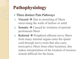
Approach to Acute Abdomen Pain 
Runal Shah 
MEM-PGY1 
KDAH, 
Mumbai
Pathophysiology 
 Three distinct Pain Pathways 
1) Visceral  Due to stretching of fibers 
innervating the walls of hollow or solid 
2) Somatic  Caused by irritation of parietal 
peritoneum fibers 
3) Referred  Peripheral afferent nerve fibers 
from many internal organs enter the spinal 
cord through nerve roots that also carry 
nociceptive fibers from other locations, this 
makes interpretation of the location of noxious 
stimuli difficult for the brain.
Quick Assessment 
 “S” of SAMPLE 
1) Pain score + External look 
2) Character of Pain (?Radiating to) 
3) Site of Pain (Localized/Diffuse) 
4) Associated Symptoms 
5) Aggravated/Relieving Factors 
• “A” for Allergy 
 History of Asthma/Allergy
 “M” for Medications 
Current meds list 
Meds Over the counter 
 “P” for Past History 
 “L” for Last meal /LMP – imp in 
Females 
 “E” for Events – Outside food 
consumption / Travel history
Examination 
• INSPECT for distention, scar, mass, rash. 
• AUSCULATE for hyperactive, sluggish, absent, or 
normal bowel sounds. 
• PALPATION to look for guarding, rigidity, rebound 
tenderness, organomegaly, ascites. 
• PERCUSSION for Organ dullness, ascites 
 Pelvic Examination for Females (Per Vaginum) 
 Per Rectal Examination
Ref. Rosen 8/e
Early Interventions 
 Pain Management 
 NSAIDs vs. Opioids 
 Anti-Spasmodic 
 Symptomatic 
 Drugs 
 Invasive Interventions (RT, Flatus Tube) 
Antibiotics 
 Definitive – Surgery (Elective vs. Emergency)
Imaging in Abd. Pain ?? 
 USG Abdomen+Pelvis  Billiary Tract Ds specific 
 CT Whole Abdomen  Definitive !!Radiation Risk 
 X-Ray Erect Abdomen  Air fluid level ?
Extra Abdominal Causes of Abd. Pain 
Ref. Rosen 8/e
Disposition from A&E 
 If diagnosis is made upon CT or USG, patient can 
be admitted with same for Observation &/or 
Surgical intervention can be planned. 
 Non-specific abdominal pain 
◦ If this is the working diagnosis, patients must be 
re-examined in 24 hours. This may be done in the 
outpatient setting.
Thank You…

Approach to acute abdomen

  • 1.
    Approach to AcuteAbdomen Pain Runal Shah MEM-PGY1 KDAH, Mumbai
  • 2.
    Pathophysiology  Threedistinct Pain Pathways 1) Visceral  Due to stretching of fibers innervating the walls of hollow or solid 2) Somatic  Caused by irritation of parietal peritoneum fibers 3) Referred  Peripheral afferent nerve fibers from many internal organs enter the spinal cord through nerve roots that also carry nociceptive fibers from other locations, this makes interpretation of the location of noxious stimuli difficult for the brain.
  • 3.
    Quick Assessment “S” of SAMPLE 1) Pain score + External look 2) Character of Pain (?Radiating to) 3) Site of Pain (Localized/Diffuse) 4) Associated Symptoms 5) Aggravated/Relieving Factors • “A” for Allergy  History of Asthma/Allergy
  • 4.
     “M” forMedications Current meds list Meds Over the counter  “P” for Past History  “L” for Last meal /LMP – imp in Females  “E” for Events – Outside food consumption / Travel history
  • 5.
    Examination • INSPECTfor distention, scar, mass, rash. • AUSCULATE for hyperactive, sluggish, absent, or normal bowel sounds. • PALPATION to look for guarding, rigidity, rebound tenderness, organomegaly, ascites. • PERCUSSION for Organ dullness, ascites  Pelvic Examination for Females (Per Vaginum)  Per Rectal Examination
  • 6.
  • 7.
    Early Interventions Pain Management  NSAIDs vs. Opioids  Anti-Spasmodic  Symptomatic  Drugs  Invasive Interventions (RT, Flatus Tube) Antibiotics  Definitive – Surgery (Elective vs. Emergency)
  • 8.
    Imaging in Abd.Pain ??  USG Abdomen+Pelvis  Billiary Tract Ds specific  CT Whole Abdomen  Definitive !!Radiation Risk  X-Ray Erect Abdomen  Air fluid level ?
  • 9.
    Extra Abdominal Causesof Abd. Pain Ref. Rosen 8/e
  • 10.
    Disposition from A&E  If diagnosis is made upon CT or USG, patient can be admitted with same for Observation &/or Surgical intervention can be planned.  Non-specific abdominal pain ◦ If this is the working diagnosis, patients must be re-examined in 24 hours. This may be done in the outpatient setting.
  • 11.