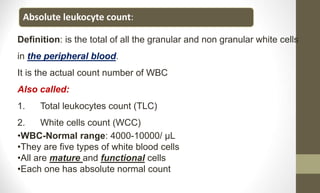
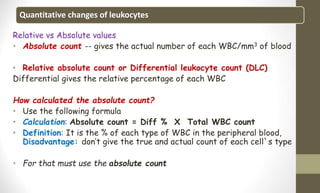
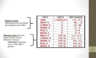
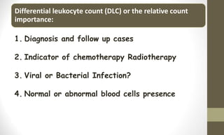
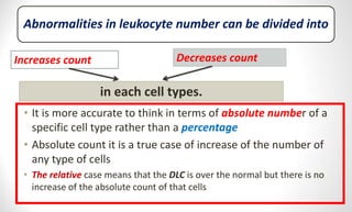
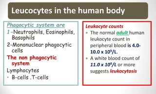
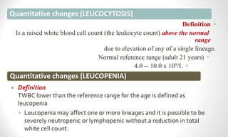
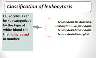
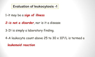
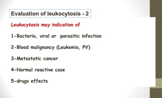
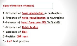
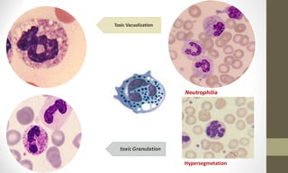
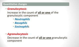
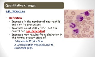
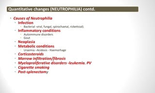
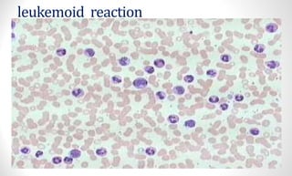
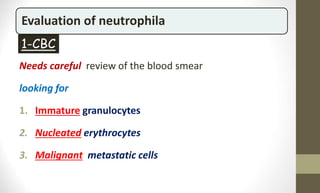
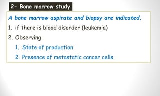
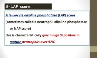
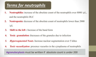
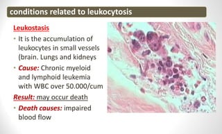
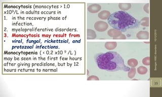
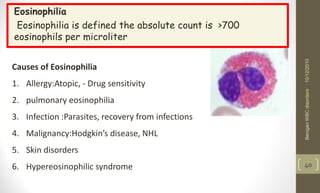
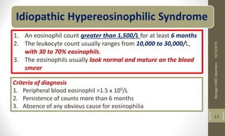
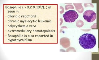
Dr.AbdulrazzaqAlagbare
MBhc-MScCP-HematologyLecturer
Abnormalities in leukocyte number
Lesson 3
Learning objectives
At the end of this lecture the student must be know
1- what means quantitative WBC abnormality
2-what is Leukocytosis
3-causes of leukocytosis
4- Minor causes of leukocytosis
5-The mechanism of leukocytosis
6-Classification of leukocytosis
7-Leukemoid reaction
8-Leukoerythroblastic reaction
9-Leukostasis
Points to know
• directly counted from an automated
instrument or by manual method
WBC on CBC
• Determine the normal range
The patients age
• more variable normal range than adults
Children
• the newborn include highest white
counts at FIRST 3 months
infant
Introduction
of
anemia‘s
4
Leukocyte indices:
1. LYM% (LY%) (lymphocyte) - relative (%) content (the norm is 25-40%) lymphocytes .
2. LYM # (LY #) (lymphocyte) - the absolute content (norma1, 2 - 3,0 h109 / l (or 1,2-3,0 x
103/mkl)) lymphocytes .
3. MXD% - relative (%) content of the mixture (the norm is 5 - 10%), monocytes , basophils ,
and eosinophils .
4. MXD # - absolute content of the mixture (norma0 ,2-0, 8 x 10⁹ / l), monocytes , basophils ,
and eosinophils .
5. NEUT% (NE%) (neutrophils) - relative (%) content of neutrophils .
6. NEUT # (NE #) (neutrophils) - the absolute content of neutrophils .
7. MON% (MO%) (monocyte) - relative (%) content of monocytes (normal 0,04-0,11).
8. MON # (MO #) (monocyte) - the absolute content of monocytes (normal 0,1-0,6 109 cells / l).
9. EO% - relative (%) content of eosinophils .
10. EO # - absolute content of eosinophils .
11. BA% - relative (%) content of basophils .
12. BA # - absolute content of basophils
13. IMM% - relative (%) content of immature granulocytes .
14. IMM # - absolute content of immature granulocytes
15. ATL% - relative (%) content of atypical lymphocytes .
16. ATL # - absolute content of atypical lymphocytes .
17. GR% - relative (%) content (the norm is 47 - 72%) of granulocytes.
18. GR # - absolute content (normal 1,2-6,8 x 10⁹ / l (or 1,2-6,8 x 10³/mkl)) granulocytes
Definition: is the total of all the granular and non granular white cells
in the peripheral blood.
It is the actual count number of WBC
Also called:
1. Total leukocytes count (TLC)
2. White cells count (WCC)
•WBC-Normal range: 4000-10000/ µL
•They are five types of white blood cells
•All are mature and functional cells
•Each one has absolute normal count
Absolute leukocyte count:
Quantitative changes of leukocytes
Relative vs Absolute values
• Absolute count -- gives the actual number of each WBC/mm3 of blood
• Relative absolute count or Differential leukocyte count (DLC)
Differential gives the relative percentage of each WBC
How calculated the absolute count?
• Use the following formula
• Calculation: Absolute count = Diff % X Total WBC count
• Definition: It is the % of each type of WBC in the peripheral blood,
Disadvantage: don’t give the true and actual count of each cell`s type
• For that must use the absolute count
Absolute value gives the
actual number of each
WBC/mm3 of blood
Calculation: absolute
count= Total WBC x
percent
Relative values
Differential gives the relative
percentage of each WBC
7
Differential leukocyte count (DLC) or the relative count
importance:
1. Diagnosis and follow up cases
2. Indicator of chemotherapy Radiotherapy
3. Viral or Bacterial Infection?
4. Normal or abnormal blood cells presence
Types of DLC:
Normal DLC
When the person is
healthy has the 5
normal white blood
cells
Abnormal DLC
The person is not healthy
And the cells may include
1- Normal + abnormal
cells
2-All are abnormal
Cells included in DLC
All nucleated cells in the peripheral blood normal or
abnormal
Details
1. Mature white blood cells in the peripheral blood.
2. Immature white blood cells in peripheral blood
3. NRBC (the nucleated RBC)
4. Abnormal cells (of metastatic cancer)
DLC over 100%
The DLC must count 100 WBC on the PBS, but there are cases needs to count
200 WBC as follow, if there:
.1
WBC >35.0 × 10⁹/L
.2
Lymphocytes > 40% or <17 %
.3
Monocytes >12%
.4
Blasts (first-time for the patient)
Critical values of WBC
Critical values the values which are out of the lower
or higher limits of normal reference.
1. WBC Low 3.0 X10⁹/L
2. High 25.0 X10⁹/L
3. Also a case of presence of blast cells as first
time for the patient
Quantitative abnormalities of White Blood Cells
Introduction
• The white blood cells are short lived (phagocytic)
• Their abnormalities are dramatic
The most important in the quantity abnormalities is
 the absolute count not the relative count only.
More important to be careful to the
• neutropenia in which the absolute neutrophils count is less
than 2.0 x10⁹/L.
Abnormalities in leukocyte number can be divided into
• It is more accurate to think in terms of absolute number of a
specific cell type rather than a percentage
• Absolute count it is a true case of increase of the number of
any type of cells
• The relative case means that the DLC is over the normal but there is no
increase of the absolute count of that cells
Increases count Decreases count
in each cell types.
Leucocytes in the human body
Phagocytic system are
1-Neutrophils, Eosinophils,
Basophils
2-Mononuclear phagocytic
cells
The non phagocytic
system
Lymphocytes
• B-cells .T-cells
Leukocyte counts
 The normal adult human
leukocyte count in
peripheral blood is 4.0-
10.0 x 10⁹/L.
 A white blood count of
11.0 x 10⁹/L or more
suggests leukocytosis
Quantitative changes (LEUCOCYTOSIS)
•
Definition
Is a raised white blood cell count (the leukocyte count) above the normal
range
due to elevation of any of a single lineage.
•
Normal reference range (adult 21 years)
•
4.0 -- 10.0 x 10⁹/L
Quantitative changes (LEUCOPENIA)
 Definition
TWBC lower than the reference range for the age is defined as
leucopenia
 Leucopenia may affect one or more lineages and it is possible to be
severely neutropenic or lymphopenic without a reduction in total
white cell count.
The mechanism of leukocytosis can be of several forms
• Decreased margination pool.
• Increase bone marrow production
• Splenoectomy  (Increase cells)
Classification of leukocytosis
•
Leukocytosis-Neutrophilia.
•
Leukocytosis-Lymphocytosis.
•
Leukocytosis-Monocytosis.
•
Leukocytosis-Eosinophilia.
Leukocytosis can
be subcategorized
by the type of
white blood cell
that is increased
in number.
1-it may be a sign of illness
2-is not a disorder, nor is it a disease
3-It is simply a laboratory finding.
4-A leukocyte count above 25 to 30 x 109/L is termed a
leukemoid reaction
Evaluation of leukocytosis -1
Leukocytosis may indication of
1-Bacteria, viral or parasitic infection
2-Blood malignancy (Leukemia, PV)
3-Metastatic cancer
4-Normal reactive case
5-drugs effects
Evaluation of leukocytosis - 2
Signs of infection (cytotoxic)
1-Presence of toxic granulation in neutrophils
1-Presence of toxic vacuolization in neutrophils
2-increase of band form over 5% "left shift
3-Presence of Dohle bodies
4-Increase of ESR
5-Positive CRP test
6- LAP test positive
Toxic Vacuolization
toxic Granulation
Neutrophilia
Hypersegmetation
Quantitative changes
• Granulocytosis
Increase in the count of all or one of the
granulocytic component
• Neutrophils
• Basophils
• Eosinophils
• Agranulocytosis
Decrease in the count of all or one granulocytic
component
Quantitative changes
NEUTROPHILIA
• Definition
• Increase in the number of neutrophils
and / or its precursors
• In adults count >8.0 x 10⁹/L but the
counts are age dependent
• Increase may results from alteration in
the normal steady state of
1-Increase Production
2-Demargination (marginal pool to
circulating pool).
Quantitative changes (NEUTROPHILIA) contd.
• Causes of Neutrophilia
• Infection
• Bacterial -viral, fungal, spirochaetal, rickettsial).
• Inflammatory conditions
• Autoimmune disorders
• Gout
• Neoplasia
• Metabolic conditions
• Uraemia –Acidosis - Haemorhage
• Corticosteroids
• Marrow infiltration/fibrosis
• Myeloproliferative disorders -leukemia. PV
• Cigarette smoking
• Post-splenectomy
Quantitative changes (NEUTROPHILIA) contd.
• Acute Neutrophilia
• Acute neutrophilia – this occurs 4-5 hours after a
pathologic stimulus
• Results from an increased flow of cells from the bone
marrow to the peripheral blood
• Bands and metamyelocytes may be seen
• =======================================
• Chronic neutrophilia – this follows acute neutrophilia
• The bone marrow starts to throw out younger and
younger cells (a shift to the left)
Stress neutrophilia
Definition: represents a shift of neutrophils from the
marginating pool into the circulating pool
• results from physical action, other types of stress, and
epinephrine injection.
This case has the following
• It is transient
• No granulocyte production increase in the B.M
Reactive Left Shift
Means less mature forms in excess of
5% band neutrophils,
Types
1-sever  include metamyelocyte –
myelocyte and band form
2-mild  include myelocyte and band
form
indicates an increase in new cell
production in this cell series
Shift to the left
Minor causes of leukocytosis include:
1-Pseudoneutrophilia (stress)
Mechanism
• No true increase of neutrophils production , but a shift from the
marginal storage pool to the circulating blood.
• 2-Asplenia can result in a moderate leukocytosis
• 3-uses of corticoid drugs
leukemoid reaction
 is a marked elevation of the white cell count resembling leukemia
 but due to a benign condition
 The WBC count is often in a range of
 over 25.0 x10 9/L to 50.0 x10 9/L –
 and sometimes can be reach to 100.0 x10 9/L
Types:
1-Neutrophilic leukemoid reaction - adult
2-Lymphocytic leukemoid reaction - mostly children
3-Eosinophilic leukemoid reaction
4-Monocytic leukemoid reaction
The patient has no
1-Anemia
2-Bleeding
3-The case is acute
leukemoid reaction
STUDY of neutrophila
1-CBC  count of Neutrophils absolute count  Blood
film study  evaluation of band form presence over 5%
 Toxic granulation in the cytoplasm and Toxic
vacuolization
2- Bone marrow study
3-Uses of some stain such as -- A leukocyte alkaline
phosphatase (LAP)
Evaluation of neutrophila
Needs careful review of the blood smear
looking for
1. Immature granulocytes
2. Nucleated erythrocytes
3. Malignant metastatic cells
1-CBC
A bone marrow aspirate and biopsy are indicated.
1. if there is blood disorder (leukemia)
2. Observing
1. State of production
2. Presence of metastatic cancer cells
2- Bone marrow study
A leukocyte alkaline phosphatase (LAP) score
(sometimes called a neutrophil alkaline phosphatase
or NAP score)
this is characteristically give a high % positive in
mature neutrophils over 97%
3-LAP score
Terms for neutrophils
1. Neutrophilia: increase of the absolute count of the neutrophils over 8000 /µL,
and the neutrophils DLC
2. Neutropenia: decrease of the absolute count of neutrophils lower than 2000
/µL
3. Shift to the left : Increase of the band form
4. Toxic granulation :Increases of the granules due to infection
5. Hypersegmented Neutr. Increase nuclear segmentation over 5 lobes
6. Toxic vacuolization: presence vacuoles in the cytoplasma of neutrophils
Agranulocytosis must be written if absolute count is under 200
Leukostasis
• It is the accumulation of
leukocytes in small vessels
(brain. Lungs and kidneys
• Cause: Chronic myeloid
and lymphoid leukemia
with WBC over 50.000/cum
Result: may occur death
• Death causes: impaired
blood flow
conditions related to leukocytosis
Monocytosis (monocytes > 1.0
x109/L in adults occurs in
1. in the recovery phase of
infection,
2. myeloproliferative disorders.
3. Monocytosis may result from
• viral, fungal, rickettsial, and
protozoal infections.
Monocytopenia ( < 0.2 x10 9 /L )
may be seen in the first few hours
after giving predislone, but by 12
hours returns to normal
10/12/2010
Benigen
WBC
disorders
39
Eosinophilia
Eosinophilia is defined the absolute count is >700
eosinophils per microliter
10/12/2010
Benigen
WBC
disorders
40
Causes of Eosinophilia
1. Allergy:Atopic, - Drug sensitivity
2. pulmonary eosinophilia
3. Infection :Parasites, recovery from infections
4. Malignancy:Hodgkin’s disease, NHL
5. Skin disorders
6. Hypereosinophilic syndrome
Eosinophilia
Idiopathic Hypereosinophilic Syndrome
1. An eosinophil count greater than 1,500/L for at least 6 months
2. The leukocyte count usually ranges from 10,000 to 30,000/L,
with 30 to 70% eosinophils.
3. The eosinophils usually look normal and mature on the blood
smear
Criteria of diagnosis
1. Peripheral blood eosinophil >1.5 x 10⁹/L
2. Persistence of counts more than 6 months
3. Absence of any obvious cause for eosinophilia
10/12/2010
Benigen
WBC
disorders
42
Basophilia ( > 0.2 X 10⁹/L ) is
seen in
• allerigic reactions
• chronic myelocytic leukemia
• polycythemia vera
• extramedullary hematopoiesis.
• Basophilia is also reported in
hypothyroidism.
10/12/2010
Benigen
WBC
disorders
43
END OF THE
LESSON
THANK
YOU
ANY
QUESTION?

Abnormalities in leukocyte number.ppt

  • 1.
  • 2.
    Learning objectives At theend of this lecture the student must be know 1- what means quantitative WBC abnormality 2-what is Leukocytosis 3-causes of leukocytosis 4- Minor causes of leukocytosis 5-The mechanism of leukocytosis 6-Classification of leukocytosis 7-Leukemoid reaction 8-Leukoerythroblastic reaction 9-Leukostasis
  • 3.
    Points to know •directly counted from an automated instrument or by manual method WBC on CBC • Determine the normal range The patients age • more variable normal range than adults Children • the newborn include highest white counts at FIRST 3 months infant
  • 4.
    Introduction of anemia‘s 4 Leukocyte indices: 1. LYM%(LY%) (lymphocyte) - relative (%) content (the norm is 25-40%) lymphocytes . 2. LYM # (LY #) (lymphocyte) - the absolute content (norma1, 2 - 3,0 h109 / l (or 1,2-3,0 x 10³/mkl)) lymphocytes . 3. MXD% - relative (%) content of the mixture (the norm is 5 - 10%), monocytes , basophils , and eosinophils . 4. MXD # - absolute content of the mixture (norma0 ,2-0, 8 x 10⁹ / l), monocytes , basophils , and eosinophils . 5. NEUT% (NE%) (neutrophils) - relative (%) content of neutrophils . 6. NEUT # (NE #) (neutrophils) - the absolute content of neutrophils . 7. MON% (MO%) (monocyte) - relative (%) content of monocytes (normal 0,04-0,11). 8. MON # (MO #) (monocyte) - the absolute content of monocytes (normal 0,1-0,6 109 cells / l). 9. EO% - relative (%) content of eosinophils . 10. EO # - absolute content of eosinophils . 11. BA% - relative (%) content of basophils . 12. BA # - absolute content of basophils 13. IMM% - relative (%) content of immature granulocytes . 14. IMM # - absolute content of immature granulocytes 15. ATL% - relative (%) content of atypical lymphocytes . 16. ATL # - absolute content of atypical lymphocytes . 17. GR% - relative (%) content (the norm is 47 - 72%) of granulocytes. 18. GR # - absolute content (normal 1,2-6,8 x 10⁹ / l (or 1,2-6,8 x 10³/mkl)) granulocytes
  • 5.
    Definition: is thetotal of all the granular and non granular white cells in the peripheral blood. It is the actual count number of WBC Also called: 1. Total leukocytes count (TLC) 2. White cells count (WCC) •WBC-Normal range: 4000-10000/ µL •They are five types of white blood cells •All are mature and functional cells •Each one has absolute normal count Absolute leukocyte count:
  • 6.
    Quantitative changes ofleukocytes Relative vs Absolute values • Absolute count -- gives the actual number of each WBC/mm3 of blood • Relative absolute count or Differential leukocyte count (DLC) Differential gives the relative percentage of each WBC How calculated the absolute count? • Use the following formula • Calculation: Absolute count = Diff % X Total WBC count • Definition: It is the % of each type of WBC in the peripheral blood, Disadvantage: don’t give the true and actual count of each cell`s type • For that must use the absolute count
  • 7.
    Absolute value givesthe actual number of each WBC/mm3 of blood Calculation: absolute count= Total WBC x percent Relative values Differential gives the relative percentage of each WBC 7
  • 8.
    Differential leukocyte count(DLC) or the relative count importance: 1. Diagnosis and follow up cases 2. Indicator of chemotherapy Radiotherapy 3. Viral or Bacterial Infection? 4. Normal or abnormal blood cells presence
  • 9.
    Types of DLC: NormalDLC When the person is healthy has the 5 normal white blood cells Abnormal DLC The person is not healthy And the cells may include 1- Normal + abnormal cells 2-All are abnormal
  • 10.
    Cells included inDLC All nucleated cells in the peripheral blood normal or abnormal Details 1. Mature white blood cells in the peripheral blood. 2. Immature white blood cells in peripheral blood 3. NRBC (the nucleated RBC) 4. Abnormal cells (of metastatic cancer)
  • 11.
    DLC over 100% TheDLC must count 100 WBC on the PBS, but there are cases needs to count 200 WBC as follow, if there: .1 WBC >35.0 × 10⁹/L .2 Lymphocytes > 40% or <17 % .3 Monocytes >12% .4 Blasts (first-time for the patient)
  • 12.
    Critical values ofWBC Critical values the values which are out of the lower or higher limits of normal reference. 1. WBC Low 3.0 X10⁹/L 2. High 25.0 X10⁹/L 3. Also a case of presence of blast cells as first time for the patient
  • 13.
    Quantitative abnormalities ofWhite Blood Cells Introduction • The white blood cells are short lived (phagocytic) • Their abnormalities are dramatic The most important in the quantity abnormalities is  the absolute count not the relative count only. More important to be careful to the • neutropenia in which the absolute neutrophils count is less than 2.0 x10⁹/L.
  • 14.
    Abnormalities in leukocytenumber can be divided into • It is more accurate to think in terms of absolute number of a specific cell type rather than a percentage • Absolute count it is a true case of increase of the number of any type of cells • The relative case means that the DLC is over the normal but there is no increase of the absolute count of that cells Increases count Decreases count in each cell types.
  • 15.
    Leucocytes in thehuman body Phagocytic system are 1-Neutrophils, Eosinophils, Basophils 2-Mononuclear phagocytic cells The non phagocytic system Lymphocytes • B-cells .T-cells Leukocyte counts  The normal adult human leukocyte count in peripheral blood is 4.0- 10.0 x 10⁹/L.  A white blood count of 11.0 x 10⁹/L or more suggests leukocytosis
  • 16.
    Quantitative changes (LEUCOCYTOSIS) • Definition Isa raised white blood cell count (the leukocyte count) above the normal range due to elevation of any of a single lineage. • Normal reference range (adult 21 years) • 4.0 -- 10.0 x 10⁹/L Quantitative changes (LEUCOPENIA)  Definition TWBC lower than the reference range for the age is defined as leucopenia  Leucopenia may affect one or more lineages and it is possible to be severely neutropenic or lymphopenic without a reduction in total white cell count.
  • 17.
    The mechanism ofleukocytosis can be of several forms • Decreased margination pool. • Increase bone marrow production • Splenoectomy  (Increase cells)
  • 18.
  • 19.
    1-it may bea sign of illness 2-is not a disorder, nor is it a disease 3-It is simply a laboratory finding. 4-A leukocyte count above 25 to 30 x 109/L is termed a leukemoid reaction Evaluation of leukocytosis -1
  • 20.
    Leukocytosis may indicationof 1-Bacteria, viral or parasitic infection 2-Blood malignancy (Leukemia, PV) 3-Metastatic cancer 4-Normal reactive case 5-drugs effects Evaluation of leukocytosis - 2
  • 21.
    Signs of infection(cytotoxic) 1-Presence of toxic granulation in neutrophils 1-Presence of toxic vacuolization in neutrophils 2-increase of band form over 5% "left shift 3-Presence of Dohle bodies 4-Increase of ESR 5-Positive CRP test 6- LAP test positive
  • 22.
  • 23.
    Quantitative changes • Granulocytosis Increasein the count of all or one of the granulocytic component • Neutrophils • Basophils • Eosinophils • Agranulocytosis Decrease in the count of all or one granulocytic component
  • 24.
    Quantitative changes NEUTROPHILIA • Definition •Increase in the number of neutrophils and / or its precursors • In adults count >8.0 x 10⁹/L but the counts are age dependent • Increase may results from alteration in the normal steady state of 1-Increase Production 2-Demargination (marginal pool to circulating pool).
  • 25.
    Quantitative changes (NEUTROPHILIA)contd. • Causes of Neutrophilia • Infection • Bacterial -viral, fungal, spirochaetal, rickettsial). • Inflammatory conditions • Autoimmune disorders • Gout • Neoplasia • Metabolic conditions • Uraemia –Acidosis - Haemorhage • Corticosteroids • Marrow infiltration/fibrosis • Myeloproliferative disorders -leukemia. PV • Cigarette smoking • Post-splenectomy
  • 26.
    Quantitative changes (NEUTROPHILIA)contd. • Acute Neutrophilia • Acute neutrophilia – this occurs 4-5 hours after a pathologic stimulus • Results from an increased flow of cells from the bone marrow to the peripheral blood • Bands and metamyelocytes may be seen • ======================================= • Chronic neutrophilia – this follows acute neutrophilia • The bone marrow starts to throw out younger and younger cells (a shift to the left)
  • 27.
    Stress neutrophilia Definition: representsa shift of neutrophils from the marginating pool into the circulating pool • results from physical action, other types of stress, and epinephrine injection. This case has the following • It is transient • No granulocyte production increase in the B.M
  • 28.
    Reactive Left Shift Meansless mature forms in excess of 5% band neutrophils, Types 1-sever  include metamyelocyte – myelocyte and band form 2-mild  include myelocyte and band form indicates an increase in new cell production in this cell series
  • 29.
  • 30.
    Minor causes ofleukocytosis include: 1-Pseudoneutrophilia (stress) Mechanism • No true increase of neutrophils production , but a shift from the marginal storage pool to the circulating blood. • 2-Asplenia can result in a moderate leukocytosis • 3-uses of corticoid drugs
  • 31.
    leukemoid reaction  isa marked elevation of the white cell count resembling leukemia  but due to a benign condition  The WBC count is often in a range of  over 25.0 x10 9/L to 50.0 x10 9/L –  and sometimes can be reach to 100.0 x10 9/L Types: 1-Neutrophilic leukemoid reaction - adult 2-Lymphocytic leukemoid reaction - mostly children 3-Eosinophilic leukemoid reaction 4-Monocytic leukemoid reaction The patient has no 1-Anemia 2-Bleeding 3-The case is acute
  • 32.
  • 33.
    STUDY of neutrophila 1-CBC count of Neutrophils absolute count  Blood film study  evaluation of band form presence over 5%  Toxic granulation in the cytoplasm and Toxic vacuolization 2- Bone marrow study 3-Uses of some stain such as -- A leukocyte alkaline phosphatase (LAP)
  • 34.
    Evaluation of neutrophila Needscareful review of the blood smear looking for 1. Immature granulocytes 2. Nucleated erythrocytes 3. Malignant metastatic cells 1-CBC
  • 35.
    A bone marrowaspirate and biopsy are indicated. 1. if there is blood disorder (leukemia) 2. Observing 1. State of production 2. Presence of metastatic cancer cells 2- Bone marrow study
  • 36.
    A leukocyte alkalinephosphatase (LAP) score (sometimes called a neutrophil alkaline phosphatase or NAP score) this is characteristically give a high % positive in mature neutrophils over 97% 3-LAP score
  • 37.
    Terms for neutrophils 1.Neutrophilia: increase of the absolute count of the neutrophils over 8000 /µL, and the neutrophils DLC 2. Neutropenia: decrease of the absolute count of neutrophils lower than 2000 /µL 3. Shift to the left : Increase of the band form 4. Toxic granulation :Increases of the granules due to infection 5. Hypersegmented Neutr. Increase nuclear segmentation over 5 lobes 6. Toxic vacuolization: presence vacuoles in the cytoplasma of neutrophils Agranulocytosis must be written if absolute count is under 200
  • 38.
    Leukostasis • It isthe accumulation of leukocytes in small vessels (brain. Lungs and kidneys • Cause: Chronic myeloid and lymphoid leukemia with WBC over 50.000/cum Result: may occur death • Death causes: impaired blood flow conditions related to leukocytosis
  • 39.
    Monocytosis (monocytes >1.0 x10⁹/L in adults occurs in 1. in the recovery phase of infection, 2. myeloproliferative disorders. 3. Monocytosis may result from • viral, fungal, rickettsial, and protozoal infections. Monocytopenia ( < 0.2 x10 9 /L ) may be seen in the first few hours after giving predislone, but by 12 hours returns to normal 10/12/2010 Benigen WBC disorders 39
  • 40.
    Eosinophilia Eosinophilia is definedthe absolute count is >700 eosinophils per microliter 10/12/2010 Benigen WBC disorders 40 Causes of Eosinophilia 1. Allergy:Atopic, - Drug sensitivity 2. pulmonary eosinophilia 3. Infection :Parasites, recovery from infections 4. Malignancy:Hodgkin’s disease, NHL 5. Skin disorders 6. Hypereosinophilic syndrome
  • 41.
  • 42.
    Idiopathic Hypereosinophilic Syndrome 1.An eosinophil count greater than 1,500/L for at least 6 months 2. The leukocyte count usually ranges from 10,000 to 30,000/L, with 30 to 70% eosinophils. 3. The eosinophils usually look normal and mature on the blood smear Criteria of diagnosis 1. Peripheral blood eosinophil >1.5 x 10⁹/L 2. Persistence of counts more than 6 months 3. Absence of any obvious cause for eosinophilia 10/12/2010 Benigen WBC disorders 42
  • 43.
    Basophilia ( >0.2 X 10⁹/L ) is seen in • allerigic reactions • chronic myelocytic leukemia • polycythemia vera • extramedullary hematopoiesis. • Basophilia is also reported in hypothyroidism. 10/12/2010 Benigen WBC disorders 43
  • 44.