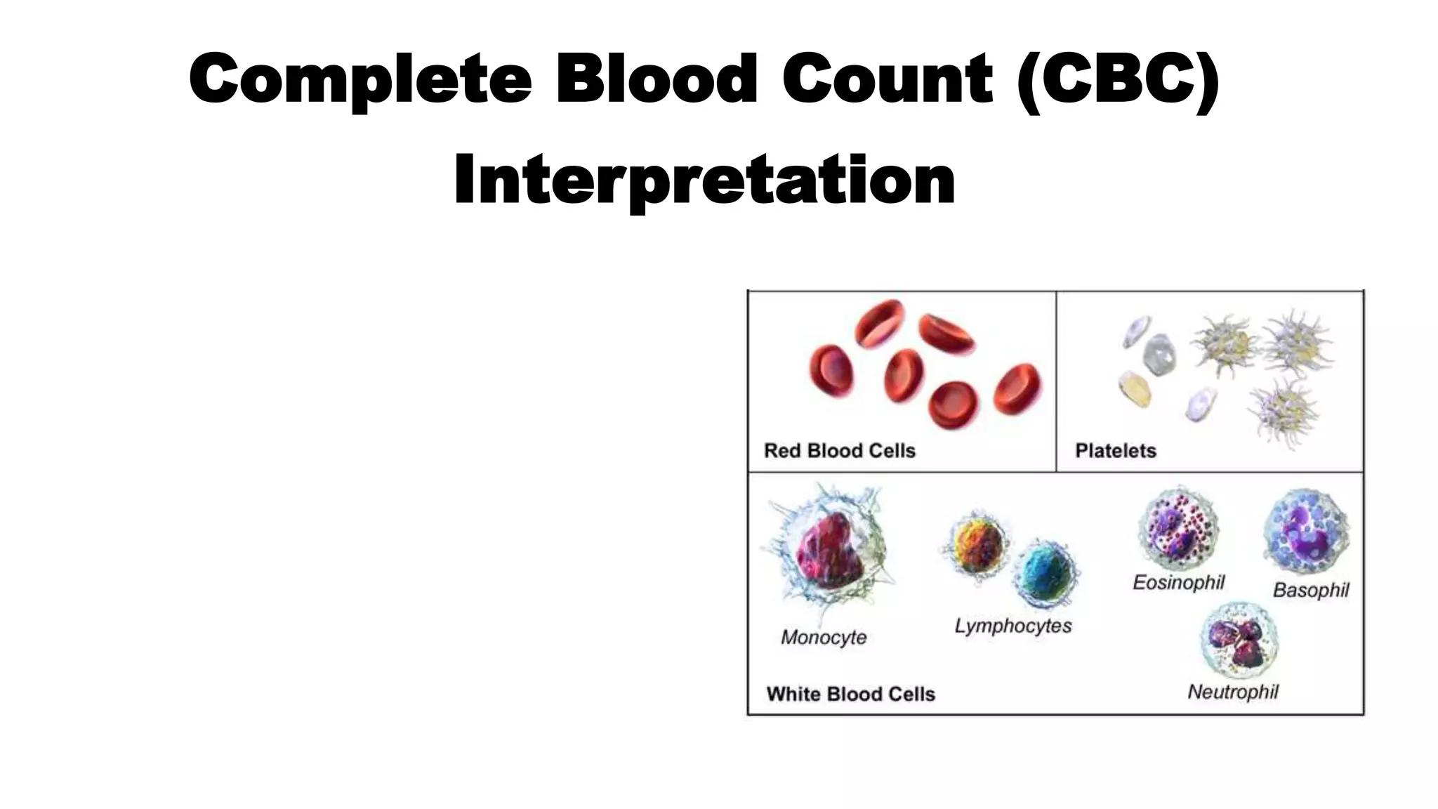
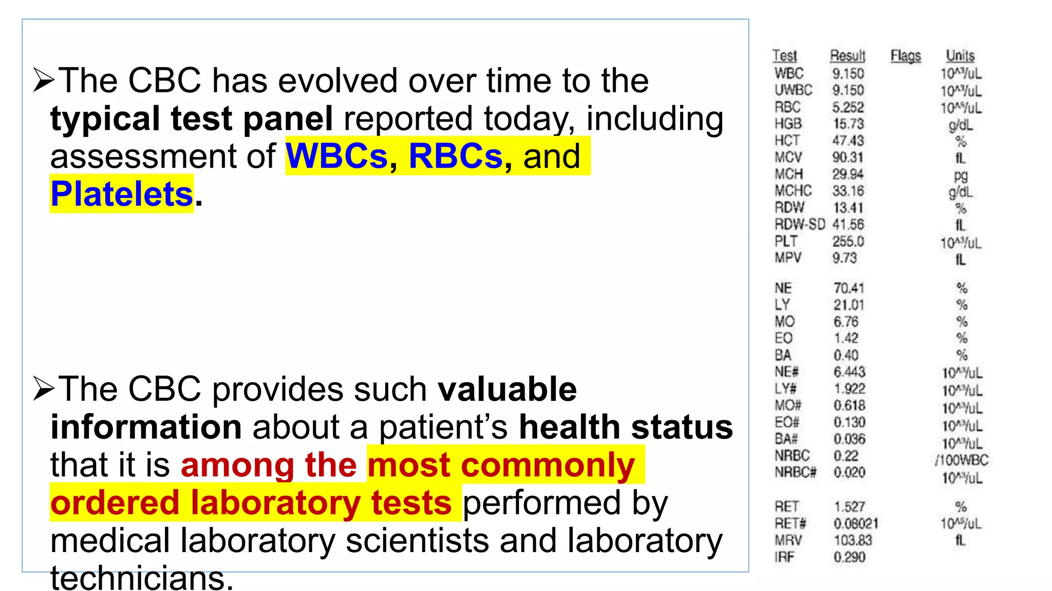
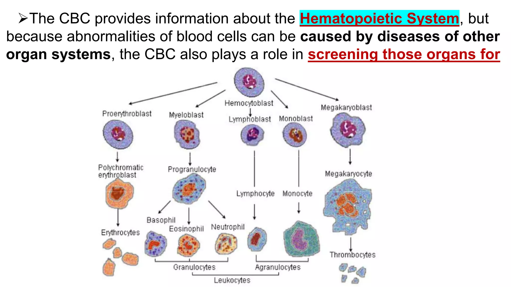
2. ďThe CBC has evolved over time to the
typical test panel reported today, including
assessment of WBCs, RBCs, and
Platelets.
ďThe CBC provides such valuable
information about a patientâs health status
that it is among the most commonly
ordered laboratory tests performed by
medical laboratory scientists and laboratory
technicians.
3. ďThe CBC provides information about the Hematopoietic System, but
because abnormalities of blood cells can be caused by diseases of other
organ systems, the CBC also plays a role in screening those organs for
disease.
4. Systematic Approach to CBC Interpretation
ďWhite Blood Cells
Step 1: Ensure that the WBC count is accurate.
Step 2: Compare the patientâs WBC count with the laboratoryâs established reference interval.
Steps 3 and 4: Examine the differential information (relative and absolute) on variations in the distribution of WBCs.
Step 5: Make note of immature cells in any cell line reported in the differential that should not appear in normal peripheral blood.
Step 6: Make note of any morphologic abnormalities and correlate film findings with the numerical values.
ďRed Blood Cells
Step 1: Examine the HGB concentration first to assess anemia.
Step 2: Examine the MCV to assess cell volume.
Step 3: Examine the MCHC to assess cell HGB concentration in RBC.
Step 4: Examine the RDW to assess anisocytosis. (Correlate both MCV and RDW with RBC histogram.)
Step 5: Examine the morphologic description and correlate with the numerical values.
Step 6: Review remaining information.
ďPlatelets
Step 1: Examine the total platelet count.
Step 2: Examine the MPV to assess platelet volume.
Step 3: Examine platelet morphology and correlate with the numerical values.
5. White Blood Cell Parameters
The WBC-related parameters of a routine
CBC include the following:
1. Total WBC count
2. WBC differential count values expressed as
percentages, called relative counts
3. WBC differential count values expressed as
the actual number of each type of cell (e.g.,
neutrophils), called absolute counts
4. WBC morphology
6. Step 1
ďąStart by ensuring that there is an accurate WBC count.
ďąCompare the WBC histogram and/or scatterplot to the respective cell counts to
make sure they correlate with one another.
ďąTodayâs automated instruments can eliminate nucleated RBCs that falsely
increase the WBC count.
ďąHowever, manual WBC results must be corrected mathematically to eliminate the
contribution of the nucleated RBCs
7. Step 2
⢠Look at the total WBC count.
⢠When the count is elevated, it is called leukocytosis.
⢠When the WBC count is low, it is called leukopenia.
⢠Increases and decreases of WBCs are associated with infections and
conditions such as leukemias.
⢠Because there is more than one type of WBC, increases and decreases
in the total count usually are due to changes in one of the subtypesâ
for example, neutrophils or lymphocytes.
⢠Determining which one is the next step.
8. Step 3
⢠Examine the relative differential counts for a preliminary assessment of which cell
lines are affected.
⢠The relative differential count is reported in percentages.
⢠The proportion of each cell type can be described by its relative number (i.e.,
percent) and compared with its reference interval.
⢠Then it is described using appropriate terminology, such as a relative neutrophilia,
which is an increase in neutrophils, or a relative lymphopenia, which is a
decrease in lymphocytes.
9. ⢠If the total WBC count or any of the relative values are
outside the reference interval, further analysis of the
WBC differential is needed.
⢠If the proportion of one of the cell types increases, then
the proportion of others must decrease because the
proportions are relative to one another.
⢠The second cell type may not have changed in actual
number at all, however. The way to assess this
accurately is with absolute differential counts.
10. ď The terms used for increases and decreases of each cell type are provided
in Table :
11. Step 4
â˘If not reported by the instrument, absolute counts can be
calculated easily using the total WBC count and the
relative differential.
â˘Multiply each relative cell count (i.e., percentage) by the
total WBC count and by so doing determine the
absolute count for each cell lineage.
12.
13. ďąOn first inspection, one may look at the WBC count and recognize that a leukocytosis is
present, but it is important to determine what cell line is causing the increased count.
ďąIn this case the cells are all within reference intervals relative to one another. There is no
indication as to which cell line could be causing the increase in total numbers of WBCs.
ďąWhen each relative number (e.g., neutrophils at 0.67 or 67%) is multiplied by the total WBC
count (13.6 10*9/L), the absolute numbers indicate that the neutrophils are elevated (9.1 10*9/L
compared with the reference interval provided).
ďąThe acronym for absolute neutrophil count is ANC.
ďąThe ANC is a very useful parameter for assessing neutropenia and neutrophilia.
ďąThe absolute lymphocyte count (3.5 10*9/L) is still within the reference interval.
ďąGiven this information, these results can be described as showing a leukocytosis with only an
absolute neutrophilia, and the overall increase in the WBC count is due to an increase only in
neutrophils.
14. ⢠When the absolute numbers of each of the individual cell types are totaled, the
sum equals the WBC count (slight differences may occur because of rounding, as
in the example).
⢠This is a method for checking whether the absolute calculations are correct.
Absolute counts may be obtained directly from automated analyzers, which count
actual numbers (i.e., produce absolute counts) and calculate relative values.
⢠Some laboratories do not report the absolute counts, so being able to calculate
them is important.
⢠As will be evident in later slides, the findings in this example point toward a
bacterial infection. Had there been an absolute lymphocytosis, a viral infection
would be likely.
15. Step 5
⢠Each cell line should be examined for immature cells. Young WBCs are not
normally seen in the peripheral blood, and they may indicate infections or
malignancies such as leukemia.
⢠For neutrophilic cells, there is a unique term that refers to the presence of
increased numbers of bands or cells younger than bands in the peripheral blood:
left shift or shift to the left.
⢠When young lymphocytic or monocytic cells are present, they can be reported
in the differential as prolymphocytes, lymphoblasts, promonocytes, or
monoblasts.
⢠When observed, young eosinophils and basophils are typically just called
immature and are not specifically staged. For example, eosinophilic
metamyelocytes are counted as eosinophils.
16. Step 6
⢠Any abnormalities of
appearance are reported in the
morphology
⢠For WBCs, abnormal
morphologic features that would
be noted include changes in
overall cellular appearance,
such as cytoplasmic toxic
granulation and nuclear
abnormalities such as
hypersegmentation.
17. ďąTo summarize the WBC parameters, begin with an accurate total
WBC count, followed by the relative differential, or preferably the
absolute counts, noting whether any abnormal young cells are
present in the blood.
ďąFinally, note the presence of any abnormal morphology or inclusions.
18. Elevated white blood cell (WBC) count
⢠Leucocytosis is defined as elevation of the white cell count (WCC) >2 SD above the mean.
ď The detection of leucocytosis should prompt immediate scrutiny of the automated WBC differential (generally accurate except in leukaemia)
and the other FBC parameters.
ď A blood film should be examined and if in doubt a manual differential count should be performed.
ď It is important to evaluate leucocytosis in terms of the age-related absolute normal ranges for neutrophils, lymphocytes,
monocytes,eosinophils, and basophils and the presence of abnormal cells: immature granulocytes, blasts, nucleated red cells, and âatypical
cellsâ.
⢠Leukaemoid reactionâleucocytosis >50 Ă 109/L defines a neutrophilia with marked âleft shiftâ (band forms,
metamyelocytes, myelocytes, and occasionally promyelocytes and myeloblasts in the blood film).
ď Differential diagnosis is CML and in children, juvenile CML.
ď Primitive granulocyte precursors are also frequently seen in the blood film of the infected or stressed neonate, and any seriously ill patient,
e.g. on the intensive therapy unit (ITU).
⢠Leucoerythroblastic blood filmâcontains myelocytes, other primitive granulocytes, nucleated red cells, and
often tear drop red cells; is due to BM invasion by tumour, fibrosis, or granuloma formation and is often an
indication for a BM biopsy.
ď Other causes include anorexia, haemolysis,and severe illness.
⢠Leucocytosis due to blastsâsuggests diagnosis of acute leukaemia and is an indication for cell typing studies
and BM examination.
⢠FBC, blood film, white cell differential count, and the clinical context in which the leucocytosis is detected will
usually indicate whether this is due to a 1° haematological abnormality or reflects a 2° response.
⢠It is clearly important to seek a history of symptoms of infection and examine the patient for signs of infection or an
19. ⢠BM examination is rarely necessary in the investigation of a patient with isolated neutrophilia.
Investigation of a leukaemoid reaction, leucoerythroblastic blood fi lm, and possible chronic
granulocytic leukaemia (CGL) or juvenile CML are fi rm indications for a BM aspirate and trephine
biopsy.
⢠BM culture, including culture for atypical mycobacteria and fungi, may be useful in patients with
persistent pyrexia or leucocytosis.
20. Neutrophilia Causes
ďą2° to acute infection is most common cause of leucocytosis.
ď Usually modest (uncommonly >30 Ă 109/L), associated with a left shift and occasionally toxic
granulation or vacuolation of neutrophils.
ďąChronic inflammation causes less marked neutrophilia often associated with monocytosis.
ďąModerate neutrophilia may occur following steroid therapy, heatstroke, and in patients with
solid tumours.
ďąMild neutrophilia may be induced by stress (e.g. immediate postoperative period) and
exercise.
ďąMay be seen following a myocardial infarction or major seizure.
ďąFrequently found in states of chronic BM stimulation (e.g. chronic haemolysis, idiopathic
thrombocytopenic purpura (ITP) and asplenia.
ďą1° haematological causes of neutrophilia are less common. CML is often the cause of
extremely high leucocyte counts (>200 Ă 109/L), predominantly neutrophils with marked left
shift, basophilia, and occasional myeloblasts.
ď The presence of the Ph chromosome on karyotype analysis are usually helpful to differentiate CML
from a leukaemoid reaction.
ďąLess common are juvenile CML, transient leukaemoid reaction in Down syndrome,
hereditary neutrophilia, and chronic idiopathic neutrophilia.
ďąNeutrophilia is often seen after treatment with granulocyte colony stimulating factor (G-
CSF).
Absolute neutrophil count >7.5 Ă 109/L
21. Lymphocytosis
⢠Lymphocytosis >4.0 à 109/L.
⢠Normal infants and young children <5 years have a higher proportion and concentration of
lymphocytes than adults.
⢠Rare in acute bacterial infection except in pertussis (may be >50 à 109/L).
⢠Acute infectious lymphocytosis also seen in children, usually associated with transient
lymphocytosis and a mild constitutional reaction.
⢠Characteristic of infectious mononucleosis but these lymphocytes are often large and atypical
and the diagnosis may be confirmed with a heterophile antibody agglutination test (Monospot;
PaulâBunnell).
⢠Similar atypical cells may be seen in patients with CMV and hepatitis A infection.
⢠Chronic infection with brucellosis, TB, 2° syphilis, and congenital syphilis may cause
lymphocytosis.
⢠Lymphocytosis is characteristic of CLL, acute lymphoblastic leukaemia (ALL), and
occasionally non-Hodgkin lymphoma (NHL). Where a 1° haematological cause is suspected,
immunophenotypic analysis of the peripheral blood lymphocytes will often confirm or exclude a
neoplastic diagnosis. BM examination is indicated if neoplasia is strongly suspected and in any
patient with concomitant neutropenia, anaemia, or thrombocytopenia, or if there are
constitutional symptoms, e.g. night sweats, weight loss.
22. Reduced WBC count
⢠It is uncommon for absolute leucopenia (WBC <4.0 à 109/L) to be
due to isolated deficiency of any cell other than the neutrophil
though in marked leucopenia several cell lines are often aff ected.
23. ⢠2 Neutropenia
⢠Defined as a neutrophil count <2.0 à 109/L. The risk of infective complications is closely related to
the absolute neutrophil count.
⢠More severe when neutropenia is due to impaired production from chemotherapy or marrow failure
rather than to peripheral destruction or maturation arrest where there is often a cellular marrow with
early neutrophil precursors and normal monocyte counts.
⢠Type of infection determined by the degree and duration of neutropenia (see Table 1.4). Ongoing
chemotherapy further i the risk of serious bacterial and fungal opportunistic infection and the
presence of an indwelling IV catheter i the incidence of infection with coagulase-negative
staphylococci and other skin commensals.
⢠Patients with chronic immune neutropenia may develop recurrent stomatitis, gingivitis, oral
ulceration, sinusitis, and peri-anal infection.
24.
25. ⢠History and physical examination provide a guide to the subsequent management
⢠of a patient with neutropenia. Simple observation is appropriate
⢠initially for an asymptomatic patient with isolated mild neutropenia who
⢠has an unremarkable history and examination. If there has been a recent
⢠viral illness or the patient can discontinue a drug which may be the cause,
⢠follow-up over a few weeks may see resolution of the abnormality.
26. ⢠Investigations
⢠BM examination if there is concomitant anaemia or thrombocytopenia,
⢠history of significant infection, or if lymphadenopathy or organomegaly
⢠on examination. Usually unhelpful in patients with an isolated neutropenia
⢠>0.5 à 109/L. However, if neutropenia persists, perform BM aspiration,
⢠biopsy and cytogenetics, and check serology for collagen diseases, antineutrophil
⢠antibodies, autoantibodies, HIV, and immunoglobulins.
27. ⢠Diff erential diagnoses
⢠Isolated neutropenia may be the presenting feature of myelodysplasia,
⢠aplastic anaemia, Fanconi anaemia, or acute leukaemia but these conditions
⢠will usually be associated with other haematological abnormalities.
⢠Post-infectious (most usually post-viral) neutropenia may last several weeks
⢠and may be followed by prolonged immune neutropenia.
28. ⢠Severe sepsisâparticularly at the extremes of life.
⢠Drugsâcytotoxic agents and many others, e.g. phenothiazines, many
⢠antibiotics, non-steroidal anti-infl ammatory drugs (NSAIDs), antithyroid
⢠agents, and psychotropic drugs. Neutrophil recovery starts within a few
⢠days of stopping off ending drug.
⢠Autoimmune neutropenia due to antineutrophil antibodies may occur
⢠in isolation or in association with haemolytic anaemia, ITP, or systemic
⢠lupus erythematosus (SLE).
⢠Feltyâs syndrome neutropenia is accompanied by seropositive rheumatoid
⢠arthritis and splenomegaly.
⢠Chronic benign neutropenia of infancy and childhood is associated with
⢠fever and infection; resolves by age 4 years, probably has immune basis.
⢠Benign familial or racial neutropenia is a feature of rare families and of
⢠certain racial groups, notably of patients of black African descent, is
⢠associated with mild neutropenia but no propensity to infection.
⢠Chronic idiopathic neutropenia is a diagnosis of exclusion, associated with
⢠severe neutropenia but often a benign course.
⢠Cyclical neutropenia is a condition usually of childhood onset and dominant
⢠inheritance characterized by severe neutropenia, fever, stomatitis,
⢠and other infections occurring at 4-week intervals.
⢠Hereditary causes (less common) include Kostmann syndrome (E p.597),
⢠ShwachmanâDiamondâOski syndrome (E p.597), ChediakâHigashi syndrome
⢠(E p.603), reticular dysgenesis, and dyskeratosis congenita.
29. ⢠Management
⢠Febrile episodes should be managed according to the severity of the neutropenia
⢠(Table 1.4) and the underlying cause (BM failure is associated with more
⢠life-threatening infections). Broad-spectrum IV antibiotics may be required
⢠and empirical systemic antifungal therapy may be required in those who fail to
⢠respond to antibiotics. Prophylactic antibiotic and antifungal therapy may be
⢠helpful in some patients with chronic neutropenia as may G-CSF. Antiseptic
⢠mouthwash is of value and regular dental care is important.
30. ⢠2 Lymphopenia
⢠Lymphopenia (<1.5 à 109/L) may be seen in acute infections, cardiac failure, pancreatitis,
tuberculosis, uraemia, lymphoma, carcinoma, SLE and other collagen diseases and after
corticosteroid therapy, radiation,
⢠chemotherapy, and antilymphocyte globulin therapy. Most common cause of chronic severe
lymphopenia in recent years is HIV infection (E HIV infection and AIDS, p.552).
⢠Chronic severe lymphopenia (<0.5 à 109/L) is associated both with opportunistic infections notably
Candida spp., Pneumocystis jiroveci, CMV, herpes zoster, Mycoplasma spp., Cryptosporidium, and
toxoplasmosis and
⢠with an i incidence of neoplasia particularly NHL, Kaposiâs sarcoma and
⢠skin and gastric carcinoma.
32. Neutropenia Causes
Congenital neutropenia syndromes Acquired neutropenia
⢠Kostmann syndrome
⢠ChediakâHigashi
⢠ShwachmanâDiamond syndrome
⢠Cyclical neutropeniaâ3â4-week periodicity; often 21d
cycle, lasts 3â6d.
⢠Miscellaneousâtranscobalamin II deficiency, reticular
dysgenesis, dyskeratosis congenita.
Absolute peripheral blood neutrophil count of <2.0 Ă 109/L.
Racial variation: black and Middle Eastern people may have neutrophil count of <1.5 Ă 109/L normally.
33. ⢠BM examination is rarely necessary in the investigation of a patient with isolated neutrophilia.
⢠Investigation of a leukaemoid reaction, leucoerythroblastic blood film, and possible chronic
granulocytic leukaemia (CGL) or juvenile CML are firm indications for a BM aspirate and trephine
bbiopsy.
⢠BM culture, including culture for atypical mycobacteria and fungi, may be useful in patients with
persistent pyrexia or leucocytosis.
37. ⢠Myeloproliferative disorders:
⢠CGL.
⢠Other chronic myeloid leukaemias.
⢠PRV.
⢠Myelofibrosis.
⢠Essential thrombocythaemia.
⢠Basophilic leukaemia.
⢠AML (rare).
⢠Hypothyroidism.
⢠IgE-mediated hypersensitivity reactions.
⢠Infl ammatory disorders, e.g. rheumatoid disease, ulcerative colitis.
⢠Drugs, e.g. oestrogens.
⢠Infection, e.g. viral.
⢠Irradiation.
⢠Hyperlipidaemia.
Basophilia Causes
Peripheral Blood Basophils >0.1 x 10⁹/L
38. Basopenia Causes
⢠As part of generalized leucocytosis, e.g.
infection, infl ammation.
⢠Thyrotoxicosis.
⢠Haemorrhage.
⢠Cushingâs syndrome.
⢠Allergic reaction.
⢠Drugs, e.g. progesterone.
peripheral blood basophils <0.1 x 10⁹/L
39. Monocytosis Causes
Common
⢠Malaria, trypanosomiasis, typhoid (commonest worldwide causes).
⢠Post-chemotherapy or stem cell transplant esp. if GM-CSF used.
⢠Tuberculosis.
⢠Myelodysplasia (MDS).
Less common
⢠Infective endocarditis.
⢠Brucellosis.
⢠Hodgkin lymphoma.
⢠AML (M4 or M5).
peripheral blood monocytes >0.8 x 10⁹/L
40. Monocytopenia causes
⢠Autoimmune disorders, e.g. SLE.
⢠Hairy cell leukaemia.
⢠Drugs, e.g. glucocorticoids, chemotherapy.
peripheral blood monocytes <0.2 x 10⁹/L
41. Summarizing Red Blood Cell Parameters
Step1
ďąExamine the hemoglobin (or hematocrit) for anemia or polycythemia.
ďAnemia is the more common condition.
ďąIf the RBC morphology is relatively normal, three times the hemoglobin
approximates the hematocrit. this is called the rule of three
ďąHemoglobin concentration (HGB) is a more reliable indicator of anemia than
is the hematocrit, because the hematocrit can be influenced by the size of the
RBCs.
42. Step 2
⢠The next RBC parameter that should be evaluated is the MCV
⢠This value provides the average RBC volume.
43. Step 3
⢠Examine the MCHC to evaluate how well the cells are filled with hemoglobin
⢠If the MCHC is within the reference interval, the cells are considered normal or
normochromic and display typical central pallor of one-third the volume of the cell.
⢠If the MCHC is less than the reference interval, the cells are called hypochromic, which
literally means âtoo little color.â
⢠It is possible for the MCHC to be elevated in two situations, but this does not correlate
with hyperchromia
1. Spherocytic (MCHC 36 g/dL)
2. Analytical problems, (MCHC 60g/dL) often associated with patient specimen problems
44. Step 4
⢠The RDW is determined from the histogram of RBC volumes.
⢠when the volumes of the RBCs are about the same, the histogram is narrow
⢠If the volumes are variable (more small cells, more large cells, or both), the histogram
becomes wider
⢠Therefore the RDW provides information about the presence and degree of anisocytosis
(variation in RBC volume)
⢠What is important is increased values only, not decreased values
⢠RDW-CV reference interval is 11.5% to 14.5%
45.
46. Summarizing Platelet Parameters
Step 1
⢠Examine Platelets for increases (thrombocytosis) or decreases (thrombocytopenia)
outside the established reference interval.
⢠The platelet count should be assessed along with the WBC count and hemoglobin to
determine whether all three are decreased (pancytopenia) or increased (pancytosis).
⢠Pancytosis frequently is associated with a diagnosis of polycythemia vera
47. Step 2
⢠Compare the instrument-generated MPV with the MPV reference interval, 6.9 to 10.2 fL.
⢠An elevated MPV should correspond with increased platelet diameter, just as an elevated
MCV reflects macrocytosis.
⢠In platelet consumption disorders such as immune thrombocytopenic purpura, an elevated
MPV, accompanied by platelets 6 mm or larger in diameter (giant platelets), reflects bone
marrow release of early âstressâ or âreticulatedâ platelets, evidence for bone marrow
compensation
48. Step 3
⢠Examine platelet morphology and platelet arrangement.
⢠Although the MPV can recognize abnormally large platelets, the
morphologic evaluation also notes this.
⢠Additional morphologic descriptors include terms for reporting
granularity, most important if missing, and in this case the platelets
are described as âhypogranularâ or âagranular.â