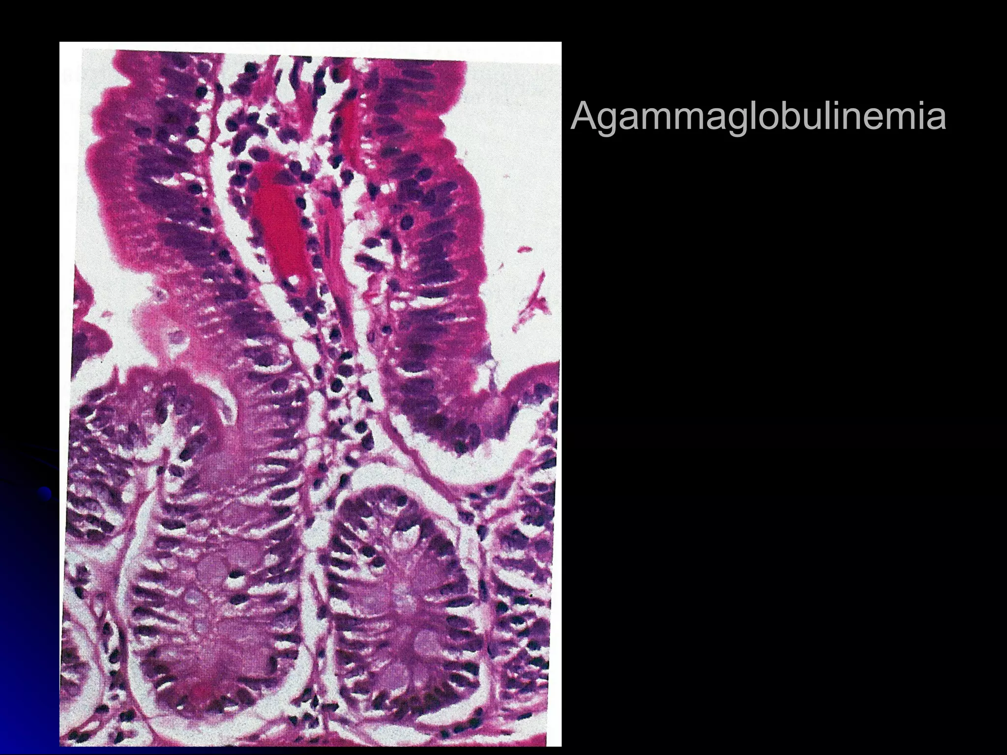
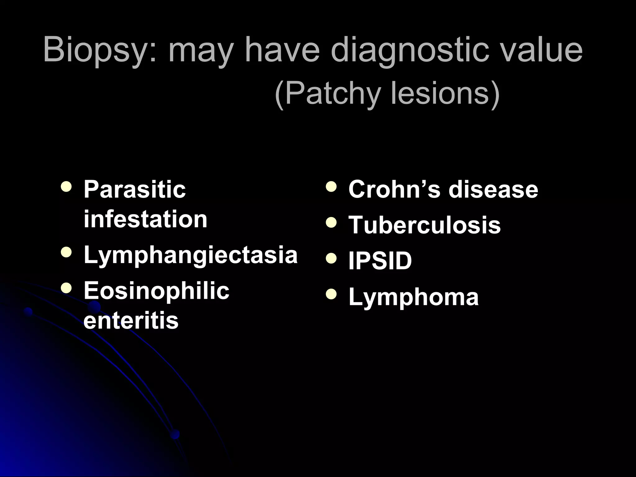
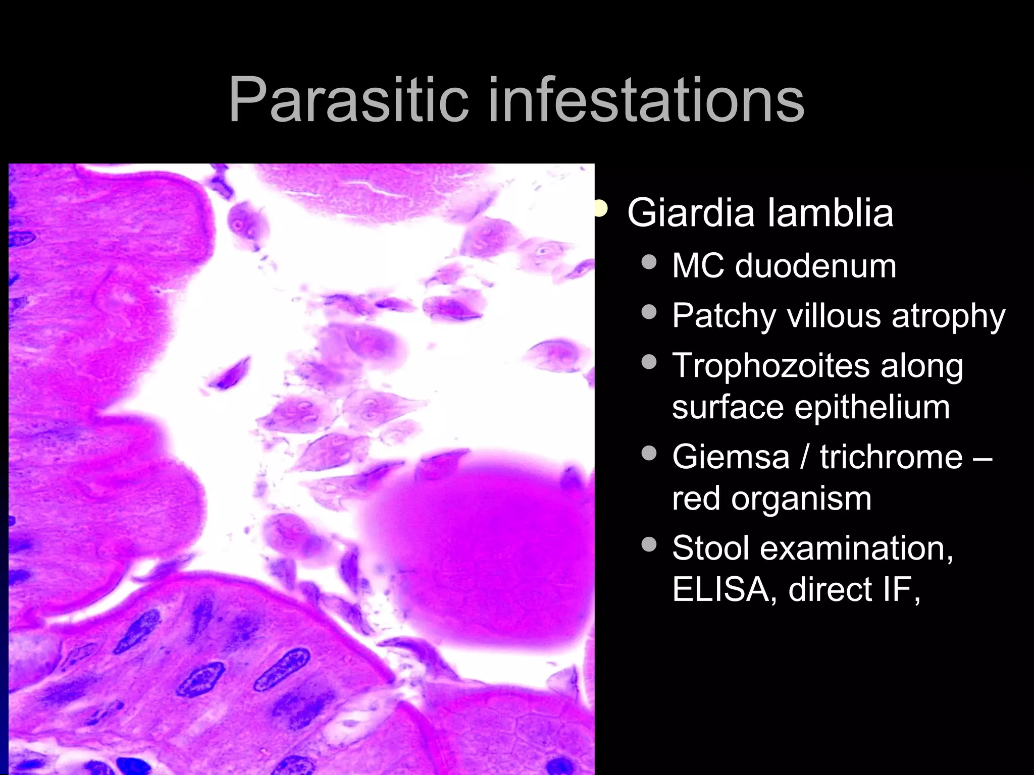
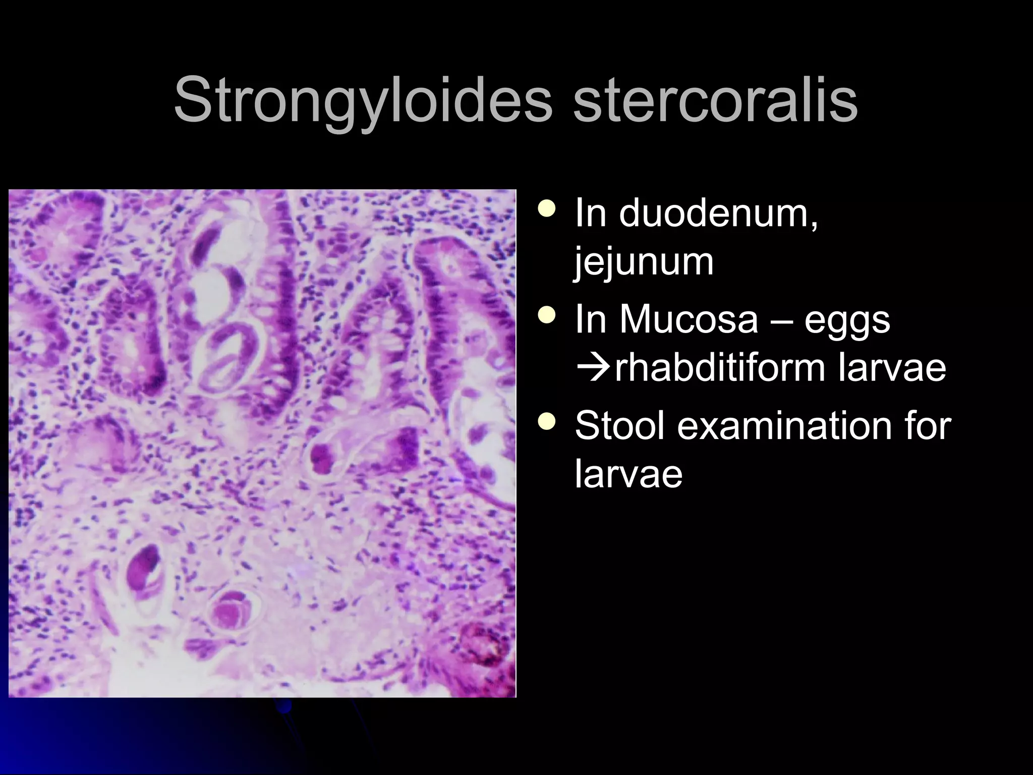
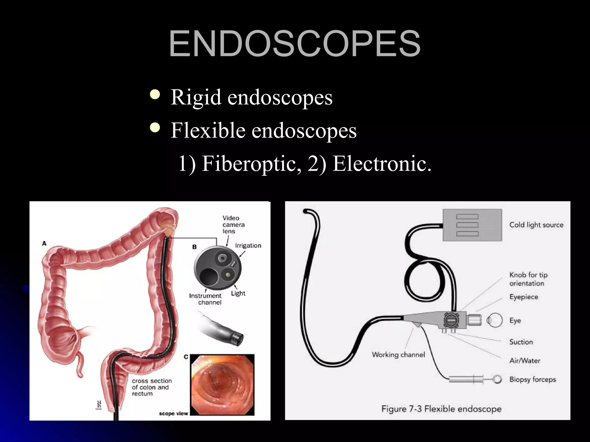
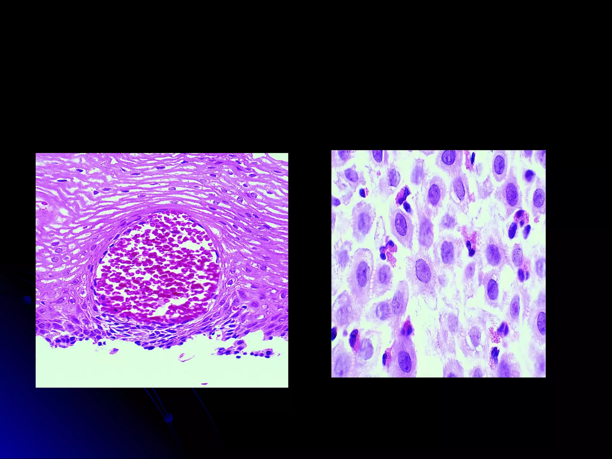
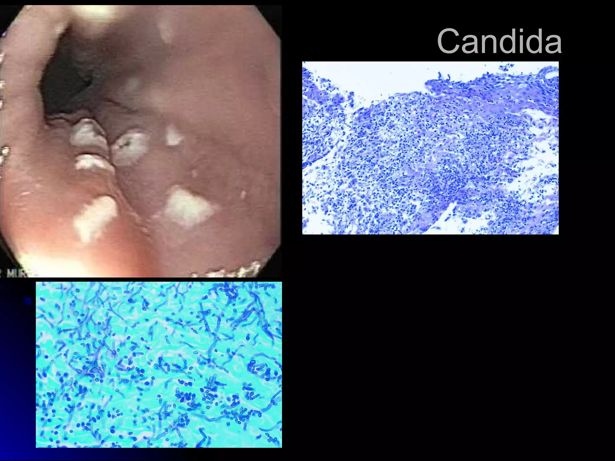
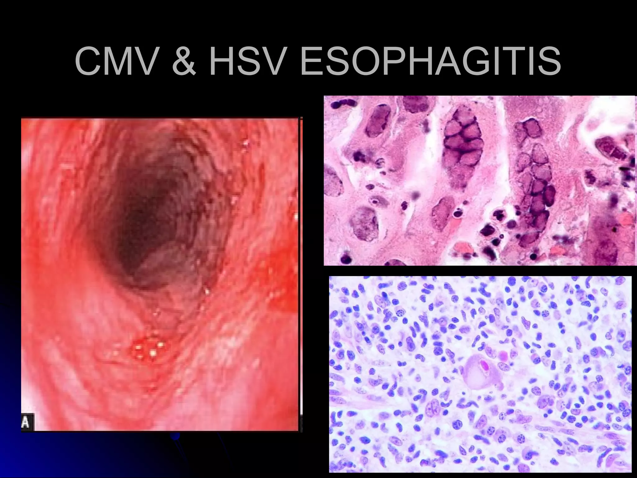
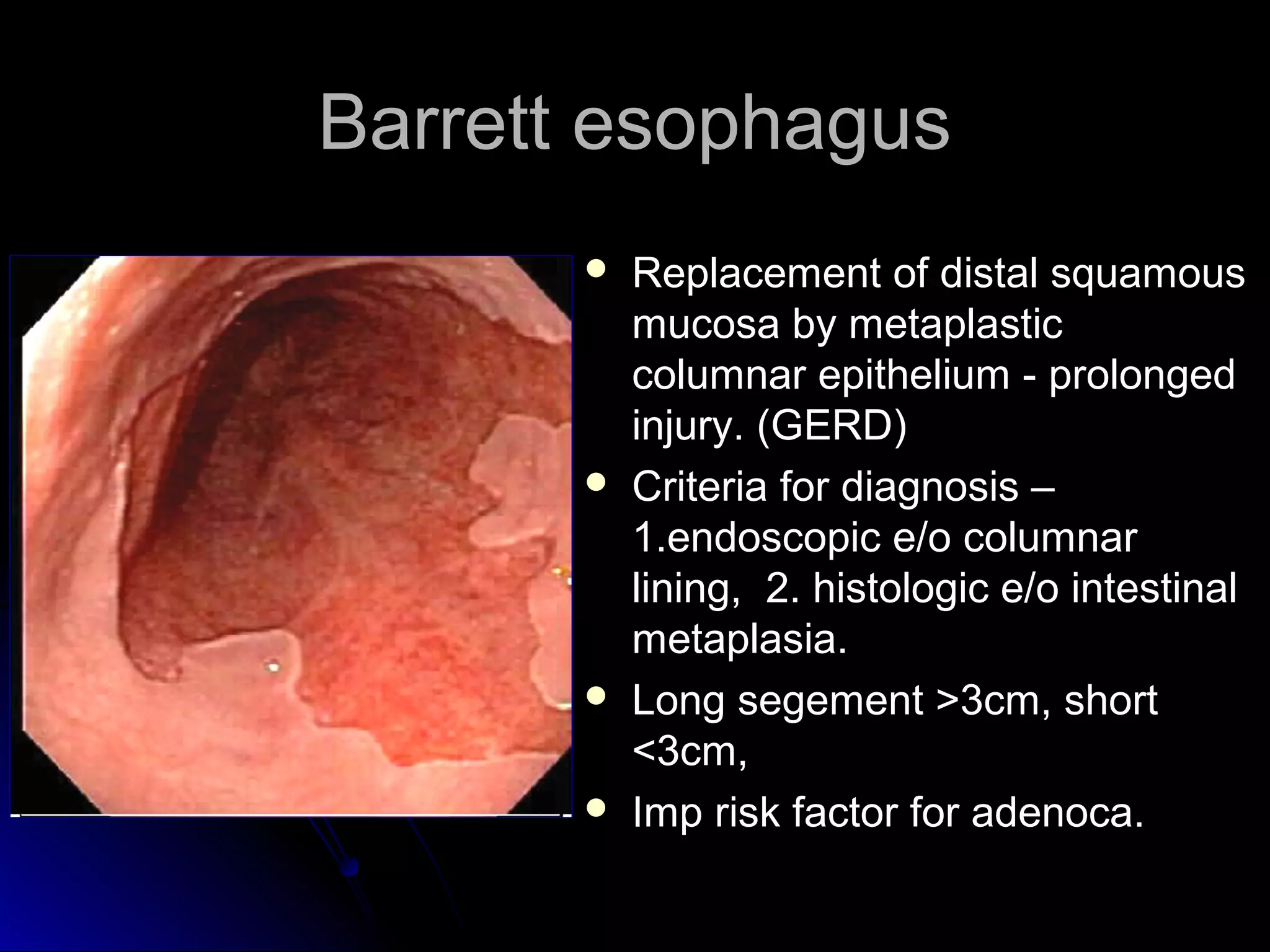
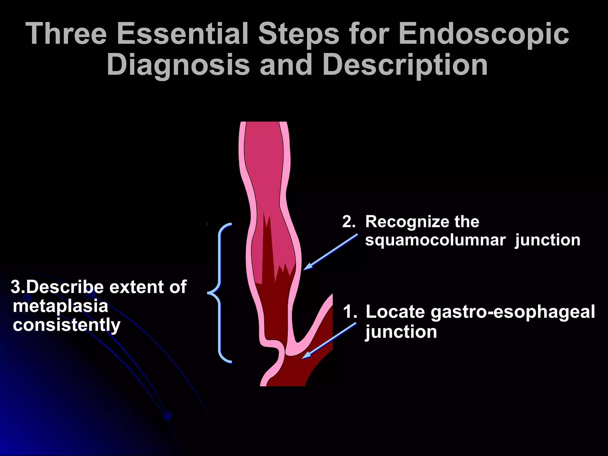
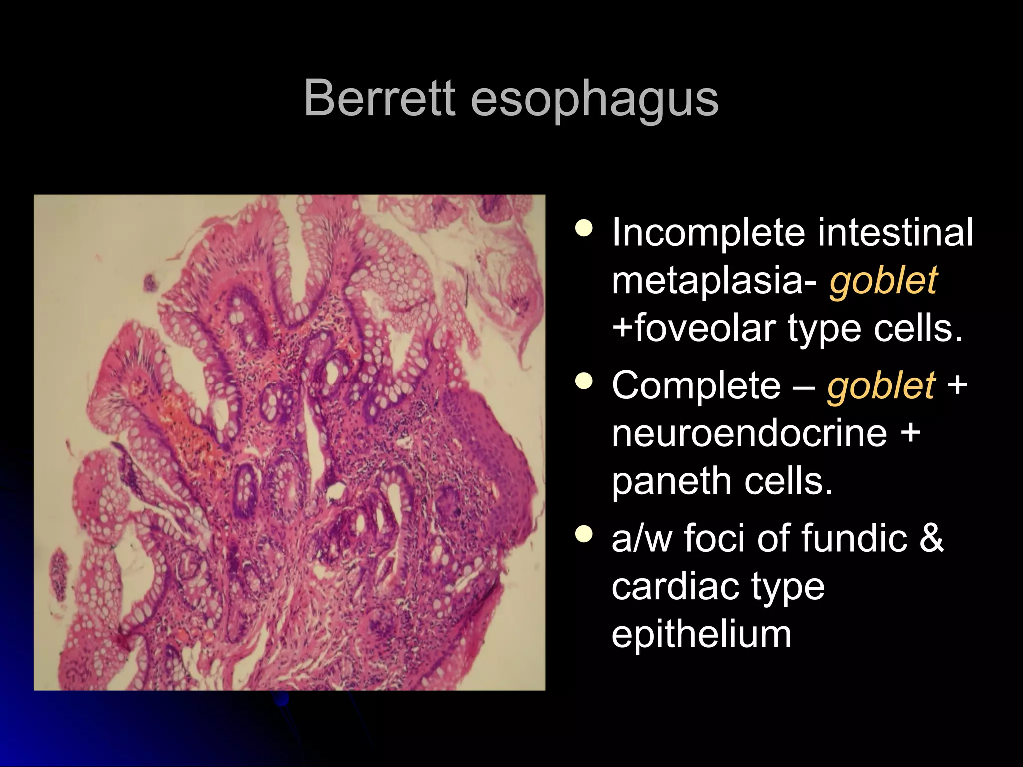
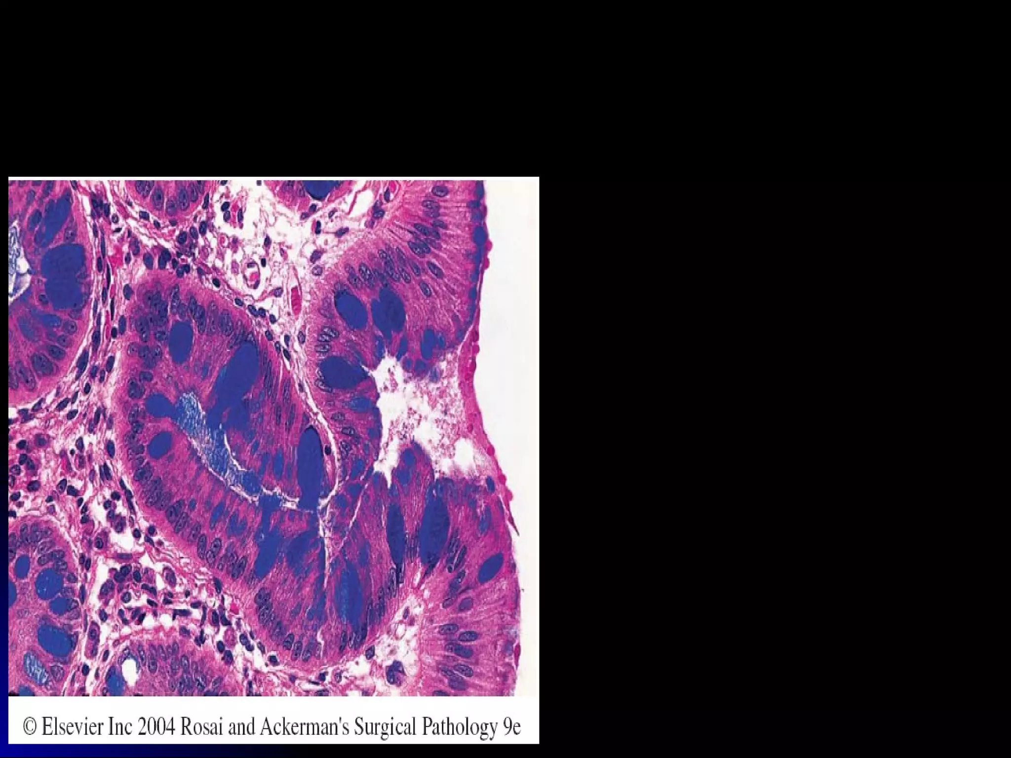
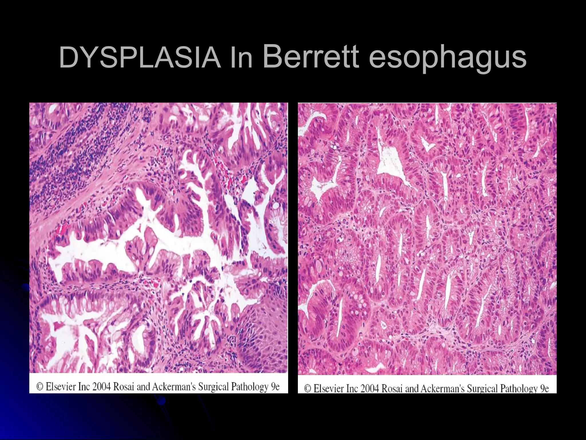
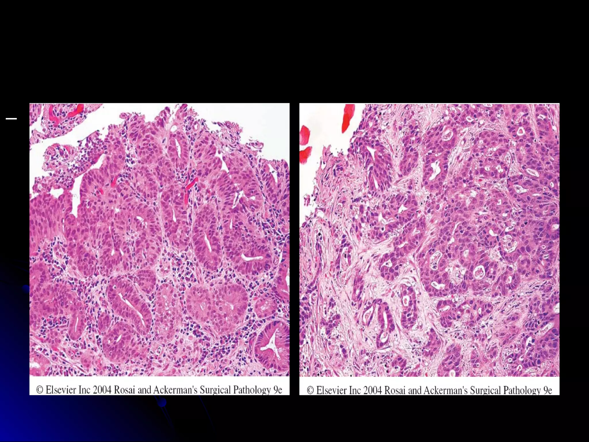
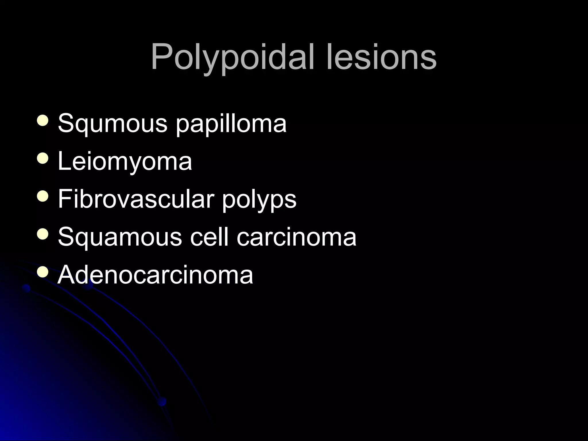
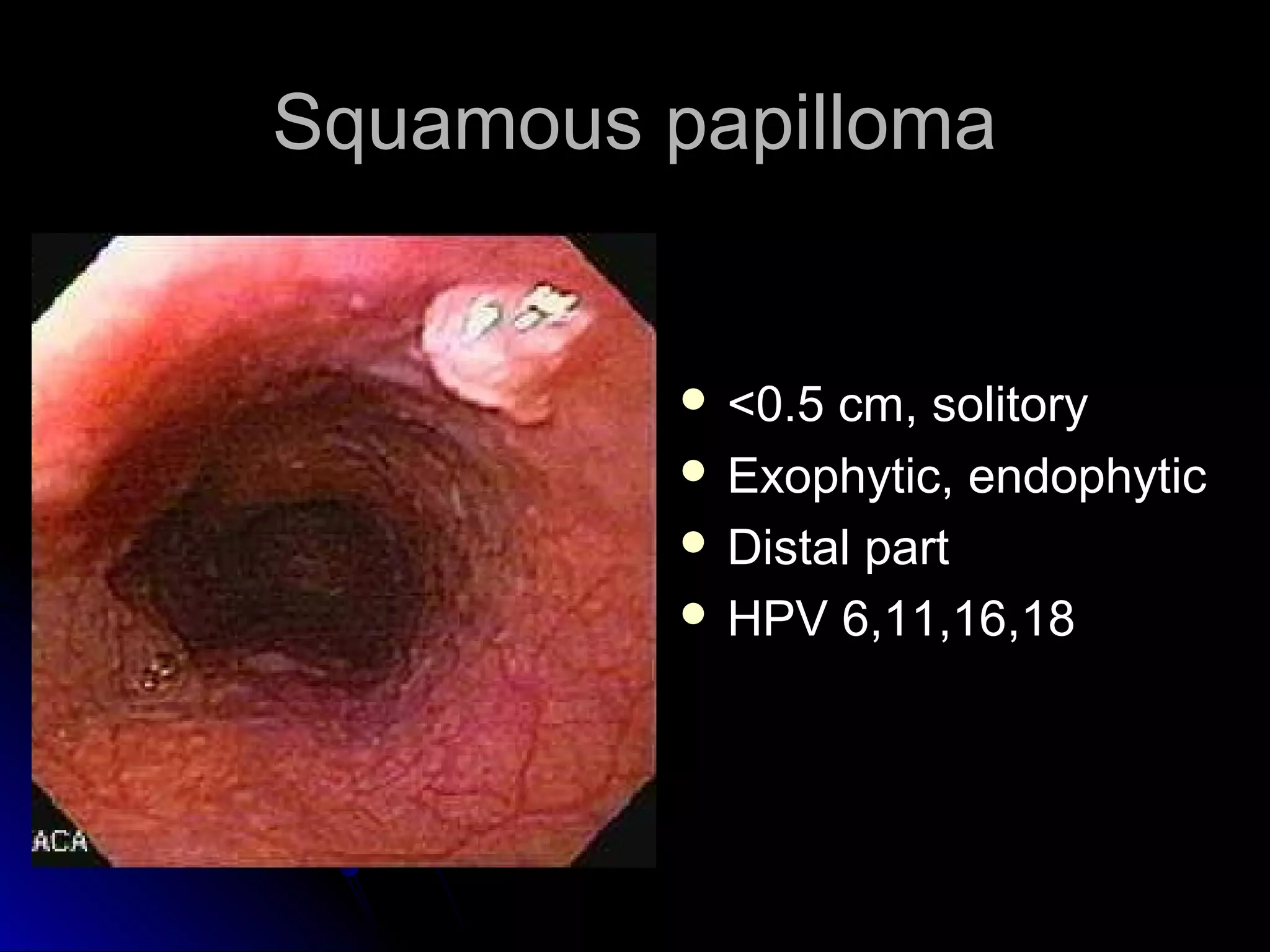
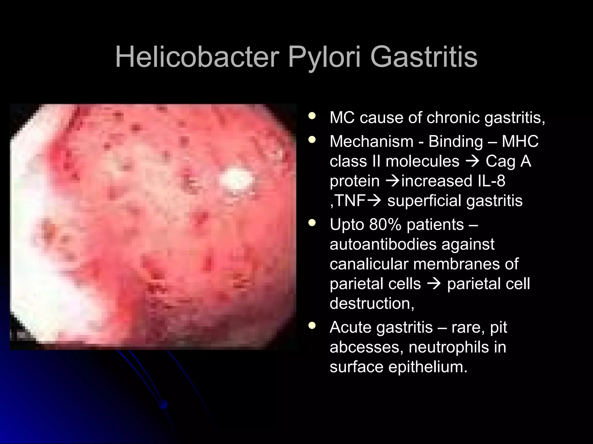
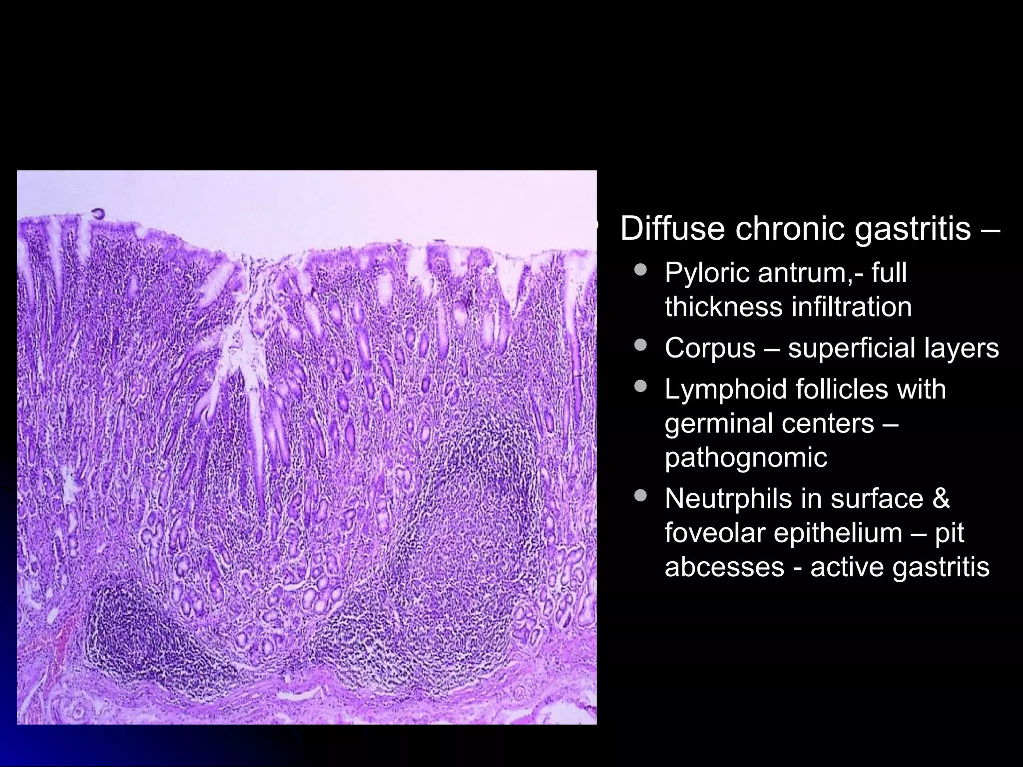
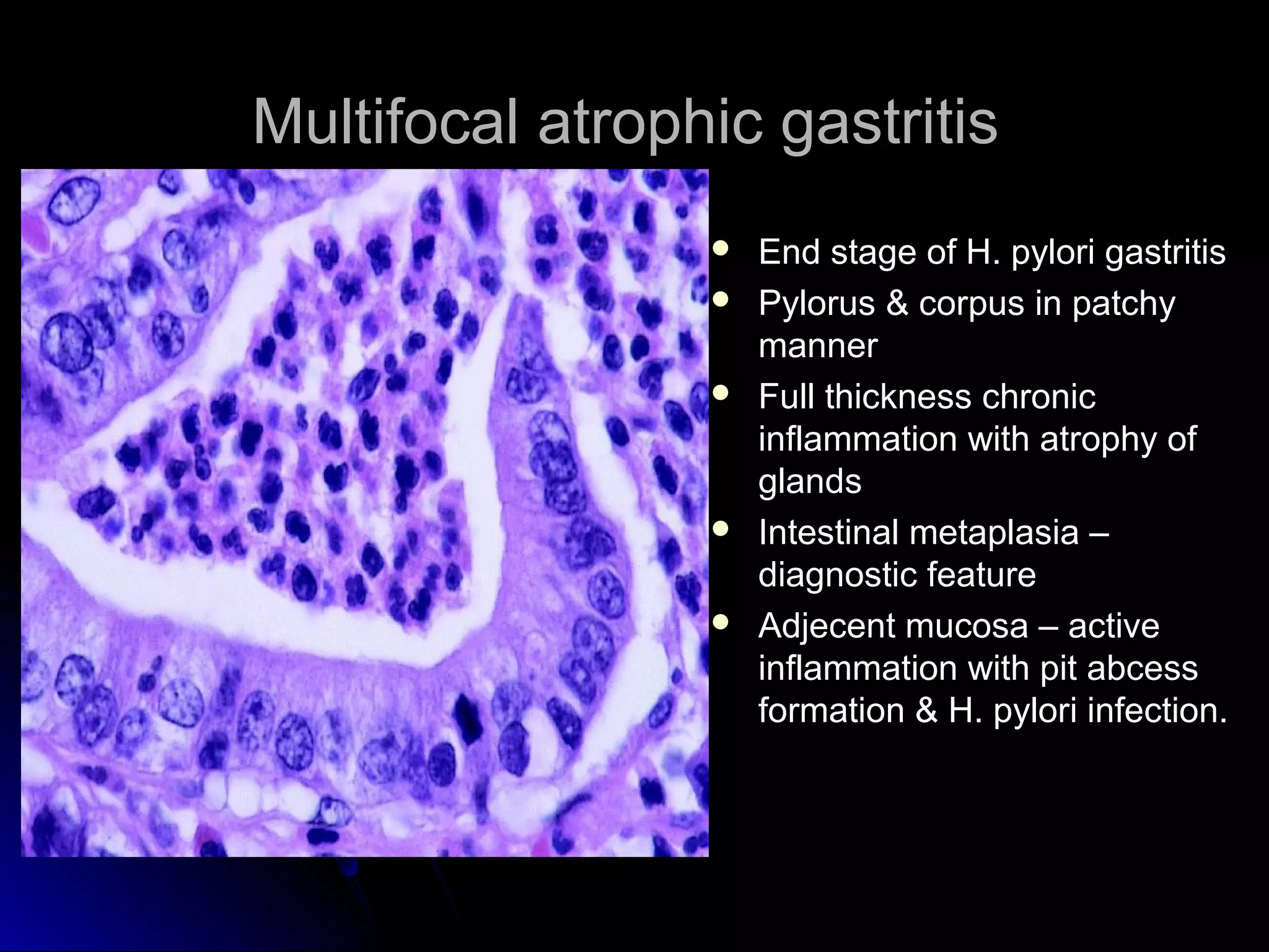
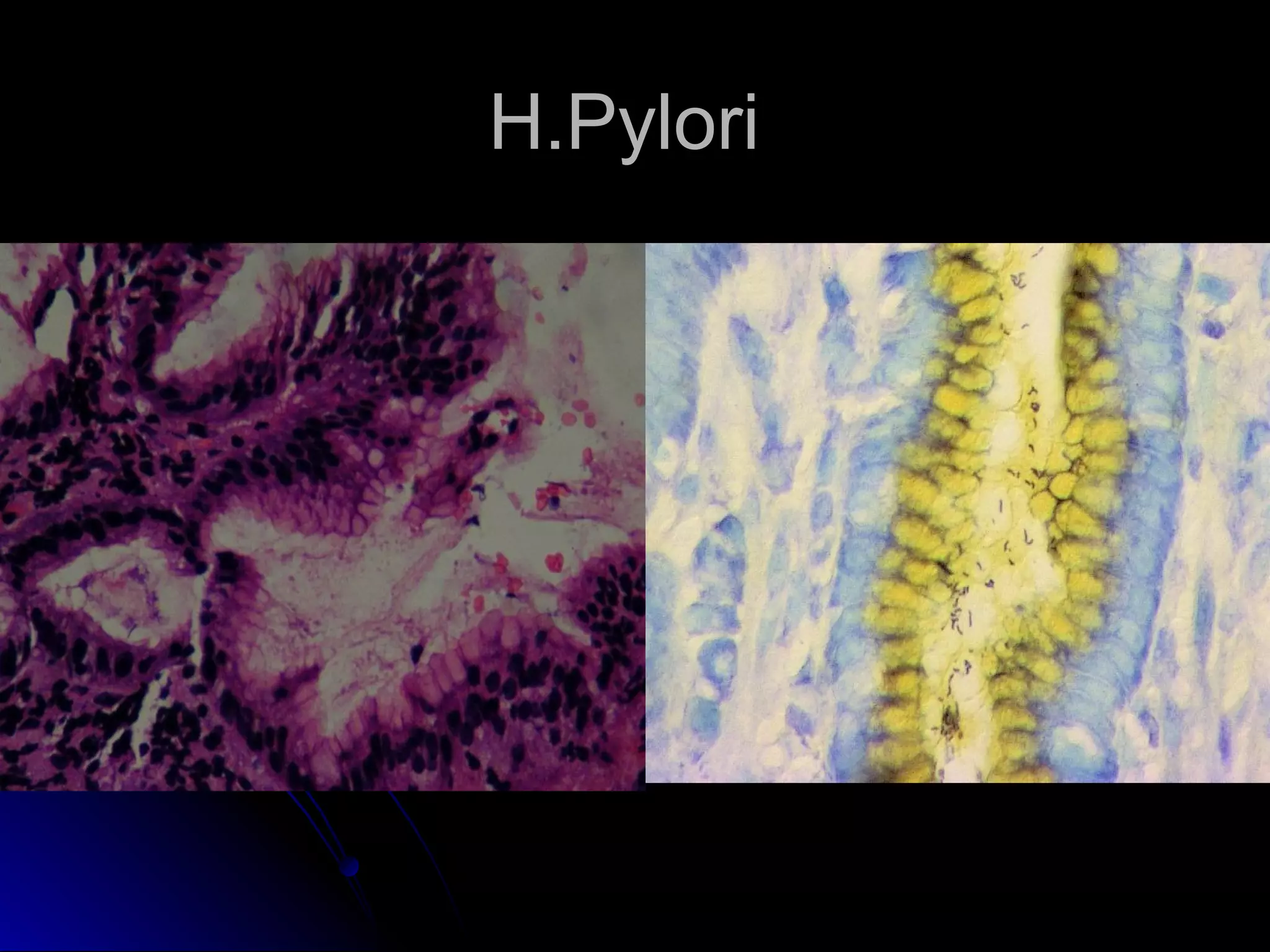
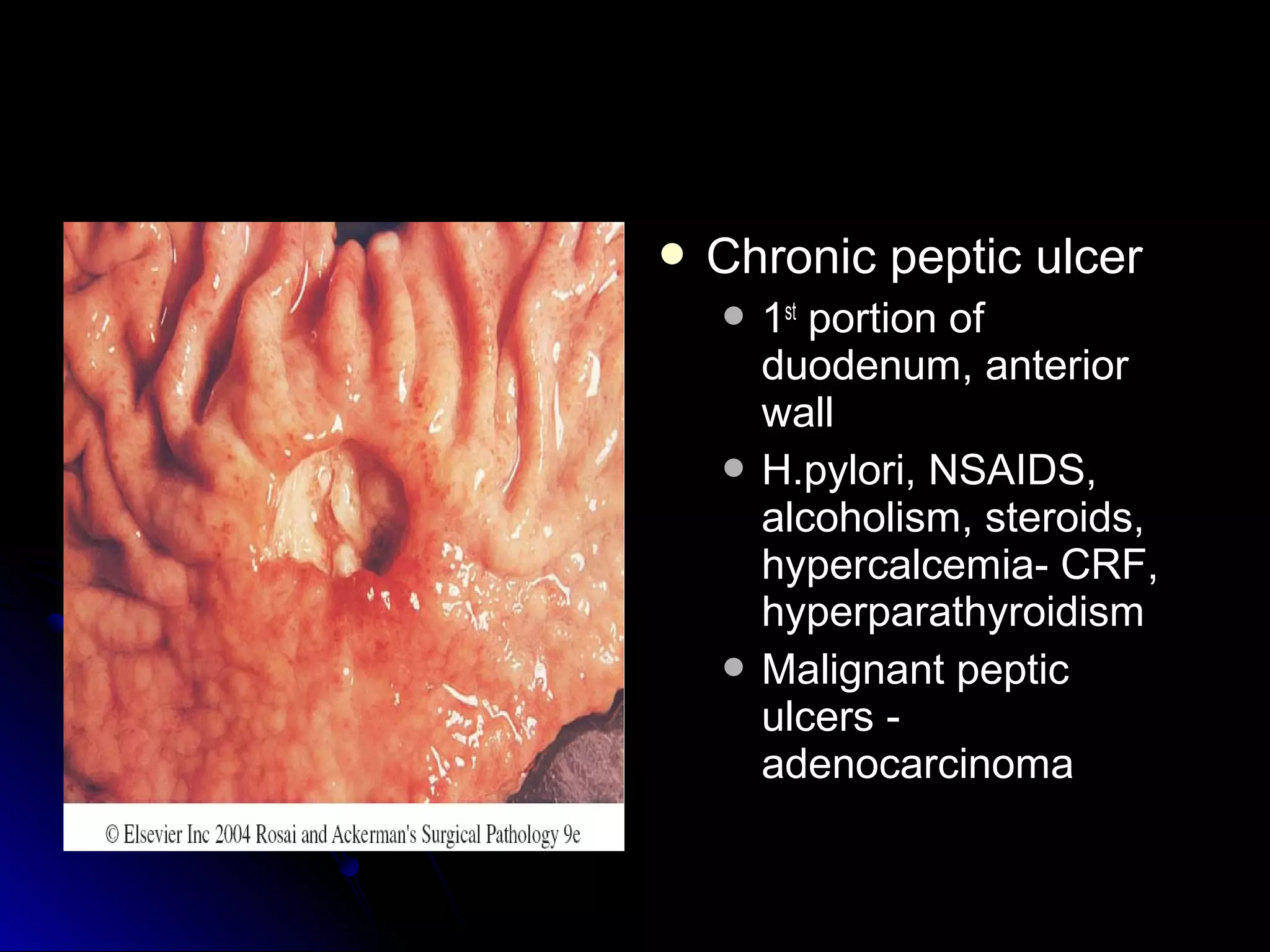
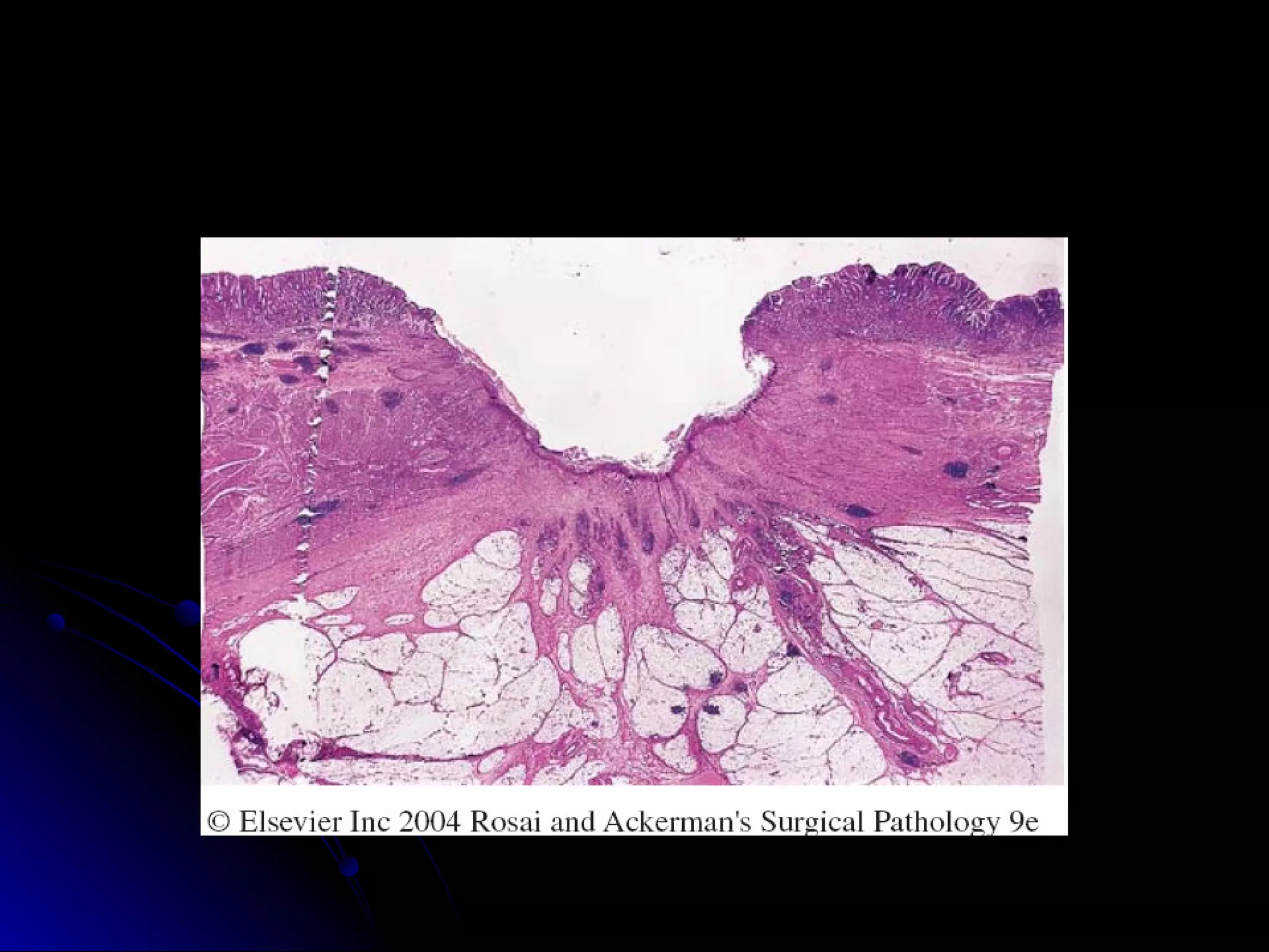
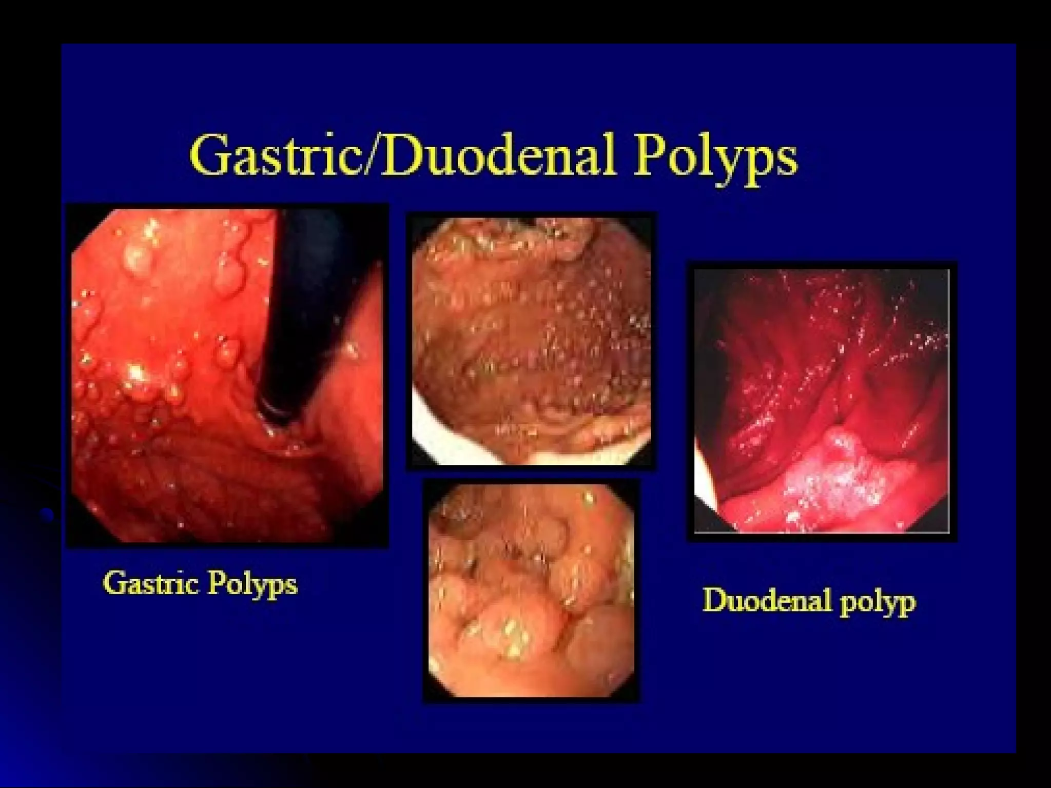
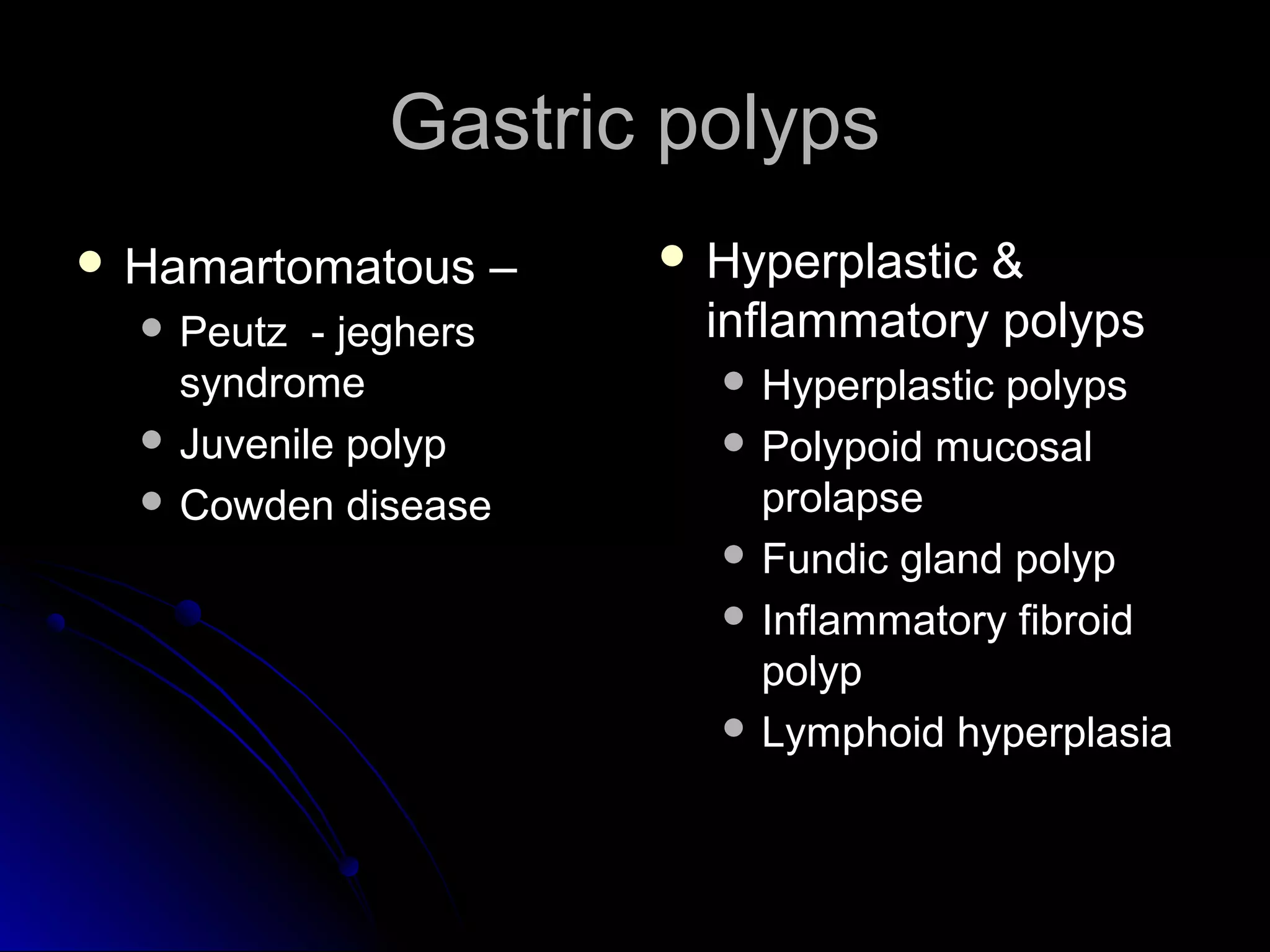
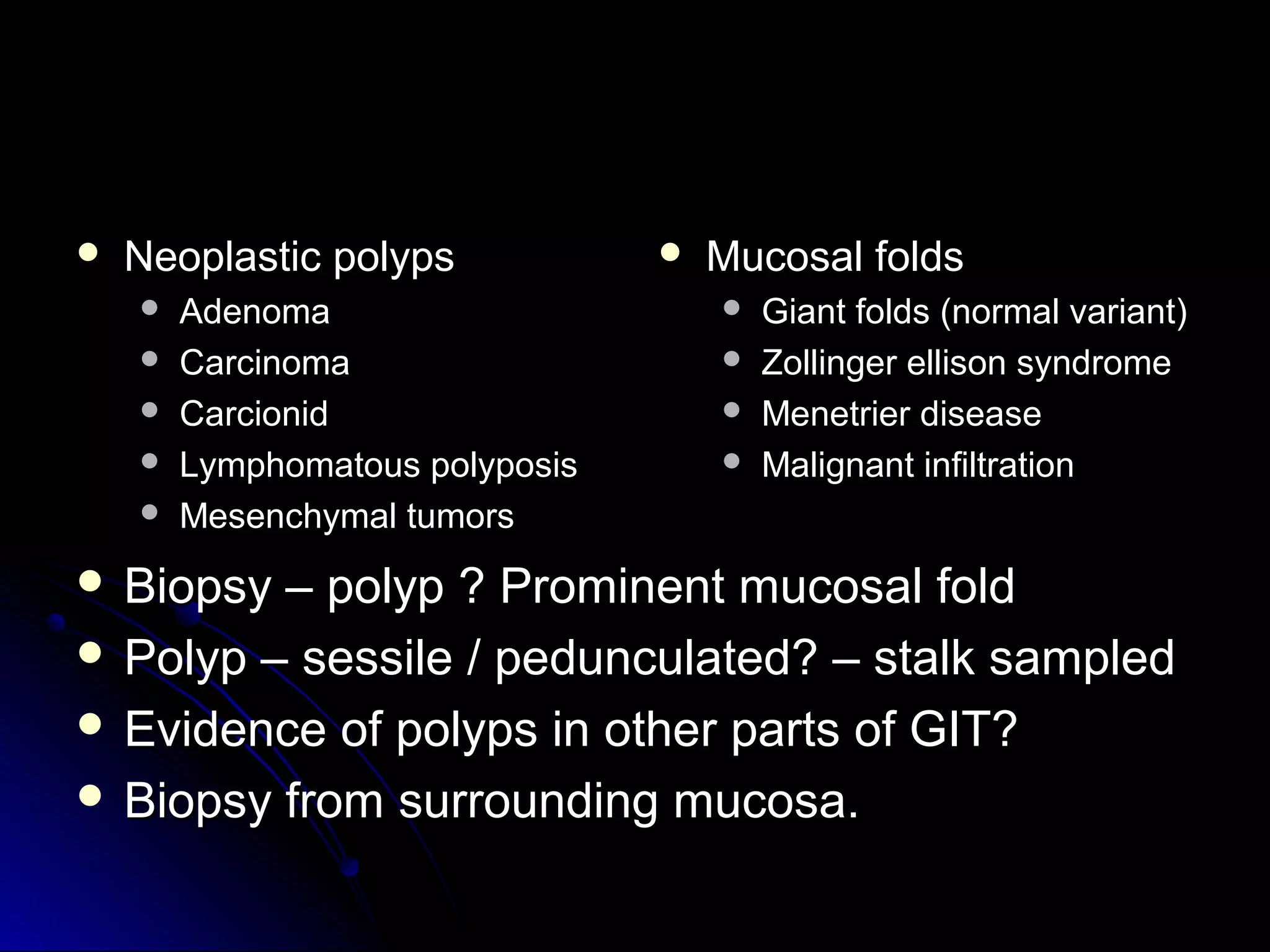
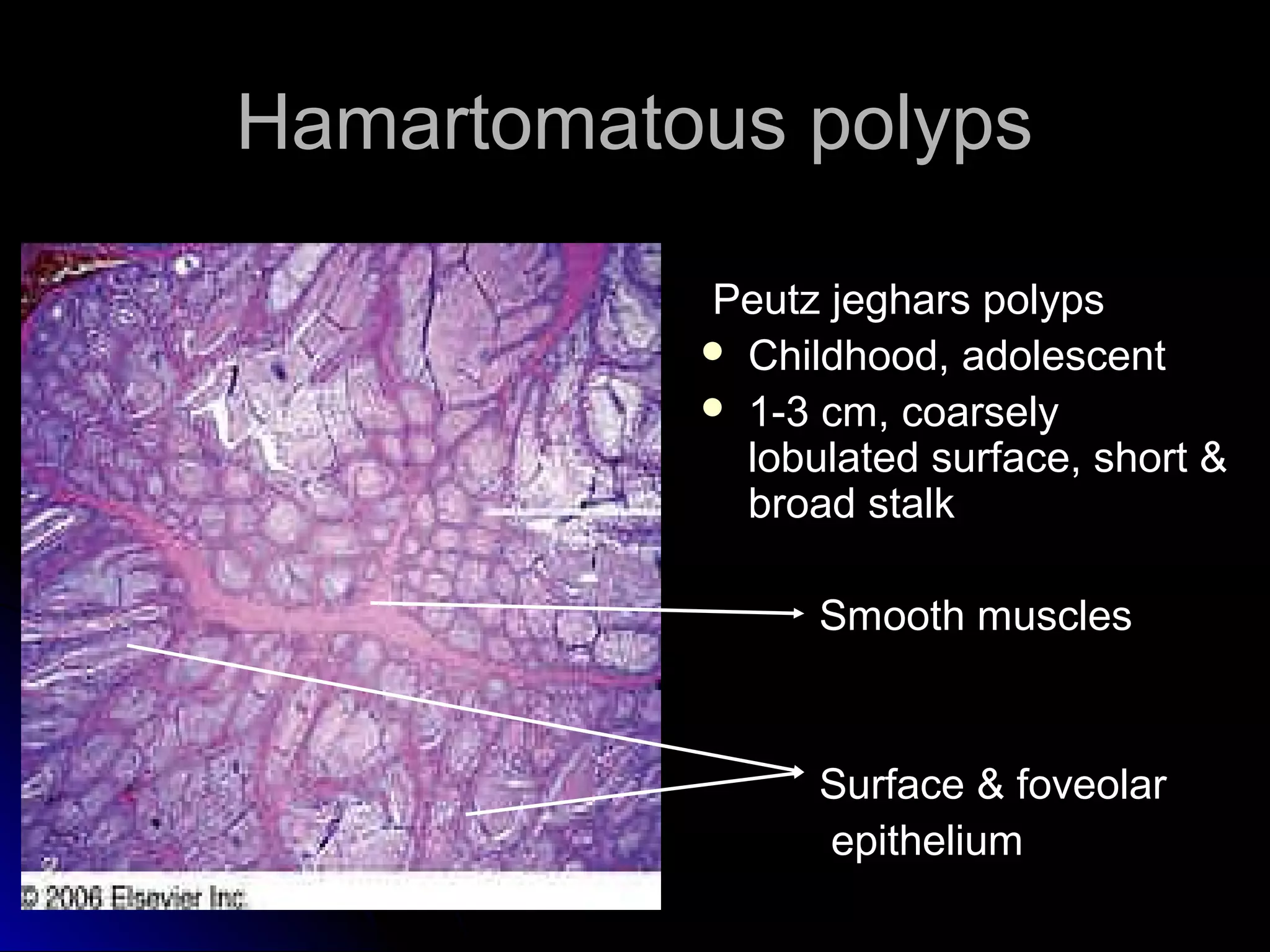
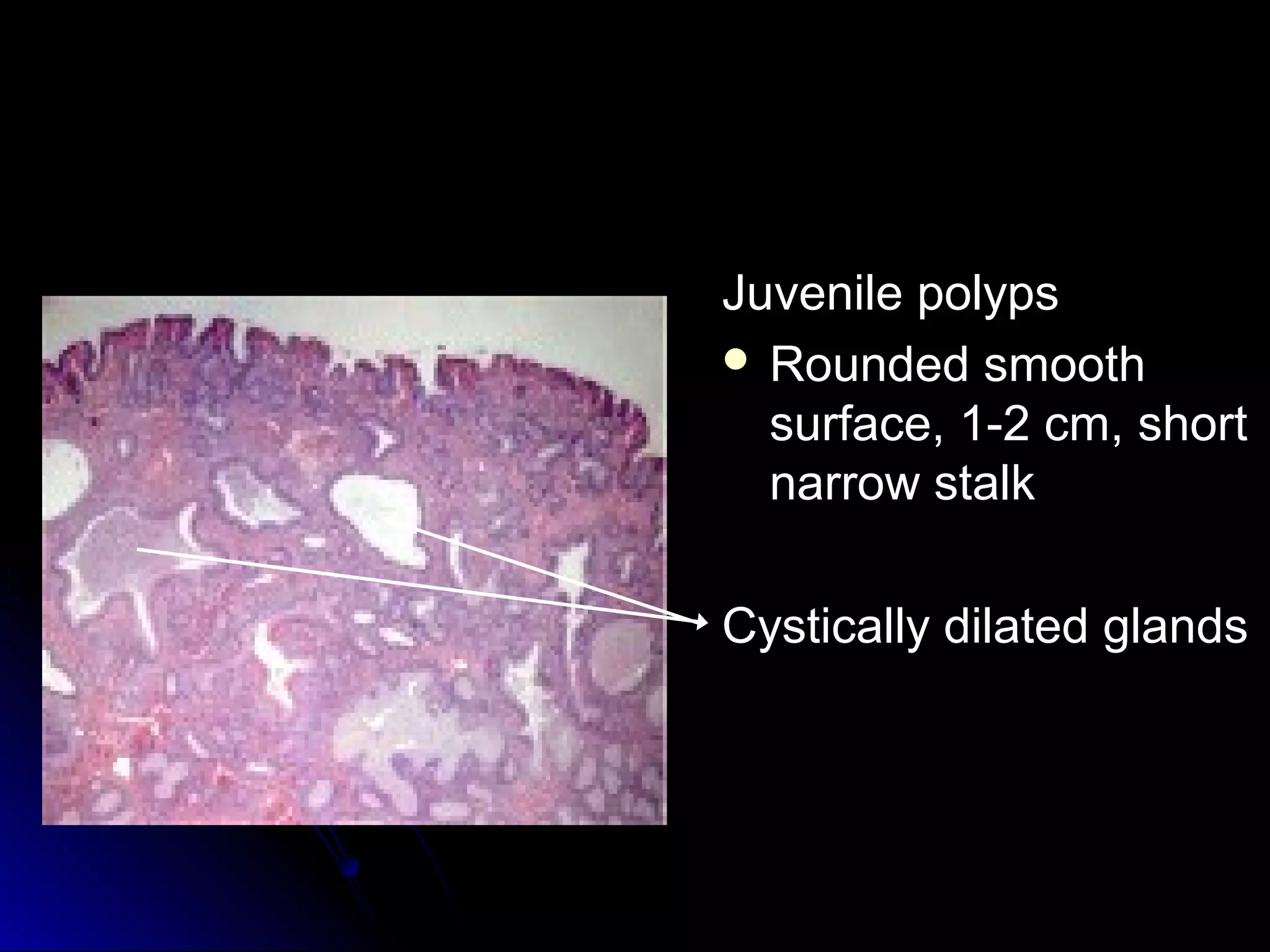
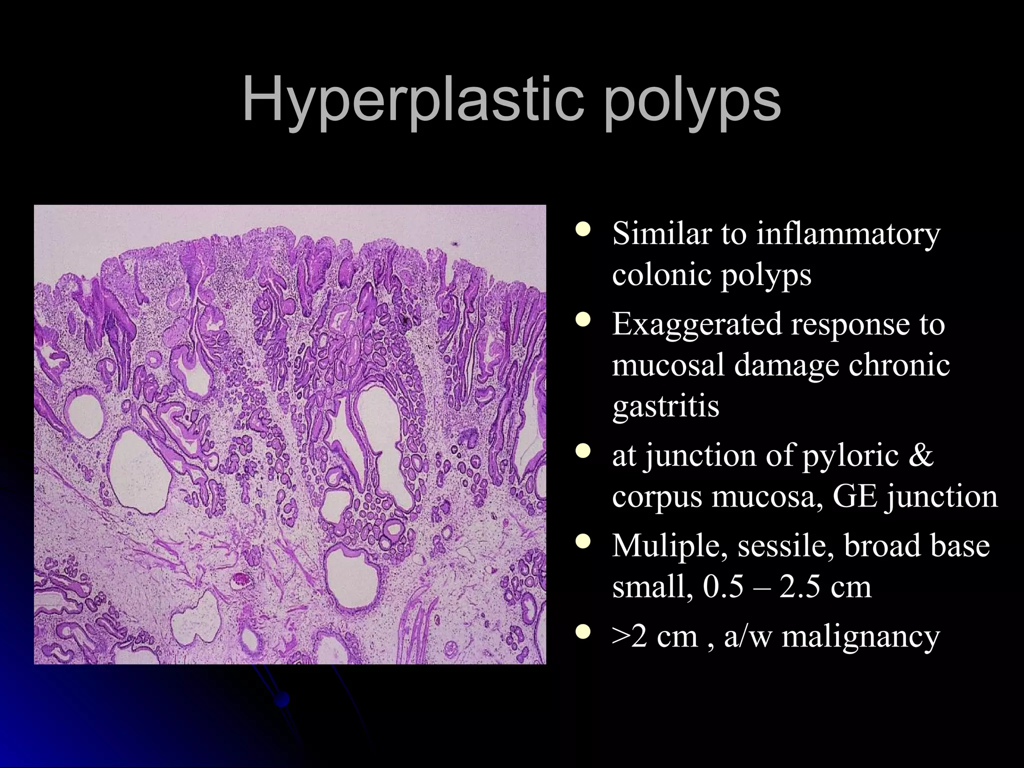
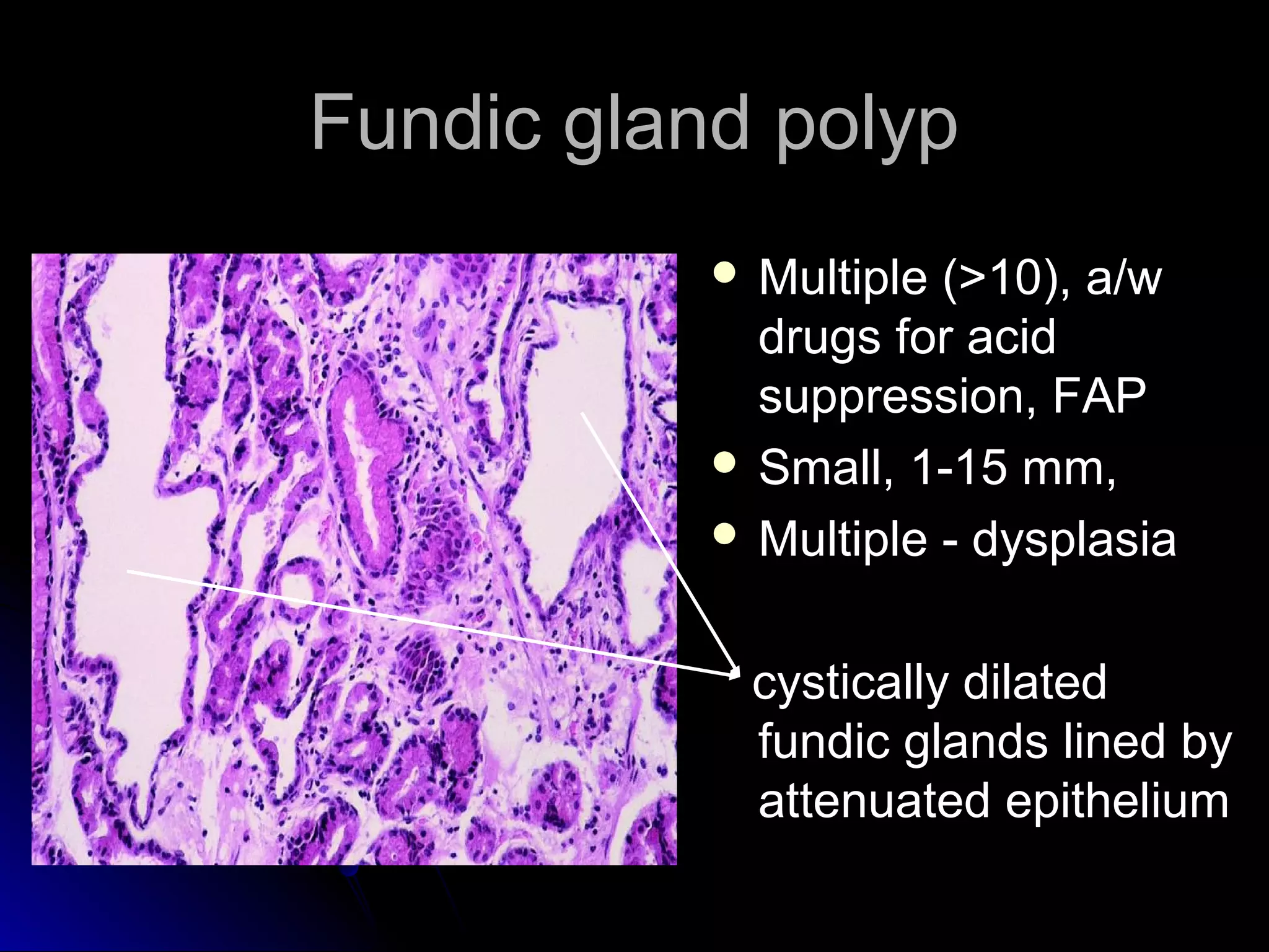
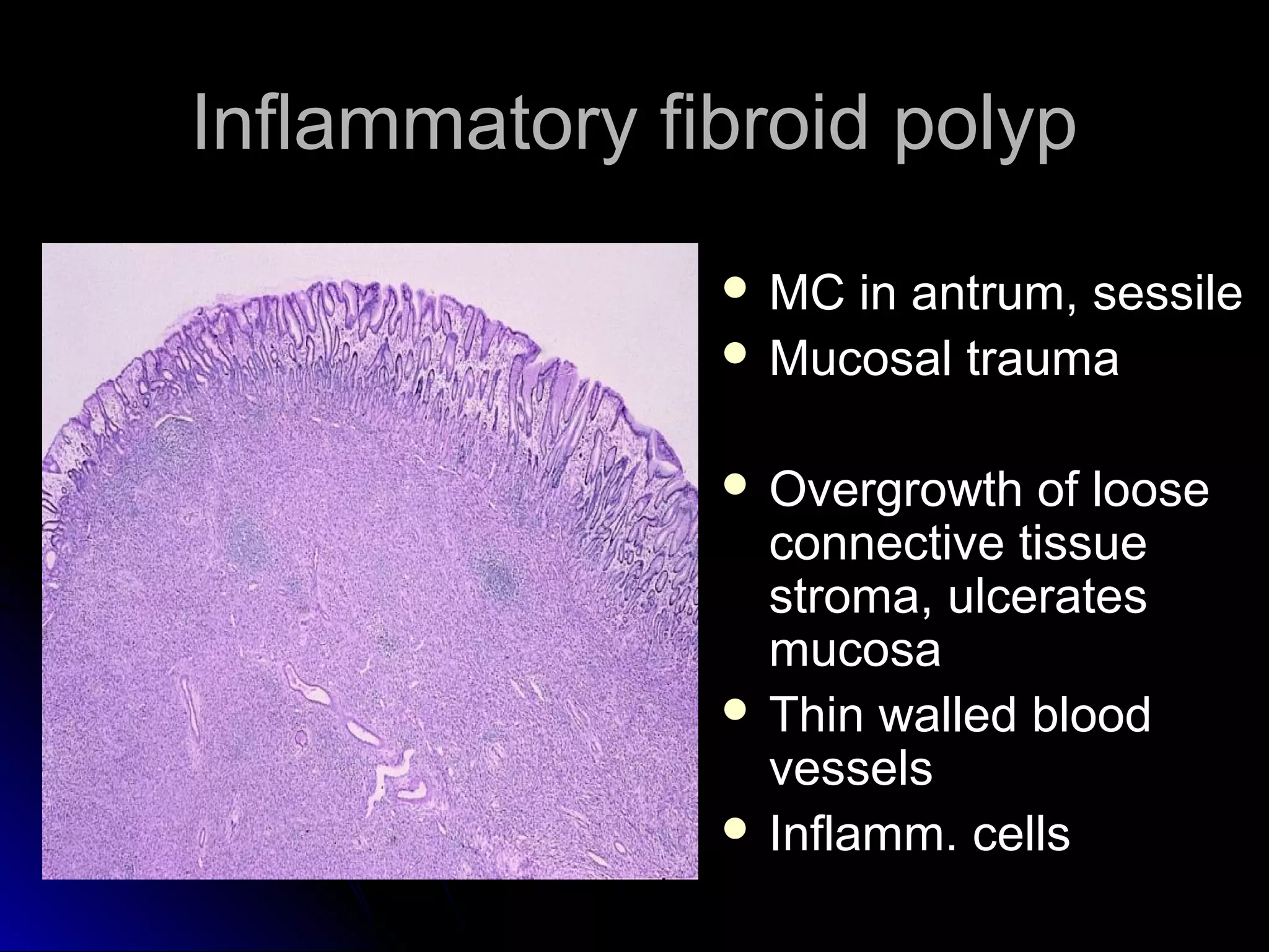
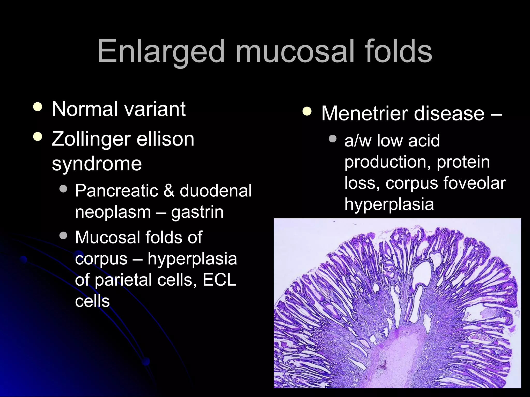
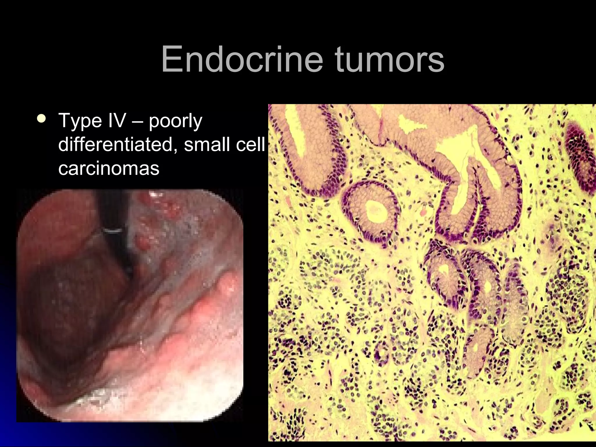
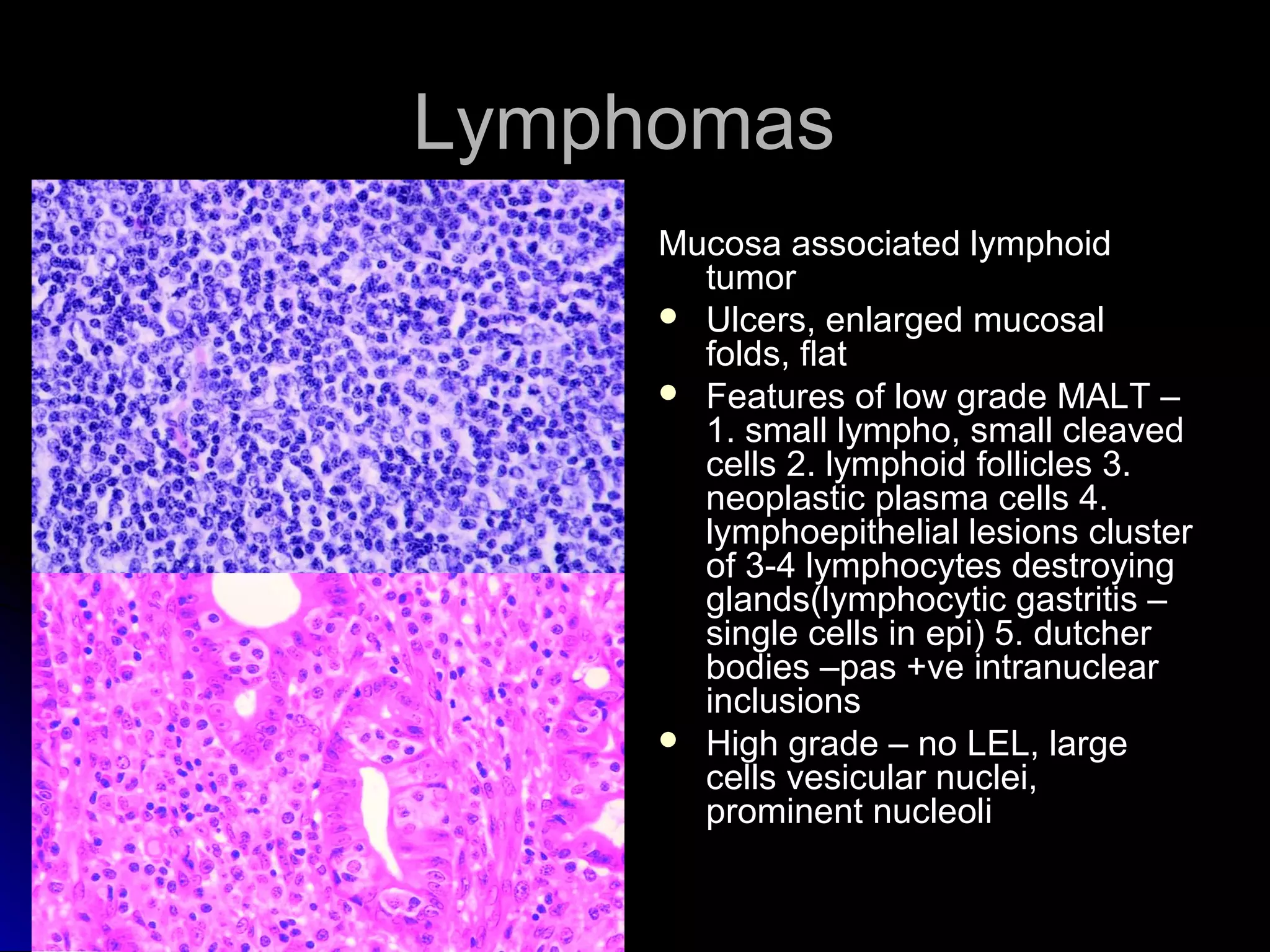
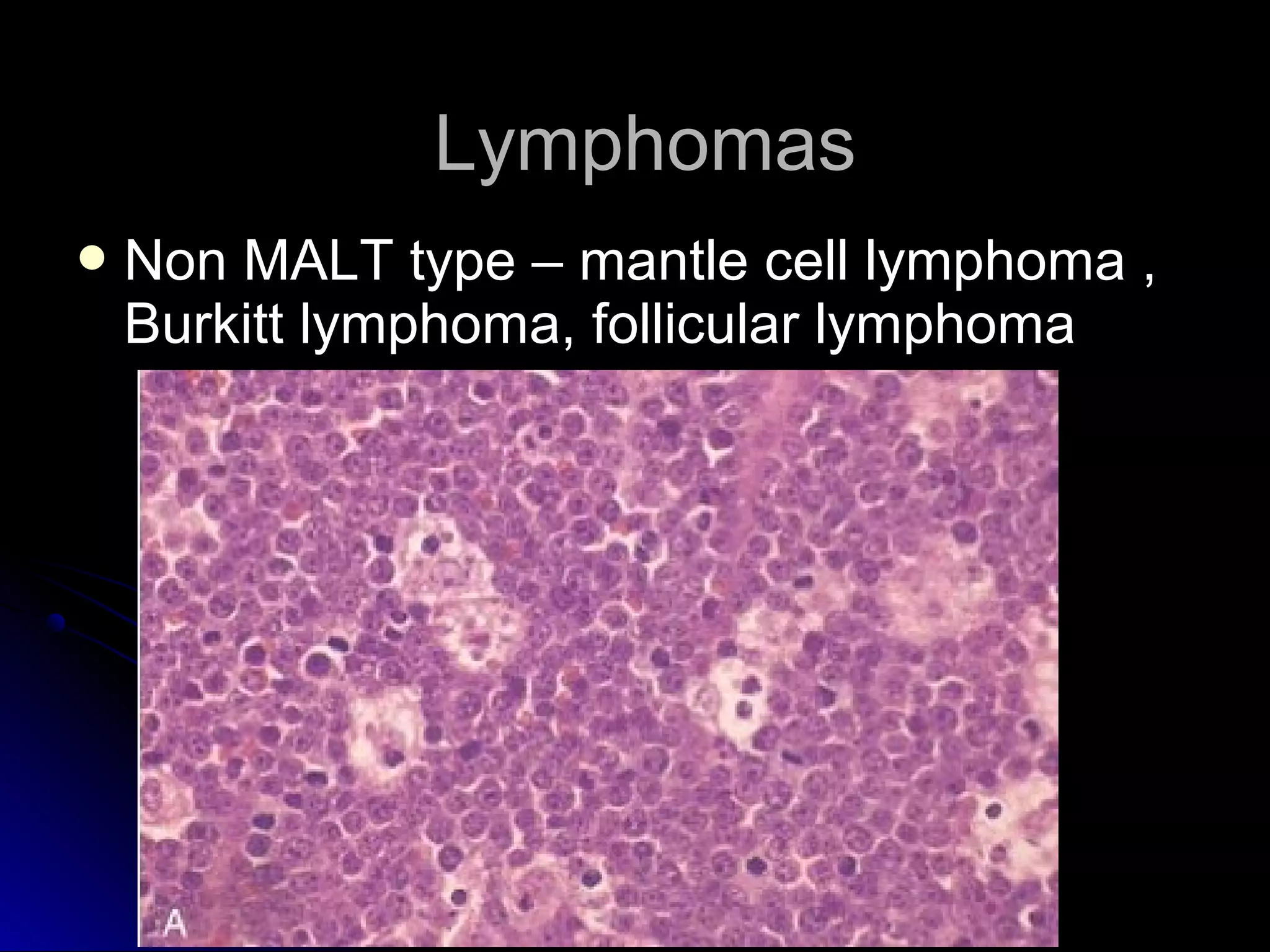
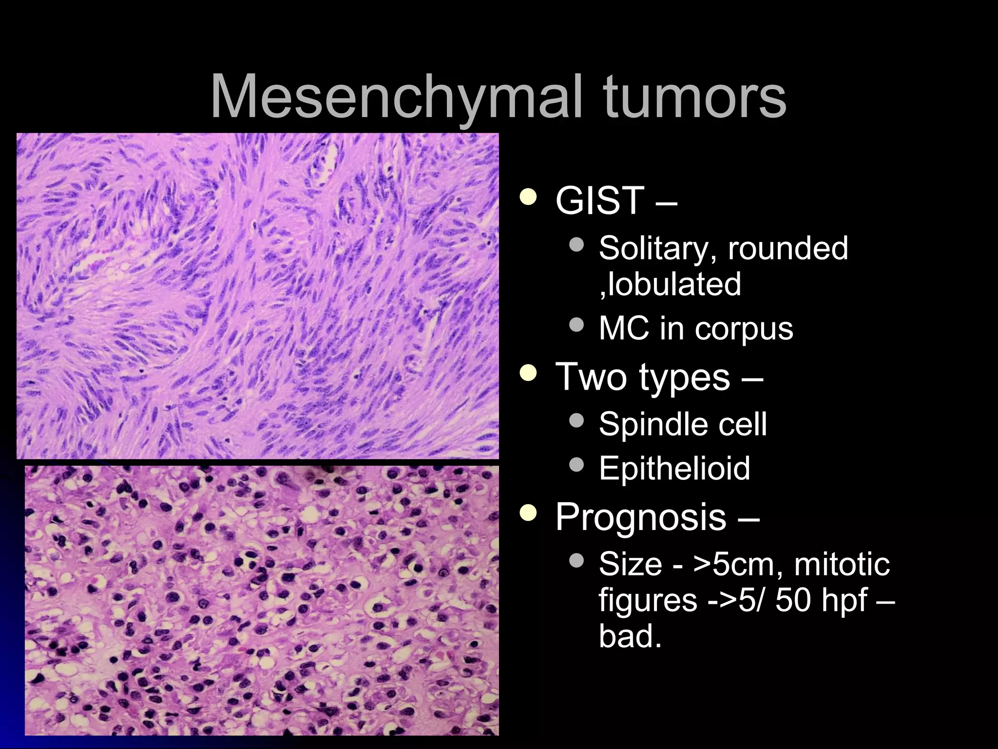
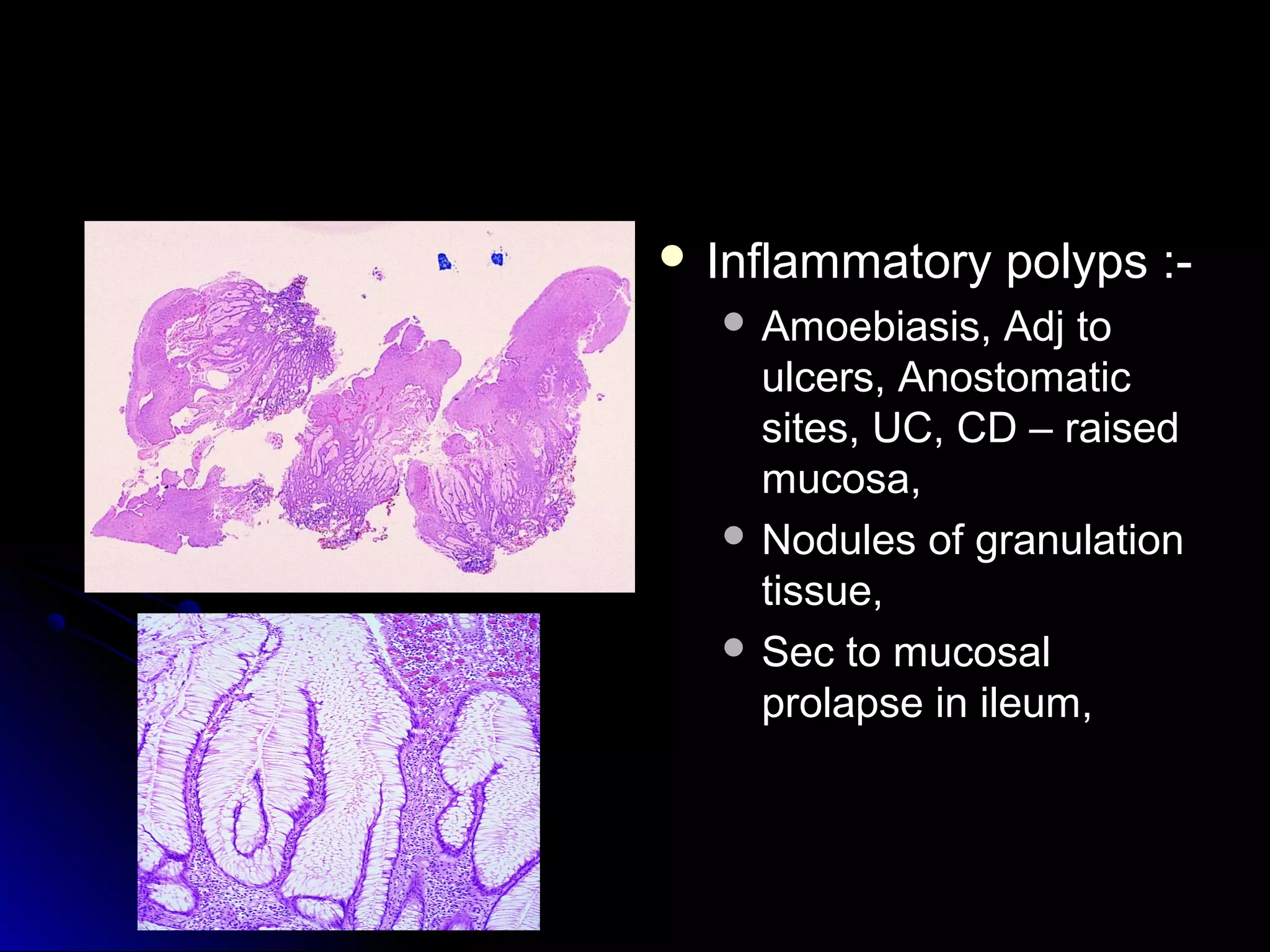
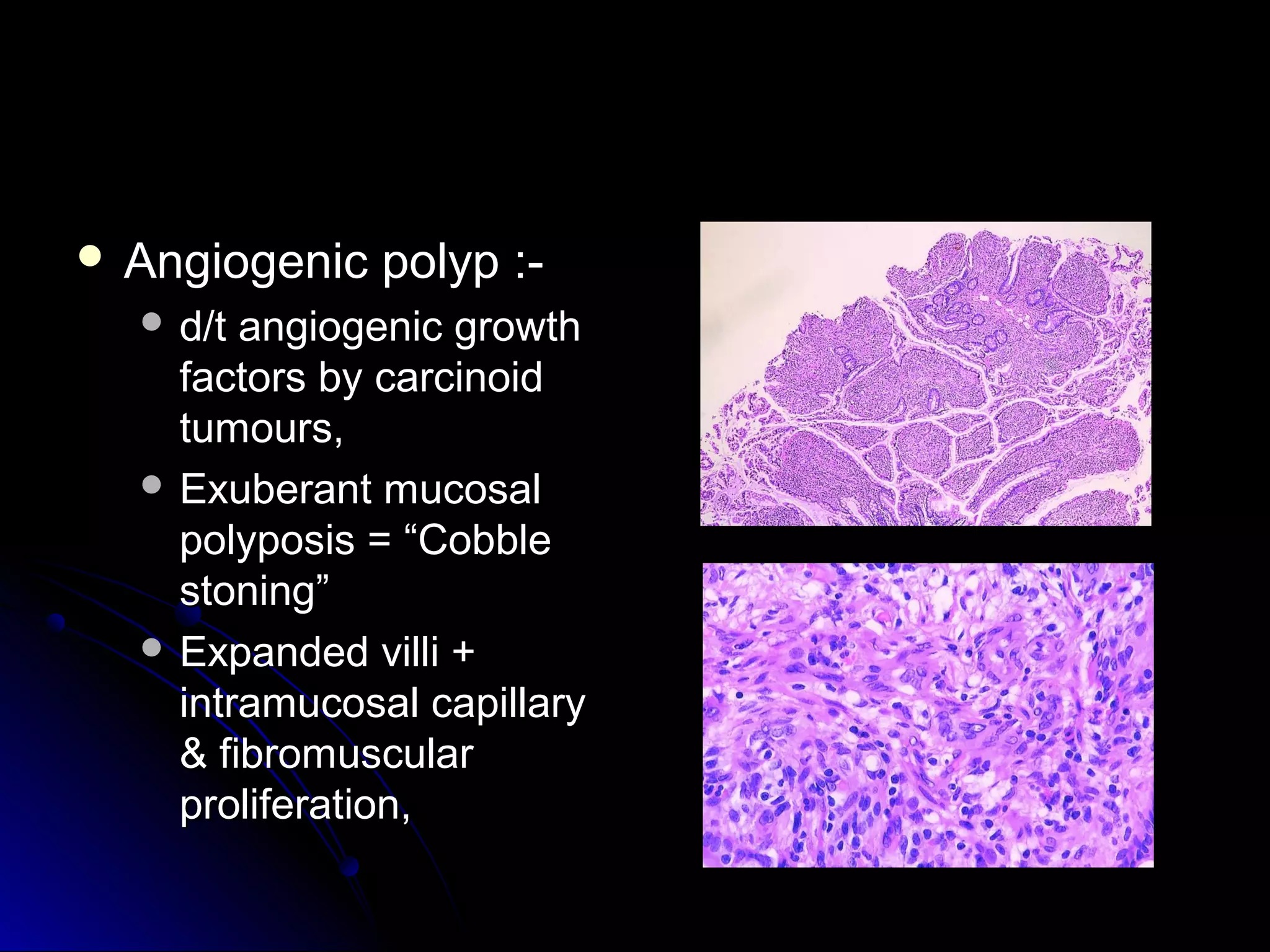
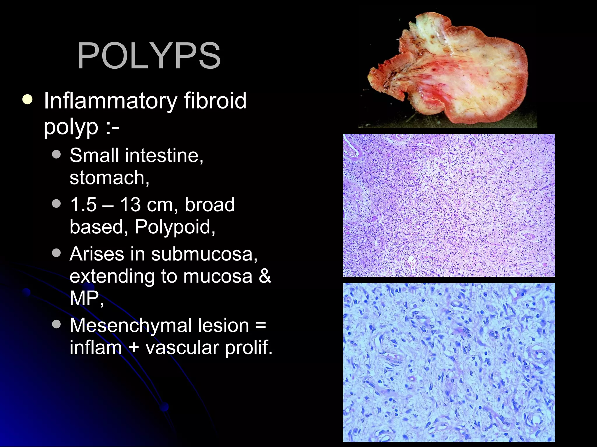
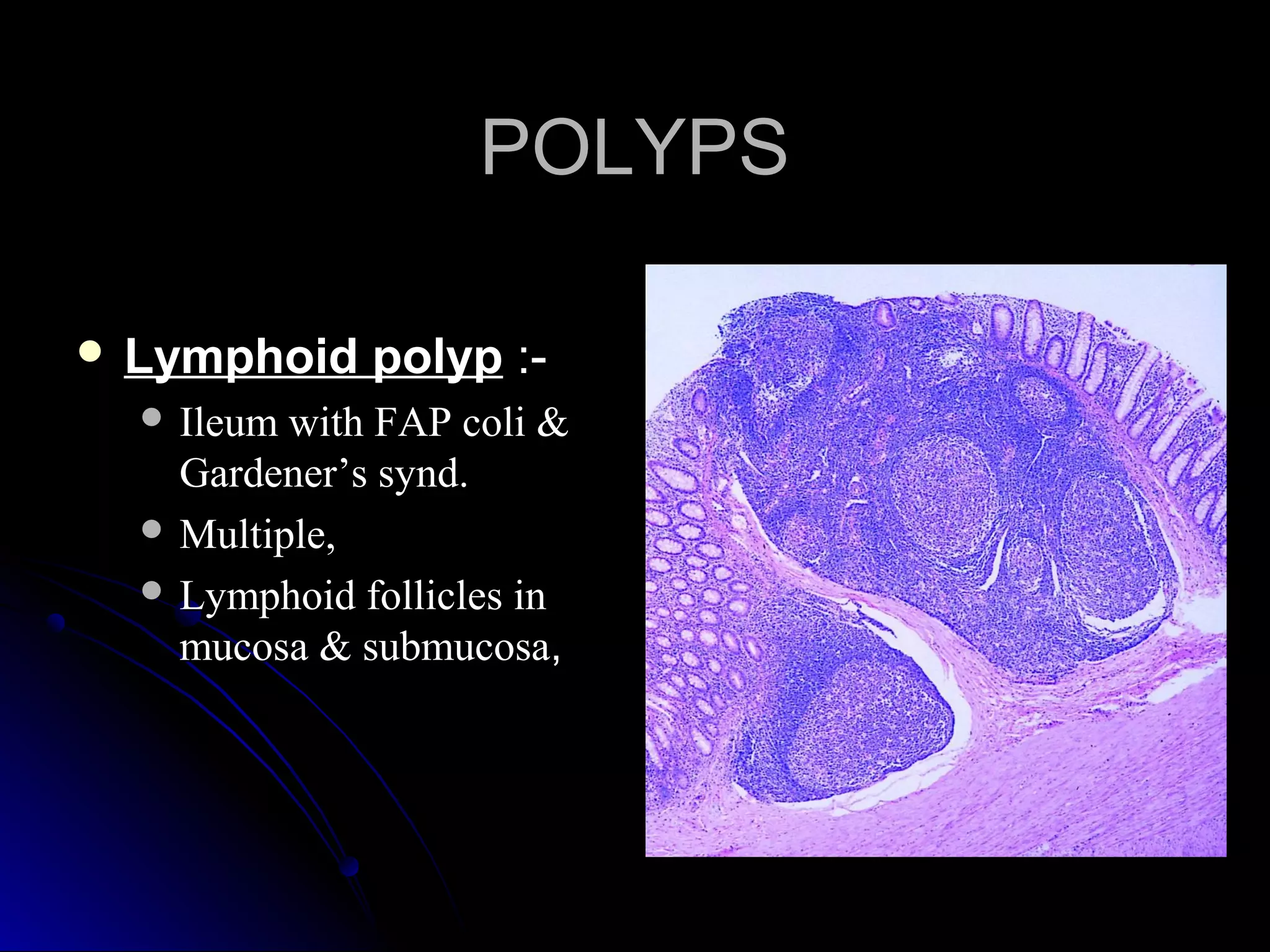
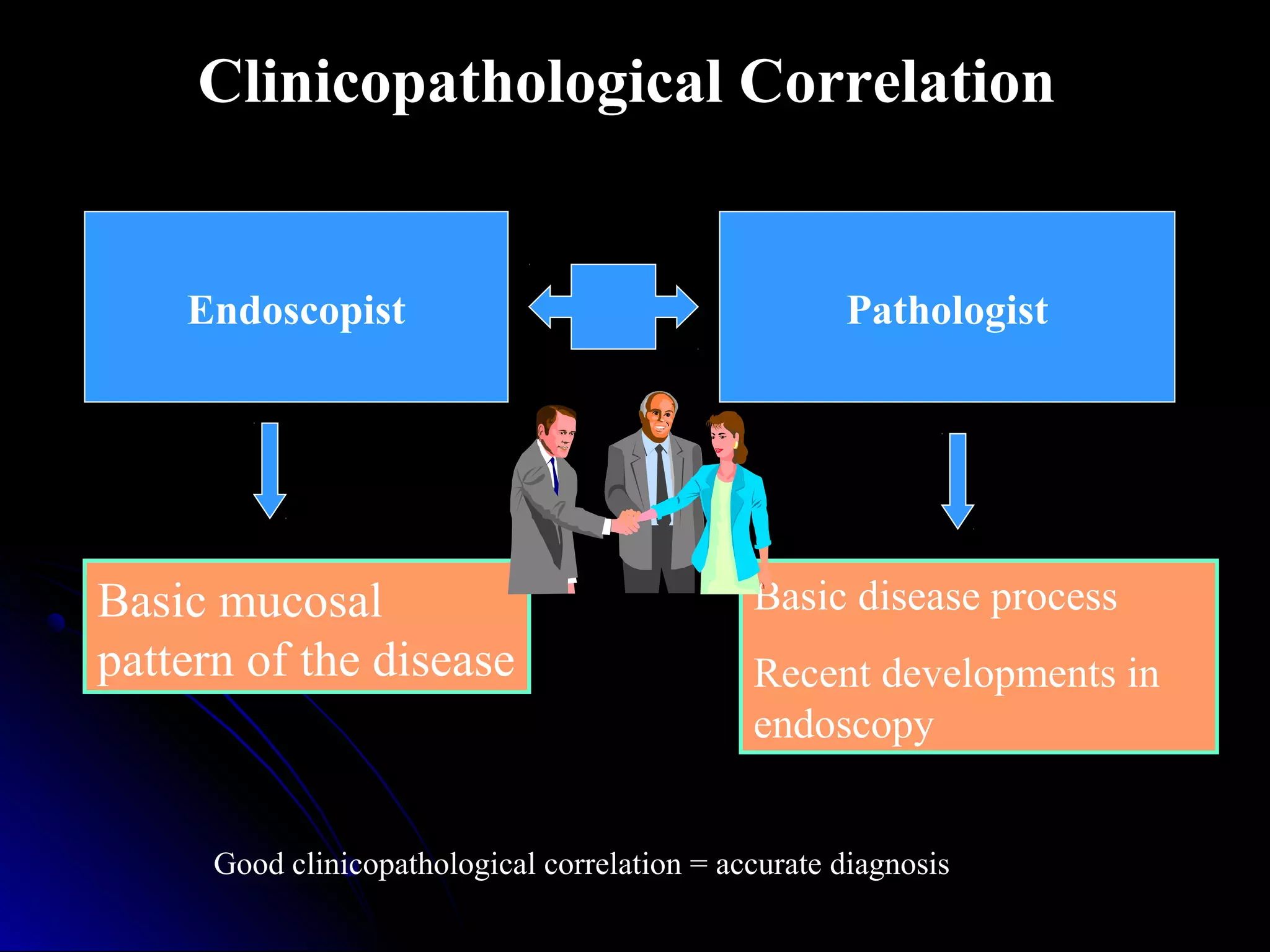
This document provides information on endoscopic gastrointestinal biopsies and their interpretation. It discusses endoscopy techniques and tools used to visualize the gastrointestinal tract and obtain biopsies. Key points include types of endoscopes, handling of biopsy specimens, processing for histological examination, common indications for endoscopy of the upper gastrointestinal tract, and histological findings and interpretations for conditions of the esophagus and stomach, including chronic gastritis, Helicobacter pylori infection, Barrett's esophagus, and polypoid lesions.
2. ENDOSCOPYENDOSCOPY
““EndoEndo” : within & “” : within & “skopeinskopein” : to view” : to view
Introduced by Rudolf Schindler in 1880.Introduced by Rudolf Schindler in 1880.
Rigid flexirigid (1920) flexibleRigid flexirigid (1920) flexible
optical axis (1980) fibreoptic biopsiesoptical axis (1980) fibreoptic biopsies
3. Direct visualization of GITDirect visualization of GIT
Taking photographsTaking photographs
Cytological specimensCytological specimens
Taking biopsiesTaking biopsies
Undertaking therapeutic procedures –Undertaking therapeutic procedures –
sclerotherapy, band ligation, polypectomy,sclerotherapy, band ligation, polypectomy,
stenting & dilatation of strictures, removalstenting & dilatation of strictures, removal
of foreign bodies.of foreign bodies.
ENDOSCOPYENDOSCOPY
5. 2 cup shaped jaws,2 cup shaped jaws,
round / elliptical, serrated / nonround / elliptical, serrated / non
serrated,serrated,
Forcep with central spike - to fix theForcep with central spike - to fix the
mucosamucosa
Other techniques for full thickness bxOther techniques for full thickness bx
specimen – FNAB thr endoscope,specimen – FNAB thr endoscope,
snare bx technique - electrocoagulationsnare bx technique - electrocoagulation
ENDOSCOPIC BIOPSY FORCEPSENDOSCOPIC BIOPSY FORCEPS
7. Automatic tissue processor or by hand processing inAutomatic tissue processor or by hand processing in
short cyclesshort cycles
Ascending grades of Alcohol - 30 mins eachAscending grades of Alcohol - 30 mins each
Xylol - 30 mins eachXylol - 30 mins each
Paraffin bath - 60 min eachParaffin bath - 60 min each
Orientation is imp while making the paraffin blockOrientation is imp while making the paraffin block
Step sections at 3-4 levelStep sections at 3-4 level
STAINS : H&E,STAINS : H&E,
SPECIAL STAINSSPECIAL STAINS :- PAS, Alcian blue, Z N, Congo:- PAS, Alcian blue, Z N, Congo
red, Masson trichrome, etc.red, Masson trichrome, etc.
BX PROCESSINGBX PROCESSING
8. Requires good communication bet endoscopist &Requires good communication bet endoscopist &
pathologist,pathologist,
REAL CHALLENGE : as Bx is very SMALLREAL CHALLENGE : as Bx is very SMALL
Pathologist must knowPathologist must know : clinical history, physical: clinical history, physical
findings, result of radiographic & lab studies, ind.findings, result of radiographic & lab studies, ind.
for scopy, endoscopic findings & site of Bx, etcfor scopy, endoscopic findings & site of Bx, etc
SoSo complete scopy reportcomplete scopy report with Bx is necessory.with Bx is necessory.
INTERPRETATION OF BIOPSIESINTERPRETATION OF BIOPSIES
32. Chronic gastritisChronic gastritis
Causes :Causes :
H. pylori gastritisH. pylori gastritis
Multifocal atrophic gastritisMultifocal atrophic gastritis
Gastritis secondary to drug therapyGastritis secondary to drug therapy
Autoimmune gastritisAutoimmune gastritis
Acute erosive gastritisAcute erosive gastritis
Granulomatous gastritisGranulomatous gastritis
Gastritis in immunosuppressed patients.Gastritis in immunosuppressed patients.
33. Helicobacter Pylori GastritisHelicobacter Pylori Gastritis
MC cause of chronic gastritis,MC cause of chronic gastritis,
Mechanism - Binding – MHCMechanism - Binding – MHC
class II moleculesclass II molecules Cag ACag A
proteinprotein increased IL-8increased IL-8
,TNF,TNF superficial gastritissuperficial gastritis
Upto 80% patients –Upto 80% patients –
autoantibodies againstautoantibodies against
canalicular membranes ofcanalicular membranes of
parietal cellsparietal cells parietal cellparietal cell
destruction,destruction,
Acute gastritis – rare, pitAcute gastritis – rare, pit
abcesses, neutrophils inabcesses, neutrophils in
surface epithelium.surface epithelium.
34. Diffuse chronic gastritis –Diffuse chronic gastritis –
Pyloric antrum,- fullPyloric antrum,- full
thickness infiltrationthickness infiltration
Corpus – superficial layersCorpus – superficial layers
Lymphoid follicles withLymphoid follicles with
germinal centers –germinal centers –
pathognomicpathognomic
Neutrphils in surface &Neutrphils in surface &
foveolar epithelium – pitfoveolar epithelium – pit
abcesses - active gastritisabcesses - active gastritis
35. Multifocal atrophic gastritisMultifocal atrophic gastritis
End stage of H. pylori gastritisEnd stage of H. pylori gastritis
Pylorus & corpus in patchyPylorus & corpus in patchy
mannermanner
Full thickness chronicFull thickness chronic
inflammation with atrophy ofinflammation with atrophy of
glandsglands
Intestinal metaplasia –Intestinal metaplasia –
diagnostic featurediagnostic feature
Adjecent mucosa – activeAdjecent mucosa – active
inflammation with pit abcessinflammation with pit abcess
formation & H. pylori infection.formation & H. pylori infection.
37. Drug therapyDrug therapy
MC causeMC cause
Alcohol, aspirin, NSAIDS, proton pump inhibitorsAlcohol, aspirin, NSAIDS, proton pump inhibitors
Ulcers are deeper & larger – direct irritation of mucosaUlcers are deeper & larger – direct irritation of mucosa
PPI – exacerbation of H.pylori gastritisPPI – exacerbation of H.pylori gastritis
- Hyperplasia of antral G & corpus ECL cells- Hyperplasia of antral G & corpus ECL cells
- multiple fundic gland polyps.- multiple fundic gland polyps.
44. Reactive gastropathyReactive gastropathy
Aspirin, NSAIDS, bile reflux, mucosal prolapse,Aspirin, NSAIDS, bile reflux, mucosal prolapse,
Mucosal edema with dilatation of mucosal capillaries,Mucosal edema with dilatation of mucosal capillaries,
Foveolar hyperplasia with loss of mucin & glandularFoveolar hyperplasia with loss of mucin & glandular
regenerative changeregenerative change
Smooth muscle fibers extending into lamina propriaSmooth muscle fibers extending into lamina propria
51. Neoplastic polypsNeoplastic polyps
AdenomaAdenoma
CarcinomaCarcinoma
CarcionidCarcionid
Lymphomatous polyposisLymphomatous polyposis
Mesenchymal tumorsMesenchymal tumors
Mucosal foldsMucosal folds
Giant folds (normal variant)Giant folds (normal variant)
Zollinger ellison syndromeZollinger ellison syndrome
Menetrier diseaseMenetrier disease
Malignant infiltrationMalignant infiltration
Biopsy – polyp ? Prominent mucosal foldBiopsy – polyp ? Prominent mucosal fold
Polyp – sessile / pedunculated? – stalk sampledPolyp – sessile / pedunculated? – stalk sampled
Evidence of polyps in other parts of GIT?Evidence of polyps in other parts of GIT?
Biopsy from surrounding mucosa.Biopsy from surrounding mucosa.
52. Hamartomatous polypsHamartomatous polyps
Peutz jeghars polypsPeutz jeghars polyps
Childhood, adolescentChildhood, adolescent
1-3 cm, coarsely1-3 cm, coarsely
lobulated surface, short &lobulated surface, short &
broad stalkbroad stalk
Smooth musclesSmooth muscles
Surface & foveolarSurface & foveolar
epitheliumepithelium
53. Juvenile polypsJuvenile polyps
Rounded smoothRounded smooth
surface, 1-2 cm, shortsurface, 1-2 cm, short
narrow stalknarrow stalk
Cystically dilated glandsCystically dilated glands
54. Hyperplastic polypsHyperplastic polyps
Similar to inflammatorySimilar to inflammatory
colonic polypscolonic polyps
Exaggerated response toExaggerated response to
mucosal damage chronicmucosal damage chronic
gastritisgastritis
at junction of pyloric &at junction of pyloric &
corpus mucosa, GE junctioncorpus mucosa, GE junction
Muliple, sessile, broad baseMuliple, sessile, broad base
small, 0.5 – 2.5 cmsmall, 0.5 – 2.5 cm
>2 cm , a/w malignancy>2 cm , a/w malignancy
61. Type I – pernicious anemiaType I – pernicious anemia hypergastrinemiahypergastrinemia
ECL cell proliferationECL cell proliferation
Multiple mucosal nodules , (> 5 mm, invading submucosa -Multiple mucosal nodules , (> 5 mm, invading submucosa -
neoplastic)neoplastic)
F>M; Body of stomachF>M; Body of stomach
Type II – a/w zollinger ellison syndrome,Type II – a/w zollinger ellison syndrome,
BodyBody
Type III – M>FType III – M>F
Solitary nodules, not a/w pernicious anemia, atrophicSolitary nodules, not a/w pernicious anemia, atrophic
gastritisgastritis
Anywhere in stomach (ECL, EC, G cells)Anywhere in stomach (ECL, EC, G cells)
Endocrine tumorsEndocrine tumors
62. Type IV – poorlyType IV – poorly
differentiated, small celldifferentiated, small cell
carcinomascarcinomas
Endocrine tumorsEndocrine tumors
63. LymphomasLymphomas
Mucosa associated lymphoidMucosa associated lymphoid
tumortumor
Ulcers, enlarged mucosalUlcers, enlarged mucosal
folds, flatfolds, flat
Features of low grade MALT –Features of low grade MALT –
1. small lympho, small cleaved1. small lympho, small cleaved
cells 2. lymphoid follicles 3.cells 2. lymphoid follicles 3.
neoplastic plasma cells 4.neoplastic plasma cells 4.
lymphoepithelial lesions clusterlymphoepithelial lesions cluster
of 3-4 lymphocytes destroyingof 3-4 lymphocytes destroying
glands(lymphocytic gastritis –glands(lymphocytic gastritis –
single cells in epi) 5. dutchersingle cells in epi) 5. dutcher
bodies –pas +ve intranuclearbodies –pas +ve intranuclear
inclusionsinclusions
High grade – no LEL, largeHigh grade – no LEL, large
cells vesicular nuclei,cells vesicular nuclei,
prominent nucleoliprominent nucleoli
64. Non MALT type – mantle cell lymphoma ,Non MALT type – mantle cell lymphoma ,
Burkitt lymphoma, follicular lymphomaBurkitt lymphoma, follicular lymphoma
LymphomasLymphomas
66. Small Bowel BiopsiesSmall Bowel Biopsies
Common IndicationsCommon Indications
Biopsy specimens are mounted with mucosalBiopsy specimens are mounted with mucosal
side up on filter paper or gelfoamside up on filter paper or gelfoam
4- 6 biopsy specimens – mandatory4- 6 biopsy specimens – mandatory
Samples fixed in 4% formaldehyde solutionSamples fixed in 4% formaldehyde solution
Chronic Diarrhea – malabsorption,Chronic Diarrhea – malabsorption,
Chronic Abdominal painChronic Abdominal pain
Occult GI BleedingOccult GI Bleeding
PolypsPolyps
67. Biopsy : The diagnostic test
(Diffuse lesions)
Whipple’s diseaseWhipple’s disease
AbetalipoproteinemiaAbetalipoproteinemia
AgammaglobinemiaAgammaglobinemia
Collagenous colitisCollagenous colitis
69. AbetalipoproteinemiaAbetalipoproteinemia
Lack of apoprotein BLack of apoprotein B
Accumulation ofAccumulation of
triglycerides intriglycerides in
enterocytesenterocytes
tips of villi showtips of villi show
intracytoplasmic lipidintracytoplasmic lipid
dropletsdroplets
DD – megaloblasticDD – megaloblastic
anemia, CS, TSanemia, CS, TS
75. Eosinophilic enteritisEosinophilic enteritis
MC in childrens & young adultsMC in childrens & young adults
Patchy Mucosal involvement – malabsorption ,Patchy Mucosal involvement – malabsorption ,
diarrhoea; submucosa, muscularis propria –diarrhoea; submucosa, muscularis propria –
obstructionobstruction
1.1. Absence of associated other inflammatory cellsAbsence of associated other inflammatory cells
2.2. Focal mucosal architectural distortion – cryptFocal mucosal architectural distortion – crypt
abcesses,abcesses,
3.3. Infiltration of muscularis mucosaeInfiltration of muscularis mucosae
76. Causes –Causes –
ParasitesParasites
IBDIBD
NHLNHL
a/w peripherala/w peripheral
eosinophiliaeosinophilia
Never associated withNever associated with
chronicity orchronicity or
metaplastic changesmetaplastic changes
77. Crohn ’s diseaseCrohn ’s disease
Mc involves terminal ileum,Mc involves terminal ileum,
Immune response to luminal flora / their productsImmune response to luminal flora / their products
Complecations :-Complecations :-
Fibrosing strictures,Fibrosing strictures,
Fistulas,Fistulas,
Protein losing enteropathy,Protein losing enteropathy,
Malabsorption,Malabsorption,
Steatorrhoea,Steatorrhoea,
78. 1.1. Small apthoid ulcers / serpiginous ulcers withSmall apthoid ulcers / serpiginous ulcers with
2.2. Skip areas,Skip areas,
3.3. Narrowing of lumen,Narrowing of lumen,
4.4. Transmural involvement,Transmural involvement,
5.5. Fissures &Fissures &
6.6. GranulomasGranulomas
79.
80. TUBERCULOSISTUBERCULOSIS
Primary or secondary,Primary or secondary,
MC – ileocaecalMC – ileocaecal
junctions,junctions,
Ulcerative /Ulcerative /
Hyperplastic,Hyperplastic,
Narrowing withNarrowing with
obstruction,obstruction,
MICRO :-MICRO :-
ICT - Caseating granulomasICT - Caseating granulomas
ICM – Large areas ofICM – Large areas of
caseation withoutcaseation without
granulomasgranulomas
81. LYMPHOMASLYMPHOMAS
IPSID (IPSID (αα--chain dis.) :-chain dis.) :-
Special type of MALToma,Special type of MALToma,
MC – Ileum,MC – Ileum,
Solitory, polypoid,Solitory, polypoid,
ulcerative or infiltrative,ulcerative or infiltrative,
Thickened folds with smallThickened folds with small
nodules,nodules,
MC – low grade,MC – low grade,
Immune response to cont.Immune response to cont.
Ag stimulationAg stimulation
82. MALT Lymphoma :-MALT Lymphoma :-
MC – Ileum,MC – Ileum,
Same as gastric MALToma,Same as gastric MALToma,
Lympho-epithelial lesions less commonLympho-epithelial lesions less common
Burkitt’s lymphomaBurkitt’s lymphoma :-:-
Sporadic,Sporadic,
Ileo-caecal inv.,Ileo-caecal inv.,
Small, non-cleaved, monomorphic sized cells,Small, non-cleaved, monomorphic sized cells,
Round nuclei, multiple nucleioli & abundantRound nuclei, multiple nucleioli & abundant
basophilic cytoplasm,basophilic cytoplasm,
‘‘Starry-sky app.’Starry-sky app.’
LYMPHOMASLYMPHOMAS
83. Biopsy: abnormal but notBiopsy: abnormal but not
diagnosticdiagnostic
Celiac sprueCeliac sprue
Tropical sprueTropical sprue
Protein energy malnutrition (Kwashiorkor)Protein energy malnutrition (Kwashiorkor)
Folate deficiencyFolate deficiency
Vitamin B12 deficiencyVitamin B12 deficiency
Bacterial over growth syndromeBacterial over growth syndrome
84. Celiac sprueCeliac sprue
Immunogenic injury d/t Gluten (wheat, rye,Immunogenic injury d/t Gluten (wheat, rye,
barley),barley),
Type II adenovirus,Type II adenovirus,
Severe in Proximal intestinal mucosa,Severe in Proximal intestinal mucosa,
85. Marsh Scoring
Grade I: Inflammation in LP:
Lymphocytes & plasma cells
normal villi & crypts
Grade II: Hypertrophy of
Crypts, mild villous atrophy
Grade III: Villous atrophy
Partial / Subtotal / Total
Grade IV: Villous atrophy
Crypt hypoplasia
Am J Gastroenterol 2003
III
III
89. Complication of CsComplication of Cs
EATCLEATCL
NS ulcerative duodenojejunoileitisNS ulcerative duodenojejunoileitis
Ca of jejunum, rarely in duo, ileum & evenCa of jejunum, rarely in duo, ileum & even
oseophagusoseophagus
90. TROPICAL SPRUETROPICAL SPRUE
Chronic diarrhoeal dis,Chronic diarrhoeal dis,
SteatorrhoeaSteatorrhoea
Bacterial inf. – E. coli, Haemophilus,Bacterial inf. – E. coli, Haemophilus,
Mild-mod villous shortening,Mild-mod villous shortening,
Increased no of chr inflam in LP & epithelium,Increased no of chr inflam in LP & epithelium,
Crypt hyperplasia,Crypt hyperplasia,
91. POLYPSPOLYPS
Hyperplastic polyps :-Hyperplastic polyps :-
Sessile,small domeSessile,small dome
shaped,shaped,
MC – Rectum,MC – Rectum,
Serrated app of glands,Serrated app of glands,
Goblet + Absorptive cellsGoblet + Absorptive cells
+nt,+nt,
Bland nuclei – round toBland nuclei – round to
oval, basally placed,oval, basally placed,
Thickened subepithelialThickened subepithelial
collagencollagen
95. Inflammatory polyps :-Inflammatory polyps :-
Amoebiasis, Adj toAmoebiasis, Adj to
ulcers, Anostomaticulcers, Anostomatic
sites, UC, CD – raisedsites, UC, CD – raised
mucosa,mucosa,
Nodules of granulationNodules of granulation
tissue,tissue,
Sec to mucosalSec to mucosal
prolapse in ileum,prolapse in ileum,
99. Adenomatous PolypsAdenomatous Polyps
Tubular adenoma :-Tubular adenoma :-
Less common than LI,Less common than LI,
Periampullary region,Periampullary region,
Small pedunculated,Small pedunculated,
Single or multiple,Single or multiple,
100. Villous adenomaVillous adenoma :-:-
Rectum & recto-sigmoidRectum & recto-sigmoid
colon,colon,
Sessile, up to 10 cm,Sessile, up to 10 cm,
Velvety cauliflower-like,Velvety cauliflower-like,
Tubulo-villousTubulo-villous :-:-
Intermediate,Intermediate,
Intermixed pattern,Intermixed pattern,
Risk of malignancyRisk of malignancy αα
villous componentvillous component
Adenomatous PolypsAdenomatous Polyps
103. Endoscopist Pathologist
Basic mucosal
pattern of the disease
Basic disease process
Recent developments in
endoscopy
Clinicopathological Correlation
Good clinicopathological correlation = accurate diagnosis
Best to use a forcep with central spike to fix the mucosa
Basal zone expansion (>15%)
Elongated lamina propria papillae(>2/3rd) epith. Thickness
Eo >6 in Bx
Lympho <10 – N, here >20
MID or distal esophagus
White plaques on edematous erythematous, friable ulcerated mucosa
Plaques - Fungal pseudohyphae, spores in tissue ulcer slough demonstration - definitive diag.
Pas methanamine silver stain
Grocott’s stain
Diagrams from sternberg
- IC patients
SMALL VESICLES -shallow ulcers
Cowdry type A - dense eosinophilic intranuclear inclu.seperated from thick nuclear memb. By clear halo.
HSV -IC , enlarged mesenchymal cells intranuclear inclusions, coarse cytoplasmic granules +ce of macrophages in perivascular distribution - diag clue
True absorptive enterocytes not observed, mucin secreting goblet cells + columnar cells with absorptive & secretory US features.
LP – chr.inflaman, muscularsis – thickened
DD esophageal glandular epith – 1. sampled gastric mucosa 2. cardiac like mucosa in distal 1-2 cm 3.superficial gastric glands in upper eso 4.heterotopic gastric fundic mucosa
Risk factors – 1. Dietary – def. of vit., trace elements, fungal contamination of foodstuffs, high contents of nitrites, nitrosomines,2. lifestyle – hot beverages, alcohol, tobacco,3.achalasia, chronic esophagitis,4.epidermolysis bullosa, celiac disease.
Dysphagia, wt.loss, aspiration thr. Cancerous TEF.
Rapid mets – mucosa & submucosa rich in lymphatics, absence of serosa.
Histoplasma – silver stains – budding yeast forms
MAIC – PAS –faintly positive bacillary form, AFB +ve bacilli
Org. predeliction for lamina propria, messentric lymph nodes, cardiac valve, CNS
IPSID – LP infiltrate with mature cell withh alpha heavy chain (Alpha chain dis.)