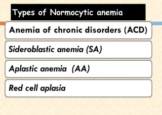
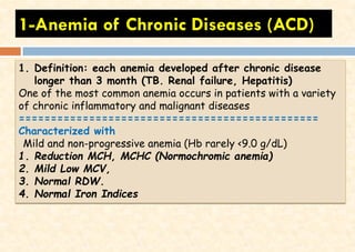
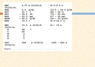
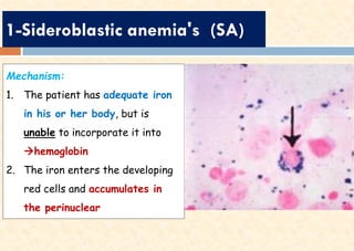
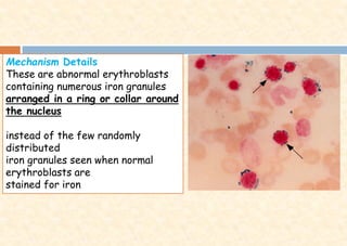
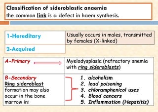
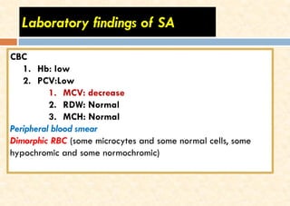
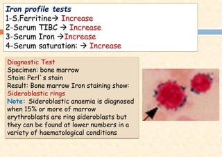
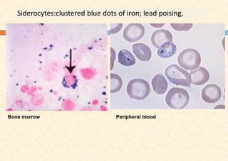
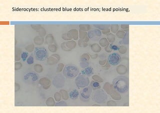
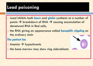
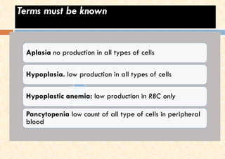
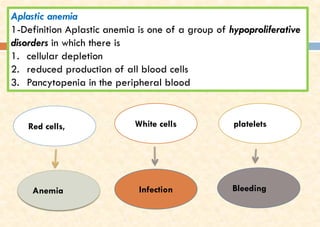
Best Rate (Guwahati ) Call Girls Guwahati ⟟ 8617370543 ⟟ High Class Call Girl...
Normocytic Anemias.pdf
1. Dr. Abdulrazzaq Othman Alagbare
MD. Clinical Pathologist – lecturer of haemato-Oncology
Normocytic anemia
2. Types of Normocytic anemia
Anemia of chronic disorders (ACD)
Sideroblastic anemia (SA)
Aplastic anemia (AA)
Red cell aplasia
3. 1-Anemia of Chronic Diseases (ACD)
1. Definition: each anemia developed after chronic disease
longer than 3 month (TB. Renal failure, Hepatitis)
One of the most common anemia occurs in patients with a variety
of chronic inflammatory and malignant diseases
===============================================
Characterized with
Mild and non-progressive anemia (Hb rarely <9.0 g/dL)
1. Reduction MCH, MCHC (Normochromic anemia)
2. Mild Low MCV,
3. Normal RDW.
4. Normal Iron Indices
4. Three mechanisms have been identified
1. Decreased release of iron from macrophages to plasma, → impaired of
Intracellular Fe metabolism
2. Reduced red cell lifespan ( increase of serum urea, creatinine in CRF)
3. An inadequate erythropoietin response to anemia ( effect of toxins such as
tumor necrosis factor (TNF)-, interferon-)
Conditions Leading to Anemia of Inflammation orAnemia of
Chronic Disease
1. Infections (Hepatitis HBAs, HCV)
2. Renal failure
3. inflammatory states (RA, TB)
4. cancers
5. Diagnosis of ACD
1. Each case of ACD investigated partially
2. No diagnostic test
3. Diagnosis made after all other forms of anemia have been
ruled out
4. The anemia is corrected by successful treatment of the
underlying disease
CBC parameters
1. Low: Hb about 9g/dl
2. Low MCH
3. Low MCV
4. Normal RDW
Blood film study
1. RBC with normocytic normochromic appearance
Iron panel tests mostly are normal
6.
7. ANEMIAS RELATED TO IRON
OVERLOAD CONDITIONS
1-Sideroblastic anemia's (SA)
8. 1-Sideroblastic anemia's (SA)
Mechanism:
1. The patient has adequate iron
in his or her body, but is
unable to incorporate it into
→hemoglobin
2. The iron enters the developing
red cells and accumulates in
the perinuclear
9. Mechanism Details
These are abnormal erythroblasts
containing numerous iron granules
arranged in a ring or collar around
the nucleus
instead of the few randomly
distributed
iron granules seen when normal
erythroblasts are
stained for iron
10. Classification of sideroblastic anaemia
the common link is a defect in haem synthesis.
1-Hereditary Usually occurs in males, transmitted
by females (X-linked)
A-Primary Myelodysplasia (refractory anemia
with ring sideroblasts)
B-Secondary
Ring sideroblast
formation may also
occur in the bone
marrow in:
1. alcoholism
2. lead poisoning
3. chloramphenicol uses
4. Blood cancers
5. Inflammation (Hepatitis)
2-Acquired
11. Laboratory findings of SA
CBC
1. Hb: low
2. PCV:Low
1. MCV: decrease
2. RDW: Normal
3. MCH: Normal
Peripheral blood smear
Dimorphic RBC (some microcytes and some normal cells, some
hypochromic and some normochromic)
12. Iron profile tests
1-S.Ferritine→ Increase
2-Serum TIBC → Increase
3-Serum Iron →Increase
4-Serum saturation: → Increase
Diagnostic Test
Specimen: bone marrow
Stain: Perl`s stain
Result: Bone marrow Iron staining show:
Sideroblastic rings
Note: Sideroblastic anaemia is diagnosed
when 15% or more of marrow
erythroblasts are ring sideroblasts but
they can be found at lower numbers in a
variety of haematological conditions
15. Lead poisoning
Lead inhibits both haem and globin synthesis at a number of
points → breakdown of RNA → causing accumulation of
denatured RNA in Red cells,
the RNA giving an appearance called basophilic stippling on
the ordinary stain
The patient has
Anemia → hypochromic
the bone marrow may show ring sideroblasts
16. Iron overload- hemosiderosis
Definition: Excess iron in the body accumulated
and storage in different organs
Causes :
1-chronic hyper absorption (Increased iron intake)
2-RBC transfusions (Repeated red cell transfusions )
3- inherited sideroblastic anemia (inherited
abnormal genes)
17. Lab diagnosis- Iron overload
Serum iron elevated
Serum ferritin Elevated > 200 ìg/L (women) and
300 ìg/L (men)
TIBC normal reference range
Transferrin saturation Elevated (> 55% for men and >
50%
measurement of transferrin saturation is
essential for early detection of iron loading.
18. Hereditary Hemochromatosis (HH)
Mechanism
•iron absorption and storage
become unbalanced due to the
inheritance of an abnormal
gene, and not bind to the
transferrin receptor in the
normal iron delivery process
❑Common genetic disorder
❑Mostly in European ancestry
Symptoms
1. Chronic fatigue and
weakness
2. Cirrhosis of the liver
3. Hyperpigmentation
4. Diabetes
5. Impotence
6. Hair loss
19. Screening for iron status would include
Serum iron elevated
Serum ferritin elevated
TIBC normal reference range
transferrin saturation elevated
1. Serum ferritin levels above 150 μg/L
2. and transferring saturation levels of greater
than 45% are indicative of HH
3. Genetic testing (must be positive)
21. Terms must be known
Aplasia no production in all types of cells
Hypoplasia. low production in all types of cells
Hypoplastic anemia: low production in RBC only
Pancytopenia low count of all type of cells in peripheral
blood
22. Red cells, White cells platelets
Anemia Infection Bleeding
Aplastic anemia
1-Definition Aplastic anemia is one of a group of hypoproliferative
disorders in which there is
1. cellular depletion
2. reduced production of all blood cells
3. Pancytopenia in the peripheral blood
23. Causes
1-Genetic
2-Idiopathic
3-Aquired (drugs , toxins ,infections, Radiation
Chemotherapy or chemicals)
It is may be a result of two possible mechanisms:
1. an antibody directed against an antigen on stem cells
2. or an immune mechanism that is at play, in which T
lymphocytes suppress stem cell proliferation
25. Diagnostic test:
Bone marrow biopsy,
shows fibrotic marrow
Notes: A plastic anemia is
not malignant case
Pure Red Cell Aplasia
Is the failure of the bone marrow to produce only RBC, the other cells are
normal in their production, count and morphology