SlideShare a Scribd company logo
Peripheral blood smear examination
(Slide preparation and reporting)
Caesar Abu Arra
MSc Life sciences
BSc Medical lab. Sciences
Medicare Labs
Aim of blood smear
 Evaluation of anemia
 Infections
• bacteria, malaria, microfilaria..etc.
 Abnormal cells
• blasts, inclusions..etc.
 Cells morphology and count
Abu Jad Caesar
Making blood films
 Three basic steps to make blood film:
 Preparation of blood smear.
 Fixation of blood smear.
 Staining of blood smear.
Abu Jad Caesar
Preparation of blood smear
 Different types of blood smears:
 The wedge smear slide to slide method.
 The spun smear.
 Two additional types of blood smears (used for specific
purposes):
o Buffy coat smear for WBCs < 1.0×10⁹/L.
o Thick blood smears for blood parasites .
Abu Jad Caesar
Wedge blood smear
 Smears should be made within 1 hour of blood
collection from EDTA specimens stored at
room temperature to avoid distortion of cell
morphology.
 Small drop (10 µl)
 Hold the spreader slide at a 30°- 45° angle,
and draw it back against the drop of blood.
 Air dry at room temperature.
Abu Jad Caesar
Abu Jad Caesar
Wedge blood smear
Procedure notes
1. The smear should be made without delay.
Any delay results in an abnormal distribution
of the white blood cells, with many of the
large white cells accumulating at the thin
edge of the smear.
2. HCT↑ ↓ The angle of the spreader slide
HCT↓ ↑ The angle of the spreader slide
Abu Jad Caesar
large angle
low HCT
small angle
high HCT
Abu Jad Caesar
Procedure notes
Spin method
 Automated method
 Placeadrop of blood in thecenter of aglassslide.
 Spin atahighspeed in aspecialcentrifugecytospin.
 Blood spreads uniformly.
Abu Jad Caesar
Characteristics of a Good Smear
 Thick at one end, thinning out to a smooth
rounded feather edge.
 Should occupy 2/3 of the total slide area.
 Should not touch any edge of the slide.
Abu Jad Caesar
tail body head
Abu Jad Caesar
Characteristics of a Good Smear
Common causes of a poor blood smear
 Drop of blood too large or too small.
 Spreader slide pushed across the slide in a
jerky manner.
 Failure to keep the spreader slide at a 30°
angle with the slide.
 Failure to push the spreader slide completely
across the slide.
Abu Jad Caesar
Common causes of a poor blood smear
 Holes in film: Slide contaminated with fat or
grease
 Cellular degenerative changes:
delay in fixing
 Inadequate fixing time
Methanol contaminated with water.
Abu Jad Caesar
Biologic causes of a poor smear
 Cold agglutinin - RBCs will clump together.
Warm the blood at 37° C for 5 minutes.
 Lipemia - holes will appear in the smear.
Nothing you can do to correct this.
 Rouleaux - RBC’s will form into stacks as coins.
Nothing you can do to correct this.
Abu Jad Caesar
Staining of blood smear
 Romanowsky stain:
Any Combination of:
Eosin Y (Acidic or anionic dye):
o Binds to cationic sites (cytoplasm & Hb)
Methylene blue (Basic, cationic or Azure B dye):
o Binds to anionic sites (nucleus and RNA)
Abu Jad Caesar
Staining of blood smear
 Buffer:
 Used to maintain an adequate pH.
 0.05M Na2PO4 (pH 6.4) or dH2O (pH 6.4-6.8)
Abu Jad Caesar
pH of the phosphate buffer
 If the pH is too acidic:
RBC :Pinker
WBC nuclei : very pale staining (very pale purple)
 If the pH is too basic:
RBC :Grayish blue
WBC nuclei :very deeply purple
Abu Jad Caesar
pH of the phosphate buffer
Abu Jad Caesar
Staining Troubleshooting
 Too Acid Stain:
Insufficient staining time
Prolonged buffering or washing
Old stain
 Correction:
 Lengthen staining time
 Check stain and buffer pH
 Shorten buffering or wash time
Abu Jad Caesar
Staining Troubleshooting
 Too Alkaline Stain:
Thick blood smear
Prolonged staining
Insufficient washing
Alkaline pH of stain components
 Correction :
 Check pH
 Shorten stain time
 Prolong buffering time
Abu Jad Caesar
Abu Jad Caesar
 Macroscopic view:
 Quality of the smear
 The microscopic view:
 Progressing from low power to high power:
Blood film examination - preliminary
Abu Jad Caesar
Blood film examination - preliminary
Abu Jad Caesar
 Low-Power (10x) Scan
 Determine the overall staining quality of the blood
smear.
 Determine if there is a good distribution of the cells.
o Discussed later
 Find an optimal area for the detailed examination and
enumeration of cells
Blood film examination - preliminary
Abu Jad Caesar
 High-Power (40x) Scan
 Determine the WBC estimate.
o WBCs are counted in 10 fields and averaged X 2000
o The estimate could be reported according to the table below:
Blood film examination - preliminary
Abu Jad Caesar
• High-Power (40x) Scan
 Grading scale for WBC count (X103 cell/µl)
Blood film examination - preliminary
Low WBC High WBC
Mild 3-4.5 Mild 10-15
Moderate 2-3 Moderate 15-30
Severe <2 Marked 30-100
Extreme >100
Abu Jad Caesar
 Oil Immersion (100x) Examination
 Perform a 100 WBC differential count.
 Correct WBC count that has greater than 10 NRBCs per
100 WBCs.
o Report as: WBC count was corrected due to presence of
NRBCs.
Blood film examination - preliminary
Abu Jad Caesar
 Oil Immersion (100x) Examination
 to correct a WBC count (/µl):
Uncorrected WBC (/ ) X l
Blood film examination - preliminary
Abu Jad Caesar
 Oil Immersion (100x) Examination
 Evaluate the RBCs for:
o Anisocytosis
o Poikilocytosis
o Inclusions
o Hypochromasia
o Polychromasia.
Blood film examination - preliminary
Abu Jad Caesar
 Oil Immersion (100x) Examination
 Perform a platelet estimate and evaluate platelet
morphology:
o Platelets are counted in 10 fields and averaged:
• X 15  Automated smear.
• X 20  Wedge smear is used.
Blood film examination - preliminary
Abu Jad Caesar
 Oil Immersion (100x) Examination
 Grading scale for platelets count (X103 cell/µl)
Blood film examination - preliminary
Abu Jad Caesar
Low Platelets High Platelets
Mild 100-150 Mild 500-700
Moderate 50-100 Moderate 700-900
Severe <50 Severe 900-1000
Extreme >1000
 Oil Immersion (100x) Examination
 The absolute WBC count (/µl) may be determined:
Relative count (%) X Total WBC count (/µl)
Blood film examination - preliminary
Abu Jad Caesar
 Relative vs. Absolute value:
 Relative value:
o Measures the percentage of corresponding WBC type in
peripheral blood.
Blood film examination - preliminary
Abu Jad Caesar
 Relative vs. Absolute value:
 Absolute value:
o Measures the total count of corresponding WBC type /µl
in peripheral blood.
o Gives more meaningful information than the percentage.
o It is the preferred reporting method for the WBC
differential.
• Reported as absolute cytosis or absolute cytopenia.
Blood film examination - preliminary
Abu Jad Caesar
 Relative vs. Absolute value:
Example: leukopenic patient and increased susceptibility
to infection and sepsis:
 WBC=5000 Relative Neutrophils=50%
o Absolute= 2500 cell/µl Normal
 WBC=2000 Relative Neutrophils=85%
o Absolute= 1700 cell/µl Low
Blood film examination - preliminary
Abu Jad Caesar
Abu Jad Caesar
 Examination of blood films should include:
 RBC
Size, Shape, color
Hemoglobin distribution
Arrangement and distribution
Inclusions
nucleated RBCs
Examination of blood films
Abu Jad Caesar
 Examination of blood films should include:
 WBC
 Total counts
 Differential counts
 Abnormal and immature WBC
Examination of blood films
Abu Jad Caesar
 Examination of blood films should include:
Platelets
 Counts should be verified by estimation
 Size
 Clumping
Examination of blood films
Abu Jad Caesar
 Examination of blood films should include:
Parasites
Examination of blood films
Abu Jad Caesar
 Pancytopenia
Deficiency in all the three cell lines RBC, WBC, and platelets.
 Aplastic anemia
 When report, recommend bone marrow assessment.
 Bicytopenia
Deficiency in two of the three cell lines
 Viruses and drugs
Terms
Abu Jad Caesar
PARASITES
Abu Jad Caesar
 Thick film When parasites are scanty
 Thin film  Identification of species
PARASITES
Abu Jad CaesarAbu Jad Caesar
Plasmodium falciparum
Trypanosoma spp.
Microfilaria
Megakaryopoiesis
Abu Jad Caesar
Platelets
 1 to 4 μm in diameter and vary in shape.
 Reddish-purple granules
 Life span 9-12 days
 One megakaryocyte can release several thousand platelets
(4000).
 Report as platelets are adequate with normal-sized ones
OR platelets are normal
Abu Jad Caesar
Platelets
 Large and Giant platelets
Large platelets (4-7 µm)
oITP
Abu Jad Caesar
Platelets
 Large and Giant platelets
Giant platelet (7-20 µm)
oPlatelets seems to be size of RBC.
oBernard Soulier syndrome
Abu Jad Caesar
Platelets
 Large and Giant platelets
NOTE:
o When estimate, Large platelets and platelet aggregates
NOT counted.
Report as Occasional, Few, Many
Abu Jad Caesar
Platelets
 Platelet anisocytosis:
Small large giant platelets will be seen
 Report as: Platelets anisocytosis ranging in size from
tiny to large giant platelets were seen.
Abu Jad Caesar
Platelets
 Thrombocytosis
 Post infection and Inflammation
 Report as:
o Thrombocytosis (with grading).
o Platelets were estimated and approved to be about ( /µl)
 In case of microcytosis (RBC and PLT diameters are
similar) report as :
o Thrombocytosis due severe microcytosis.
Abu Jad Caesar
Platelets
 Thrombocytopenia:
Could be due to:
o Decreased production Aplastic anemia
o Increased destruction ITP
Abu Jad Caesar
Platelets
 Thrombocytopenia:
 In case of no aggregates report as:
o Thrombocytopenia (with grading).
o No platelet clumps are noted OR Negative for
microaggregates.
o Platelets were estimated and approved to be about ( /µl)
Abu Jad Caesar
Platelets
 Pseudothrombocytopenia:
In vitro EDTA dependent phenomena
oPlatelet clumping
Abu Jad Caesar
Platelets
 Pseudothrombocytopenia:
In vitro EDTA dependent phenomena
oPlatelet satellitism
• Platelets adhere to neutrophil surface
Abu Jad Caesar
Platelets
 Pseudothrombocytopenia:
Repeat on new samples (EDTA and sodium citrated)
Scan for platelet clumping in thin and thick portion of
smear
Abu Jad Caesar
Platelets
 Pseudothrombocytopenia:
Reporting criteria:
Two samples were drawn to rule out EDTA- induced
pseudothrombocytopenia:
o EDTA smear revealed platelet clumps / Platelet
satellitism
o Na+ citrated smear revealed normal platelets count and
distribution.
Conclusion: Platelets are adequate
Abu Jad Caesar
Platelets
Morphology of Normal RBC
 Biconcave disc
 Diameter 7 ~ 8 μm
 Central pallor occupy 3rd of total size
 Approx same as nucleus of mature lymphocyte
 Stained with eosin component of Romanowsky dyes
Abu Jad Caesar
RBC
Abu Jad Caesar
Erythropoiesis
RBC Abnormality
 Examination of blood films for RBC’s should
include:
 Anisocytosis
 Poikilocytosis
 Hemoglobin distribution
 Arrangement and Distribution
 Inclusions
 NRBCs
Abu Jad Caesar
RBC Abnormality
Abu Jad Caesar
Arrangement and Distribution
 Normal arrangement and distribution:
 The thin portion of the smear where RBC’s are slightly
separated from one another or at most, barely touching,
with no overlap.
 The thin area should represent at least 3rd of the entire
film.
Abu Jad Caesar
Arrangement and Distribution
 Normal arrangement and distribution:
 The reviewer should avoid the thicker portion of the
slide where cells are overlapping and the edges of smear
where cells may be distorted in size, shape, and color.
o An exception is to be made when scanning for platelet
clumping
Abu Jad Caesar
Arrangement and Distribution
 Rouleaux
 RBC’s appear as stacks of coins (linear)
 Due to ↑ Globulins or fibrinogens
 seen in:
o Multiple myeloma
o Macroglobulinemia
Abu Jad Caesar
Arrangement and Distribution
 Agglutination
 More irregular and round clumping than linear rouleaux
 Seen in:
o Cold agglutinin
o Anti RBC antibodies
o Autoimmune HA
o Macroglobulinemia
Abu Jad Caesar
Inclusions
 Basophilic stippling (Punctate basophilia)
 Aggregates of Ribrosomes.
 Multiple blue black inclusions (coarse or punctate )
 Seen in:
o Lead and arsenic poisoning
o Thalassemias
o Alcoholism
o Megaloblastic anemias
Abu Jad Caesar
Inclusions
 Howell Jolly bodies
 Large round inclusions which are remnant of nuclear
chromatin
 Appear singly or doubly in an eccentric position on the
cell periphery
 Deep purple
Abu Jad Caesar
Inclusions
 Howell Jolly bodies
 Seen in:
o Postsplenectomy
o Megalobalstic anaemia
o Haemolytic anaemia
Abu Jad Caesar
Inclusions
 Pappenheimer Bodies
 Ferric compound complexed with protein
 Small dark blue bodies of uniform size.
Abu Jad Caesar
Inclusions
 Pappenheimer Bodies
 Unlike basophilic stippling:
o Basophilic stippling appears homogeneously over the cell
whereas Pappenheimers tend to appear as single or clusters
in the cells periphery.
 Unlike Howell–Jolly bodies:
o Howell–Jolly bodies appear to be
larger
Abu Jad Caesar
Inclusions
 Pappenheimer Bodies
 Seen in:
o Sideroblastic erythropoiesis
o Postsplenectomy
o Hemolytic anemia
o Thalassemia
Abu Jad Caesar
Inclusions
 Heinz bodies
 Result of denatured or precipitated hemoglobin
 Purple, blue, large, single or multiple inclusions
attached to the inner surface of the red blood cell.
Abu Jad Caesar
Inclusions
 Heinz bodies
 Stained by supravital stains
 Not Stained by Romanowsky stain.
 Seen in:
o Postsplenectomy
o oxidant stress of drugs and
chemicals
Abu Jad Caesar
Inclusions
 Cabot rings
 Represent a part of the mitotic spindle, remnants of
microtubules, or a fragment of the nuclear membrane.
 Have no internal structure
Abu Jad Caesar
Inclusions
 Cabot rings
 Red or reddish purple
 Could be appear in a figure-of-eight conformation
Abu Jad Caesar
Inclusions
 Cabot rings
 Seen rarely in
o Megaloblastic anemias
o Postsplenectomy
Abu Jad Caesar
Poikilocytosis
 Shape variation
 Mention the abnormal shapes rather than the term
poikilocytosis.
 Grading scale:
Abu Jad Caesar
Poikilocytosis
 Spherocytes
 Loss of biconcavity
o ↓ Surface-to-volume ratio
o ↑ Osmotic fragility
 Smaller diameter
 Dense-staining
o No central pallor area
Abu Jad Caesar
Poikilocytosis
 Spherocytes
 Seen in:
o Hereditary spherocytosis
o Immune hemolytic anemia
Abu Jad Caesar
Poikilocytosis
 Target cell
 Codocytes
 ↑ membrane cholesterol and phospholipid and ↓ cellular
hemoglobin.
 ↑ surface-to-volume ratio
Abu Jad Caesar
Poikilocytosis
 Target cell
 Seen in:
o Obstructive liver disease
o Iron deficiency anemia
o Thalassemia
o Post-transfusion
Abu Jad Caesar
Poikilocytosis
 Leptocyte
 Synonymous with target cell, BUT:
o Red cells with large unstained central area.
Abu Jad Caesar
Poikilocytosis
 Leptocyte
 Seen in:
o IDA
o Thalassemia
Abu Jad Caesar
Poikilocytosis
 Elliptocyte and ovalocyte
 Elliptocytes
o Pencil, rod, or cigar shaped
o Hb appears to be concentrated on both ends of the cell
o Seen in IDA
Abu Jad Caesar
Poikilocytosis
 Elliptocyte and ovalocyte
 Ovalocyte
o More egg-shaped
o Have a greater tendency to vary in their Hb content
o Seen in megaloblastic anemia
Abu Jad Caesar
Poikilocytosis
 Elliptocyte and ovalocyte
Abu Jad Caesar
Poikilocytosis
 Teardrop cell
 Dacrocytes
 Physiologic mechanism is unknown
 Seen in:
o IDA
o Pernicious anemia
Abu Jad Caesar
Poikilocytosis
 Stomatocyte
 RBC with narrow slit-like area of central pallor
 Normal size, but are not biconcave
 Seen in:
o Acute alcoholism
o Hemolytic anemia
o Malignancies
Abu Jad Caesar
Poikilocytosis
 Acanthocyte
Thorn Cells, Spur Cells
3 to 12 irregular, uneven length spicules distributed
along the periphery of the cell membrane
Abu Jad Caesar
Poikilocytosis
 Acanthocyte
 Seen in:
o Severe hepatic disease
o Autoimmune HA
o Postsplenectomy
o Vitamin E deficiency
Abu Jad Caesar
Poikilocytosis
 Echinocyte
 Burr Cells, crenated cells
 10 to 30 regular spicules, evenly placed over the surface
of RBC
 Considered pathologic and should be reported.
Abu Jad Caesar
Poikilocytosis
 Echinocyte
 Seen in:
o Liver disease
o Renal disease
o Severe burns
Abu Jad Caesar
Poikilocytosis
 Fragmented cells
 Several types
1- Schistocytes
2- Helmet Cells (bite cell) usually 2 projections
3- Keratocytes
Abu Jad Caesar
Poikilocytosis
 Fragmented cells
 Sometimes counted as platelets
o Estimate platelets and report thrombocytosis due to presence
of RBC fragments
 Seen in : DIC, TTP, HUS
Abu Jad Caesar
Poikilocytosis
 Fragmented cells
 Several types
4- Prekeratocyte (Blister cell) resemble a women’s handbag
o RBC’s containing a peripherally located vacuole.
o Keratocytes are RBC’s that have been caught on fibrin strands in
circulation, and rather than splitting, the cell hangs over the fibrin
fusing two sides of the cell together, creating a vacuole.
o Seen in:
 Oxidative stress G6PD deficiency
Abu Jad Caesar
Anisocytosis
 Size variation
 Grading scale:
Abu Jad Caesar
Anisocytosis
 Size variation
 Normocytic
 Microcytic
 Macrocytic
Abu Jad Caesar
Anisocytosis
 Normocyte
 MCV 80-100 fl
 Diameter 7 to 8 μm
Abu Jad Caesar
Anisocytosis
 Microcytes
 MCV <80 fl
 Diameter <7 μm
 Results from a defect in hemoglobin formation
 Seen in:
o IDA
o Thalassemia
Abu Jad Caesar
Anisocytosis
 Macrocytes
 MCV >100 fl
 Diameter >9 μm
 Result from impaired DNA synthesis.
 Seen in:
 Megaloblastic anemia (↓ B12 and folic acid).
 Aplastic anaemia
Abu Jad Caesar
Anisocytosis
 Anisocytosis is a feature of most anemias.
o Report as anisocytosis (with Qual. or Quan. Grading):
Abu Jad Caesar
RDW: 16-18  Slight
RDW: 18-22  Moderate
RDW: >22 Marked or severe
Anisocytosis
 Mixture of large and small with normal MCV and high
RDW usually due:
Recent blood transfusion
Anemia or recovery stages of anemia.
o Report as Anisocytosis with dimorphic RBC population
(with grading):
Abu Jad Caesar
RDW: 16-18  Slight
RDW: 18-22  Moderate
RDW: >22 Marked or severe
Hemoglobin Distribution
 MCHC used in conjunction with MCV used
do describe anemias
Normochromia
Hypochromia
Hyperchromia
Polychromasia
Anisochromia
Abu Jad Caesar
Hemoglobin Distribution
 Normochromia: MCHC 32-36%
 Normal intensity of staining.
 The central pallor < 3µm (<3rd of total size)
Abu Jad Caesar
Hemoglobin Distribution
 Hypochromia : MCHC < 32%
 Unusually palely RBC (↓Hb content)
 The central pallor > 3µm (>1/3 rd of total size)
 Seen in:
o IDA
o Thalassemia
Abu Jad Caesar
Hemoglobin Distribution
 Hypochromia : MCHC < 32%
 Grading:
Abu Jad Caesar
Hemoglobin Distribution
 Hyperchromia : MCHC > 36%
 Decreased or absent central pallor
 Seen in:
o Sperocytosis (↓surface-to-volume ratio)
o hemolytic anemias (hemolysis caused by burns)
Abu Jad Caesar
Hemoglobin Distribution
 Hyperchromia : MCHC > 36%
 Even though true hyperchromia does exist it is not
reported as such:
o It is reported in terms of the cell abnormalities resulting
from the increased volume of hemoglobin and the
decreased surface area (e.g spherocytes).
Abu Jad Caesar
Hemoglobin Distribution
 Polychromasia
 Premature RBC in circulation called Polychromatophilic
cells actually reticulocytes.
 Gray-blue in color and usually larger than normal RBC
o Result of the residual RNA involved in Hb synthesis.
Abu Jad Caesar
Hemoglobin Distribution
 Polychromasia
 Seen in:
o Acute and chronic blood loss.
o Recovering from anemias
Abu Jad Caesar
Hemoglobin Distribution
 Polychromasia
 Grading excellent indicator of therapeutic effectiveness
when patient is given iron or vitamin therapy as treatment
of anemia
Abu Jad Caesar
Hemoglobin Distribution
 Anisochromia
 Hypochromic cells and normochromic cells in the
same film
 Seen in:
o Development or recovering from anemias
o Post-transfusion
Abu Jad Caesar
Hemoglobin Distribution
 MCHC used in conjunction with MCV used do
describe anemias
Abu Jad Caesar
White blood cells
 WBC categories:
 Granulocytes (PMN):
o Neutrophils, Eosinophils and basophiles
o Granules in their cell cytoplasm.
o Multilobed nucleus
 Agranulocytes (MNC):
o Lymphocytes and monocytes.
o Do not have granules (contain non specific azurophilic
granules)
o Nonlobular nuclei.
Abu Jad Caesar
Neutrophil
Abu Jad Caesar
Neutrophil
 Terms
 Shift to the left
o Release of younger granulocytes (specifically bands and
metamyelocytes from the bone marrow).
Abu Jad Caesar
Neutrophil
 Terms
 Shift to the right
o An abnormal cell maturation situation that occurs when
hypersegmented cells are seen.
• It is indicative of vitamin B12 and/or folate deficiency.
Abu Jad Caesar
Neutrophil
 Cytoplasm
o Pink
 Nucleus
o Dark purple blue
o Dense chromatin
o 3-5 lobes
Abu Jad Caesar
Neutrophil
 Report as:
o Mature/normal appearing neutrophils.
o In case of leukocytosis neutrophilic leukocytosis
o Absolute / relative neutrophilia or neutropenia
Abu Jad Caesar
Neutrophil
 Neutrophilia
o Acute bacterial infection.
o Many inflammatory processes.
 Neutropenia
o Typhoid fever
o Brucellosis
o Viral diseases.
Abu Jad Caesar
Abnormal neutrophil
 Abnormal neutrophil morphology:
Band or stab forms
Hypersegmentation (≥ 6 lobes)
Vacuolization
Toxic granulation
Dohle bodies
Nuclear pyknosis and karyorrhexis
Abu Jad Caesar
Band or Stab
 Cytoplasm
o Pink
 Nucleus
o U shaped nucleus of uniform thickness.
o Purplish red
o Dense chromatin
Abu Jad Caesar
Band or Stab
 Normally form 5-10% of WBC’s
 Increased bands:
o Acute infection (usually bacterial)
o Non infectious inflammatory disease
Abu Jad Caesar
Band or Stab
 Include it when calculate absolute neutrophil
 Report as:
o Many/Few neutrophilc bands OR with shift to the
left
Abu Jad Caesar
Hypersegmentation
 Neutropils with ≥ 6 lobes
 Normally < 3% of WBC’s
 Seen in megaloblastic anemia
Abu Jad Caesar
Hypersegmentation
 Report as:
o Few/many hypersegmented neutrophils OR with shift to
the right.
o Further hematological examinations are recommended
(B12 & folic acid).
Abu Jad Caesar
Vacuolization
 Vacuoles in neutrophil contain ingested microorganisms.
 Seen in:
o Severe infection (leukemoid reaction)
o Non infectious inflammation
Abu Jad Caesar
Toxic granulation
 ↑ staining density and number of granules
 Seen in:
o Bacterial infection and leukemoid reaction
o Other inflammation
Abu Jad Caesar
Döhle bodies
 Pale blue inclusions at the periphery of the cytoplasm
 Strands of RER that have aggregated
o Bacterial infection and leukemoid reaction
o Inflammation
Abu Jad Caesar
Nuclear pyknosis and karyorrhexis
 Degenerative changes in nucleus of neutrophil :
 Pyknosis: Condensation and shrinkage of cells through degeneration.
 Karyorrhexis: A necrotic stage with fragmentation of the nucleus,
whereby chromatin is distributed irregularly throughout the cytoplasm.
Abu Jad Caesar
Nuclear pyknosis and karyorrhexis
 Due to:
 Prolonged time in circulation.
 Increased numbers in inflammatory and neoplastic disorders
 Old blood samples.
 New fresh sample is preferred.
Abu Jad Caesar
Nuclear pyknosis and karyorrhexis
 Could be mistaken with NRBC’s.
Abu Jad Caesar
Eosinophil
Abu Jad Caesar
Eosinophil
 Cytoplasm
o Full of orange-red granules
 Nucleus
o Blue purple
o Coarsely granular chromatin
o 2 Lobes (like a pair of glass)
Abu Jad Caesar
Eosinophil
 Eosinophilia
o Parasitic infection
o Allergic Disorders
o Eosinophilic leukemia
Abu Jad Caesar
Basophil
Abu Jad Caesar
Basophil
 Cytoplasm
o Pale blue with coarse violet-blue granules
 Nucleus
o Blue purple
o Coarsely granular chromatin
o 2 Lobes
Abu Jad Caesar
Basophil
 Basophilia
o Allergic Reactions
o Infections
Smallpox
Chicken pox
Influenza
Abu Jad Caesar
Monocyte
Abu Jad Caesar
Monocyte
 Cytoplasm
o Pale gray-blue
o Vacuoles and granules may be observed
 Nucleus
o Blue-purple
o large irregularly, folded, kidney shaped, ….etc
Abu Jad Caesar
Monocyte
Often mistaken for a large lymphocyte
Abu Jad Caesar
Monocyte
 Monocytosis
o Tuberculosis
o Brucellosis
o Malaria
Abu Jad Caesar
Lymphocyte
Abu Jad Caesar
Lymphocyte
 Cytoplasm
o Light sky blue
 Nucleus
o Dark blue
o Dense chromatin
o Round
Abu Jad Caesar
Lymphocyte
 Report as:
o Mature-appearing lymphocytes with homogenous chromatin
pattern (Resting lymphocytes).
o In case of leukocytosis Lymphocytic leukocytosis
Abu Jad Caesar
Lymphocyte
 Lymphocytosis
o Lymphocytic leukemia
o Hepatitis
o Mumps
o Syphilis
 Lymphocytopenia
o Severe bacterial infection
Abu Jad Caesar
Lymphocyte
Abu Jad Caesar
Abnormal lymphocyte
 Abnormal lymphocyte morphology:
Reactive lymphocytes
Smudge cells (Basket cell)
Turk cell (Transformed lymphocyte or immunoblast)
Atypical Lymphocytes (Malignant-appearing cell)
All should be reported
Abu Jad Caesar
Reactive lymphocyte
 Normally < 10% of the total lymphocytes
 Nucleus:
o Slightly large with more open chromatin
 Cytoplasm:
o Abundant and irregular (↓N:C)
Abu Jad Caesar
Reactive lymphocyte
 Seen in:
o Infectious mononucleosis
o Viral infections
 The term atypical should not be used interchangeably with
reactive lymphocyte.
Abu Jad Caesar
Smudge cell
 Normally < 1%
 Remnants of cells that lack:
o Identifiable cytoplasmic membrane and Nuclear structure.
 Associated with abnormally fragile lymphocytes (mostly
CLL)
 Should be reported.
Abu Jad Caesar
Turk cell
 Reactive lymphocyte with large nucleolus, abundant and
deeply basophilic cytoplasm
 Seen in bacterial and viral infection
 Round nucleus
Abu Jad Caesar
Atypical Lymphocyte
 Malignant-appearing cells
 Discussed later
Abu Jad Caesar
Leukemia
 Two major types according to cell maturity:
 Acute
o The malignant cells are immature (stem cells, blasts, or other
immature precursors.
 Chronic
o The cells are predominantly mature.
Abu Jad Caesar
Leukemia
 Two major types according to cell lineage:
Myeloid (Myelogenous)
o Granulocytic leukemia
o Monocytic leukemia
o Megakaryocytic leukemia
o Erythrocytic leukemia
Lymphoid (Lymphocytic)
Abu Jad Caesar
Leukemia
 So, four major types of leukemias:
 Acute myeloid leukemia (AML)
 Chronic myeloid leukemia (CML)
 Acute lymphoblastic leukemia (ALL)
 Chronic lymphocytic leukemia (CLL)
Abu Jad Caesar
Acute vs. Chronic
Abu Jad Caesar
WBC Reporting
 WBC Reporting should includes:
Leukocytosis/Leukocytopenia.
Maturation stage
Absolute value of major cell.
Chromatin (fine, coarse, clumped)
Nucleolus ??
Amount of cytoplasm ??.
Abu Jad Caesar
WBC Reporting
 WBC Reporting should includes:
For Leukemia, recommend flow cytometry for
immunological markers
For CML Recommend Philadelphia chromosome
(BCR–ABL fusion genepositive >95%)
Abu Jad Caesar
Granulocytopoiesis
Abu Jad Caesar
Granulocytopoiesis
Abu Jad Caesar
Granulocytopoiesis
Abu Jad Caesar
Granulocytopoiesis
Abu Jad Caesar
Erythropoiesis
Abu Jad Caesar
Lymphopoiesis
Abu Jad Caesar
Monopoiesis
Abu Jad Caesar
Thrombopoiesis
Abu Jad Caesar
CML
 CML is characterized by three phases
Chronic phase
Accelerated phase
Blast phase
Abu Jad Caesar
CML
 Chronic phase
 Neutrophilic leukocytosis
o From segmented neutrophils to occasional blasts (≤10%)
But mainly mature
 Basophilia/eosinophilia
Abu Jad Caesar
CML
 Chronic phase
 Thrombocytosis (~50% of cases).
 Normocytic anemia
 ↑ uric acid , LDH, Vitamin B12
Abu Jad Caesar
CML
 Accelerated phase
 Worsening of anemia
 Basophils (>20%).
 Shift to the more immature myeloid forms (blasts 10-19%).
Abu Jad Caesar
CML
 Accelerated phase
 Persistent thrombocytopenia ( 100 X 109/L)
 Persistent thrombocytosis ( 1000 X 109/L)
Abu Jad Caesar
CML
 Blast phase
 Blast crisis (≥20%)
 Conversion of CML to AML
Abu Jad Caesar
CML
Abu Jad Caesar
Leukemoid reaction
 Moderate or marked leukocytosis
Usually
o >30000 cell/µl
Absolute neutrophilia
Shift to the left
o Mostly neutrophilic metamyelocytes and band forms.
o Immaturity is similarly observed in the early stages of
CML
Abu Jad Caesar
Leukemoid reaction
 Not a result of a leukemic disease
Infectious disease
o Bacterial (most common)
o Toxoplasma and viral (less common)
Other inflammatory process
o Uremia, acute gout and burns
Drug and chemical intoxication
Abu Jad Caesar
Leukemoid reaction
 Reactive changes of neutrophils
 Usually appear as follow arrangement
1- Toxic granulation
o Nonspecific reactive changes
2- Döhle bodies
o Nonspecific reactive changes
3- Cytoplasmic vacuolization
o Strongly indicates a serious bacterial infection
Abu Jad Caesar
Leukemoid reaction
 Reactive changes of neutrophils
 Usually appear as follow arrangement
1- Toxic granulation
o Nonspecific reactive changes
2- Döhle bodies
o Nonspecific reactive changes
3- Cytoplasmic vacuolization
o Strongly indicates a serious bacterial infection
Abu Jad Caesar
Leukemoid reaction
Abu Jad Caesar
 Report as:
WBC immaturity.
Neutrophils reactive changes
Features in keeping with marked response to infectious
or inflammatory processes (Leukemoid reaction).
Abu Jad Caesar
Leukemoid reaction
 Slight leukocytosis with neutrophilia
With or without the shift to the left.
Toxic granulation
 Report as:
Features in keeping with mild response to infection
or inflammatory process.
Abu Jad Caesar
Mild infection
 Mild to severe normocytic normochromic anemia
 ↓ platelet counts
 WBC count is variable (↓ to ↑↑↑)
 Auer rods in blasts cytoplasm (for AML)
 The blood smear usually reveals blasts or other immature
cells
 Circulating NRBC are occasionally seen
Abu Jad Caesar
AML & ALL
Abu Jad Caesar
AML & ALL
Abu Jad Caesar
AML & ALL
Abu Jad Caesar
For AML
AML & ALL
Abu Jad Caesar
AML & ALL
For ALL
Abu Jad Caesar
AML & ALL
Abu Jad Caesar
AML & ALL
Abu Jad Caesar
AML & ALL
Abu Jad Caesar
AML & ALL
Abu Jad Caesar
AML & ALL
Abu Jad Caesar
AML & ALL
Abu Jad Caesar
AML & ALL
Abu Jad Caesar
AML & ALL
Abu Jad Caesar
AML & ALL
Abu Jad Caesar
AML & ALL
Abu Jad Caesar
AML & ALL
 Normochromic normocytic anemia
 ↑ Indirect serum bilirubin level
 Thrombocytopenia is not common.
Abu Jad Caesar
CLL
 Lymphocytic leukocytosis From mature appearing
lymphocytes to prolymphocytes (10%)
o Small lymphocytes or slightly larger than a normal
lymphocyte and have a hypercondensed nuclear
chromatin pattern
 Smudge cells are common
Abu Jad Caesar
CLL
Abu Jad Caesar
CLL
Smudge cells
 WBC Reporting
o Leukocytosis/Leukocytopenia.
o Maturation stage
o Smudge cell
o Chromatin (fine, coarse, clumped)
o Nucleolus ?? (for prolymphocyte)
o Amount of cytoplasm ??.
o For definitive diagnosis, recommend flow cytometry for
immunological markers.
Abu Jad Caesar
WBC Reporting for CLL
 WBC Reporting
o Mostly reported as: Mature-appearing lymphocyte with
hypercondensed nuclear chromatin pattern and few/many
prolymphocytes
Abu Jad Caesar
WBC Reporting for CLL
Abu Jad Caesar
CLL
 A lymphoblasts
 B lymphocytes
 C smudge cells
THANK YOU
A B U J A D C A E S A R

More Related Content

What's hot

cbc histogram.pdf
cbc histogram.pdfcbc histogram.pdf
cbc histogram.pdf
Mohammed893327
 
Gel card technology ppt nc
Gel card technology ppt ncGel card technology ppt nc
Gel card technology ppt nc
Nainshi Bhatt
 
Automated cell counters
Automated cell countersAutomated cell counters
Automated cell counters
Kasturba Medical College
 
Updated - WBC histogram
Updated - WBC histogramUpdated - WBC histogram
Updated - WBC histogram
jadcaesar
 
Reticulocyte count
Reticulocyte countReticulocyte count
Reticulocyte count
Medina College
 
Peripheral blood smear [autosaved]
Peripheral blood smear [autosaved]Peripheral blood smear [autosaved]
Peripheral blood smear [autosaved]
Ankit Singh
 
Interpretation of histograms
Interpretation of histogramsInterpretation of histograms
Interpretation of histograms
Pankaj Gupta
 
Automated cell counters: principle and types
Automated cell counters: principle and typesAutomated cell counters: principle and types
Automated cell counters: principle and types
Sivaranjini N
 
Peritonial fluid
Peritonial fluidPeritonial fluid
Peritonial fluid
Bhaikaka University
 
Pericardial fluid
Pericardial fluidPericardial fluid
Pericardial fluid
Bhaikaka University
 
Automated cell counters
Automated  cell countersAutomated  cell counters
Automated cell countersRicha Sharma
 
special and routine stains in haematology 1
special and routine stains in haematology 1special and routine stains in haematology 1
special and routine stains in haematology 1
Dr.SHAHID Raza
 
Forward and reverse grouping by Negash Alamin
Forward and reverse grouping  by Negash AlaminForward and reverse grouping  by Negash Alamin
Forward and reverse grouping by Negash Alamin
Negash Alamin
 
cbc Histogram
cbc Histogramcbc Histogram
cbc Histogram
TONY SCARIA
 
Reticulocytic count .pdf
Reticulocytic count .pdfReticulocytic count .pdf
Reticulocytic count .pdf
Mohamed Shaheen
 
Reticulocyte
ReticulocyteReticulocyte
Reticulocyte
Bhaikaka University
 
Abo blood groups
Abo blood groupsAbo blood groups
Abo blood groups
dr yogendra vijay
 
Automation in cytology.
Automation in cytology.Automation in cytology.
Automation in cytology.
Manan Shah
 
Bone marrow aspirate&biopsy preparation
Bone marrow aspirate&biopsy preparationBone marrow aspirate&biopsy preparation
Bone marrow aspirate&biopsy preparation
Malak Salam
 
Gel tech
Gel techGel tech

What's hot (20)

cbc histogram.pdf
cbc histogram.pdfcbc histogram.pdf
cbc histogram.pdf
 
Gel card technology ppt nc
Gel card technology ppt ncGel card technology ppt nc
Gel card technology ppt nc
 
Automated cell counters
Automated cell countersAutomated cell counters
Automated cell counters
 
Updated - WBC histogram
Updated - WBC histogramUpdated - WBC histogram
Updated - WBC histogram
 
Reticulocyte count
Reticulocyte countReticulocyte count
Reticulocyte count
 
Peripheral blood smear [autosaved]
Peripheral blood smear [autosaved]Peripheral blood smear [autosaved]
Peripheral blood smear [autosaved]
 
Interpretation of histograms
Interpretation of histogramsInterpretation of histograms
Interpretation of histograms
 
Automated cell counters: principle and types
Automated cell counters: principle and typesAutomated cell counters: principle and types
Automated cell counters: principle and types
 
Peritonial fluid
Peritonial fluidPeritonial fluid
Peritonial fluid
 
Pericardial fluid
Pericardial fluidPericardial fluid
Pericardial fluid
 
Automated cell counters
Automated  cell countersAutomated  cell counters
Automated cell counters
 
special and routine stains in haematology 1
special and routine stains in haematology 1special and routine stains in haematology 1
special and routine stains in haematology 1
 
Forward and reverse grouping by Negash Alamin
Forward and reverse grouping  by Negash AlaminForward and reverse grouping  by Negash Alamin
Forward and reverse grouping by Negash Alamin
 
cbc Histogram
cbc Histogramcbc Histogram
cbc Histogram
 
Reticulocytic count .pdf
Reticulocytic count .pdfReticulocytic count .pdf
Reticulocytic count .pdf
 
Reticulocyte
ReticulocyteReticulocyte
Reticulocyte
 
Abo blood groups
Abo blood groupsAbo blood groups
Abo blood groups
 
Automation in cytology.
Automation in cytology.Automation in cytology.
Automation in cytology.
 
Bone marrow aspirate&biopsy preparation
Bone marrow aspirate&biopsy preparationBone marrow aspirate&biopsy preparation
Bone marrow aspirate&biopsy preparation
 
Gel tech
Gel techGel tech
Gel tech
 

Similar to Updated - Blood film preparation and reporting

Cerebrospinal fluid sample collection
Cerebrospinal fluid  sample collectionCerebrospinal fluid  sample collection
Cerebrospinal fluid sample collection
Abdelwahab Khalid
 
pbs-fields staining-converted"""""""...pdf
pbs-fields staining-converted"""""""...pdfpbs-fields staining-converted"""""""...pdf
pbs-fields staining-converted"""""""...pdf
r8fdq7w2m9
 
1-Blood-smear.ppt
1-Blood-smear.ppt1-Blood-smear.ppt
1-Blood-smear.ppt
chithankandeepan
 
Peripheral Blood Smear Examination
Peripheral Blood Smear ExaminationPeripheral Blood Smear Examination
Peripheral Blood Smear Examination
Dr Snehal Kosale
 
1 blood-smear
1 blood-smear1 blood-smear
1 blood-smear
UMC VICTORIA HOSPITAL
 
Rbc method 2
Rbc method 2Rbc method 2
Rbc method 2
SUNIL SHAH
 
PERIPHERAL SMEAR EXAMINATION
PERIPHERAL SMEAR EXAMINATIONPERIPHERAL SMEAR EXAMINATION
PERIPHERAL SMEAR EXAMINATION
Nithin Mathew
 
Heamatology Manual
Heamatology ManualHeamatology Manual
Heamatology Manual
Prasad kumar
 
Blood_Smear_Prep__eaf7622f.pptx
Blood_Smear_Prep__eaf7622f.pptxBlood_Smear_Prep__eaf7622f.pptx
Blood_Smear_Prep__eaf7622f.pptx
JamieShaneNaco
 
Peripheral blood Smear Preparation
Peripheral blood Smear PreparationPeripheral blood Smear Preparation
Peripheral blood Smear Preparation
raihan6112
 
blood practical CBC
blood practical CBCblood practical CBC
blood practical CBC
Ali Faris
 
Rbc counting unopette
Rbc counting   unopetteRbc counting   unopette
Rbc counting unopette
Shabab Ali
 
Esr pcv blood indices 6
Esr pcv blood indices 6Esr pcv blood indices 6
Esr pcv blood indices 6
annepaka eliya raju
 
Wbc method
Wbc methodWbc method
Wbc method
SUNIL SHAH
 
Blood count
Blood countBlood count
Blood count
PoojaVishnoi7
 
BODY FLUIDS EXAMINATION.pptx FOR MBBS AND PG
BODY FLUIDS EXAMINATION.pptx FOR MBBS AND PGBODY FLUIDS EXAMINATION.pptx FOR MBBS AND PG
BODY FLUIDS EXAMINATION.pptx FOR MBBS AND PG
NehaBanseria1
 
blood products .pdf
blood products .pdfblood products .pdf
blood products .pdf
BhavadharaniR
 
RBC Methodology
RBC MethodologyRBC Methodology
RBC Methodology
Maj Lopena
 
esr.pptx
esr.pptxesr.pptx
esr.pptx
ashnajoy9
 

Similar to Updated - Blood film preparation and reporting (20)

Cerebrospinal fluid sample collection
Cerebrospinal fluid  sample collectionCerebrospinal fluid  sample collection
Cerebrospinal fluid sample collection
 
pbs-fields staining-converted"""""""...pdf
pbs-fields staining-converted"""""""...pdfpbs-fields staining-converted"""""""...pdf
pbs-fields staining-converted"""""""...pdf
 
1-Blood-smear.ppt
1-Blood-smear.ppt1-Blood-smear.ppt
1-Blood-smear.ppt
 
Peripheral Blood Smear Examination
Peripheral Blood Smear ExaminationPeripheral Blood Smear Examination
Peripheral Blood Smear Examination
 
1 blood-smear
1 blood-smear1 blood-smear
1 blood-smear
 
Rbc method 2
Rbc method 2Rbc method 2
Rbc method 2
 
PERIPHERAL SMEAR EXAMINATION
PERIPHERAL SMEAR EXAMINATIONPERIPHERAL SMEAR EXAMINATION
PERIPHERAL SMEAR EXAMINATION
 
Heamatology Manual
Heamatology ManualHeamatology Manual
Heamatology Manual
 
Blood_Smear_Prep__eaf7622f.pptx
Blood_Smear_Prep__eaf7622f.pptxBlood_Smear_Prep__eaf7622f.pptx
Blood_Smear_Prep__eaf7622f.pptx
 
Peripheral blood Smear Preparation
Peripheral blood Smear PreparationPeripheral blood Smear Preparation
Peripheral blood Smear Preparation
 
blood practical CBC
blood practical CBCblood practical CBC
blood practical CBC
 
Rbc counting unopette
Rbc counting   unopetteRbc counting   unopette
Rbc counting unopette
 
Esr pcv blood indices 6
Esr pcv blood indices 6Esr pcv blood indices 6
Esr pcv blood indices 6
 
Wbc method
Wbc methodWbc method
Wbc method
 
Blood count
Blood countBlood count
Blood count
 
Blood smear
Blood smearBlood smear
Blood smear
 
BODY FLUIDS EXAMINATION.pptx FOR MBBS AND PG
BODY FLUIDS EXAMINATION.pptx FOR MBBS AND PGBODY FLUIDS EXAMINATION.pptx FOR MBBS AND PG
BODY FLUIDS EXAMINATION.pptx FOR MBBS AND PG
 
blood products .pdf
blood products .pdfblood products .pdf
blood products .pdf
 
RBC Methodology
RBC MethodologyRBC Methodology
RBC Methodology
 
esr.pptx
esr.pptxesr.pptx
esr.pptx
 

Recently uploaded

The POPPY STUDY (Preconception to post-partum cardiovascular function in prim...
The POPPY STUDY (Preconception to post-partum cardiovascular function in prim...The POPPY STUDY (Preconception to post-partum cardiovascular function in prim...
The POPPY STUDY (Preconception to post-partum cardiovascular function in prim...
Catherine Liao
 
basicmodesofventilation2022-220313203758.pdf
basicmodesofventilation2022-220313203758.pdfbasicmodesofventilation2022-220313203758.pdf
basicmodesofventilation2022-220313203758.pdf
aljamhori teaching hospital
 
MANAGEMENT OF ATRIOVENTRICULAR CONDUCTION BLOCK.pdf
MANAGEMENT OF ATRIOVENTRICULAR CONDUCTION BLOCK.pdfMANAGEMENT OF ATRIOVENTRICULAR CONDUCTION BLOCK.pdf
MANAGEMENT OF ATRIOVENTRICULAR CONDUCTION BLOCK.pdf
Jim Jacob Roy
 
heat stroke and heat exhaustion in children
heat stroke and heat exhaustion in childrenheat stroke and heat exhaustion in children
heat stroke and heat exhaustion in children
SumeraAhmad5
 
Charaka Samhita Sutra sthana Chapter 15 Upakalpaniyaadhyaya
Charaka Samhita Sutra sthana Chapter 15 UpakalpaniyaadhyayaCharaka Samhita Sutra sthana Chapter 15 Upakalpaniyaadhyaya
Charaka Samhita Sutra sthana Chapter 15 Upakalpaniyaadhyaya
Dr KHALID B.M
 
Are There Any Natural Remedies To Treat Syphilis.pdf
Are There Any Natural Remedies To Treat Syphilis.pdfAre There Any Natural Remedies To Treat Syphilis.pdf
Are There Any Natural Remedies To Treat Syphilis.pdf
Little Cross Family Clinic
 
BRACHYTHERAPY OVERVIEW AND APPLICATORS
BRACHYTHERAPY OVERVIEW  AND  APPLICATORSBRACHYTHERAPY OVERVIEW  AND  APPLICATORS
BRACHYTHERAPY OVERVIEW AND APPLICATORS
Krishan Murari
 
KDIGO 2024 guidelines for diabetologists
KDIGO 2024 guidelines for diabetologistsKDIGO 2024 guidelines for diabetologists
KDIGO 2024 guidelines for diabetologists
د.محمود نجيب
 
micro teaching on communication m.sc nursing.pdf
micro teaching on communication m.sc nursing.pdfmicro teaching on communication m.sc nursing.pdf
micro teaching on communication m.sc nursing.pdf
Anurag Sharma
 
Prix Galien International 2024 Forum Program
Prix Galien International 2024 Forum ProgramPrix Galien International 2024 Forum Program
Prix Galien International 2024 Forum Program
Levi Shapiro
 
ARTIFICIAL INTELLIGENCE IN HEALTHCARE.pdf
ARTIFICIAL INTELLIGENCE IN  HEALTHCARE.pdfARTIFICIAL INTELLIGENCE IN  HEALTHCARE.pdf
ARTIFICIAL INTELLIGENCE IN HEALTHCARE.pdf
Anujkumaranit
 
Hemodialysis: Chapter 3, Dialysis Water Unit - Dr.Gawad
Hemodialysis: Chapter 3, Dialysis Water Unit - Dr.GawadHemodialysis: Chapter 3, Dialysis Water Unit - Dr.Gawad
Hemodialysis: Chapter 3, Dialysis Water Unit - Dr.Gawad
NephroTube - Dr.Gawad
 
Antiulcer drugs Advance Pharmacology .pptx
Antiulcer drugs Advance Pharmacology .pptxAntiulcer drugs Advance Pharmacology .pptx
Antiulcer drugs Advance Pharmacology .pptx
Rohit chaurpagar
 
Pulmonary Thromboembolism - etilogy, types, medical- Surgical and nursing man...
Pulmonary Thromboembolism - etilogy, types, medical- Surgical and nursing man...Pulmonary Thromboembolism - etilogy, types, medical- Surgical and nursing man...
Pulmonary Thromboembolism - etilogy, types, medical- Surgical and nursing man...
VarunMahajani
 
Superficial & Deep Fascia of the NECK.pptx
Superficial & Deep Fascia of the NECK.pptxSuperficial & Deep Fascia of the NECK.pptx
Superficial & Deep Fascia of the NECK.pptx
Dr. Rabia Inam Gandapore
 
The Normal Electrocardiogram - Part I of II
The Normal Electrocardiogram - Part I of IIThe Normal Electrocardiogram - Part I of II
The Normal Electrocardiogram - Part I of II
MedicoseAcademics
 
TEST BANK for Operations Management, 14th Edition by William J. Stevenson, Ve...
TEST BANK for Operations Management, 14th Edition by William J. Stevenson, Ve...TEST BANK for Operations Management, 14th Edition by William J. Stevenson, Ve...
TEST BANK for Operations Management, 14th Edition by William J. Stevenson, Ve...
kevinkariuki227
 
Ophthalmology Clinical Tests for OSCE exam
Ophthalmology Clinical Tests for OSCE examOphthalmology Clinical Tests for OSCE exam
Ophthalmology Clinical Tests for OSCE exam
KafrELShiekh University
 
HOT NEW PRODUCT! BIG SALES FAST SHIPPING NOW FROM CHINA!! EU KU DB BK substit...
HOT NEW PRODUCT! BIG SALES FAST SHIPPING NOW FROM CHINA!! EU KU DB BK substit...HOT NEW PRODUCT! BIG SALES FAST SHIPPING NOW FROM CHINA!! EU KU DB BK substit...
HOT NEW PRODUCT! BIG SALES FAST SHIPPING NOW FROM CHINA!! EU KU DB BK substit...
GL Anaacs
 
BENIGN PROSTATIC HYPERPLASIA.BPH. BPHpdf
BENIGN PROSTATIC HYPERPLASIA.BPH. BPHpdfBENIGN PROSTATIC HYPERPLASIA.BPH. BPHpdf
BENIGN PROSTATIC HYPERPLASIA.BPH. BPHpdf
DR SETH JOTHAM
 

Recently uploaded (20)

The POPPY STUDY (Preconception to post-partum cardiovascular function in prim...
The POPPY STUDY (Preconception to post-partum cardiovascular function in prim...The POPPY STUDY (Preconception to post-partum cardiovascular function in prim...
The POPPY STUDY (Preconception to post-partum cardiovascular function in prim...
 
basicmodesofventilation2022-220313203758.pdf
basicmodesofventilation2022-220313203758.pdfbasicmodesofventilation2022-220313203758.pdf
basicmodesofventilation2022-220313203758.pdf
 
MANAGEMENT OF ATRIOVENTRICULAR CONDUCTION BLOCK.pdf
MANAGEMENT OF ATRIOVENTRICULAR CONDUCTION BLOCK.pdfMANAGEMENT OF ATRIOVENTRICULAR CONDUCTION BLOCK.pdf
MANAGEMENT OF ATRIOVENTRICULAR CONDUCTION BLOCK.pdf
 
heat stroke and heat exhaustion in children
heat stroke and heat exhaustion in childrenheat stroke and heat exhaustion in children
heat stroke and heat exhaustion in children
 
Charaka Samhita Sutra sthana Chapter 15 Upakalpaniyaadhyaya
Charaka Samhita Sutra sthana Chapter 15 UpakalpaniyaadhyayaCharaka Samhita Sutra sthana Chapter 15 Upakalpaniyaadhyaya
Charaka Samhita Sutra sthana Chapter 15 Upakalpaniyaadhyaya
 
Are There Any Natural Remedies To Treat Syphilis.pdf
Are There Any Natural Remedies To Treat Syphilis.pdfAre There Any Natural Remedies To Treat Syphilis.pdf
Are There Any Natural Remedies To Treat Syphilis.pdf
 
BRACHYTHERAPY OVERVIEW AND APPLICATORS
BRACHYTHERAPY OVERVIEW  AND  APPLICATORSBRACHYTHERAPY OVERVIEW  AND  APPLICATORS
BRACHYTHERAPY OVERVIEW AND APPLICATORS
 
KDIGO 2024 guidelines for diabetologists
KDIGO 2024 guidelines for diabetologistsKDIGO 2024 guidelines for diabetologists
KDIGO 2024 guidelines for diabetologists
 
micro teaching on communication m.sc nursing.pdf
micro teaching on communication m.sc nursing.pdfmicro teaching on communication m.sc nursing.pdf
micro teaching on communication m.sc nursing.pdf
 
Prix Galien International 2024 Forum Program
Prix Galien International 2024 Forum ProgramPrix Galien International 2024 Forum Program
Prix Galien International 2024 Forum Program
 
ARTIFICIAL INTELLIGENCE IN HEALTHCARE.pdf
ARTIFICIAL INTELLIGENCE IN  HEALTHCARE.pdfARTIFICIAL INTELLIGENCE IN  HEALTHCARE.pdf
ARTIFICIAL INTELLIGENCE IN HEALTHCARE.pdf
 
Hemodialysis: Chapter 3, Dialysis Water Unit - Dr.Gawad
Hemodialysis: Chapter 3, Dialysis Water Unit - Dr.GawadHemodialysis: Chapter 3, Dialysis Water Unit - Dr.Gawad
Hemodialysis: Chapter 3, Dialysis Water Unit - Dr.Gawad
 
Antiulcer drugs Advance Pharmacology .pptx
Antiulcer drugs Advance Pharmacology .pptxAntiulcer drugs Advance Pharmacology .pptx
Antiulcer drugs Advance Pharmacology .pptx
 
Pulmonary Thromboembolism - etilogy, types, medical- Surgical and nursing man...
Pulmonary Thromboembolism - etilogy, types, medical- Surgical and nursing man...Pulmonary Thromboembolism - etilogy, types, medical- Surgical and nursing man...
Pulmonary Thromboembolism - etilogy, types, medical- Surgical and nursing man...
 
Superficial & Deep Fascia of the NECK.pptx
Superficial & Deep Fascia of the NECK.pptxSuperficial & Deep Fascia of the NECK.pptx
Superficial & Deep Fascia of the NECK.pptx
 
The Normal Electrocardiogram - Part I of II
The Normal Electrocardiogram - Part I of IIThe Normal Electrocardiogram - Part I of II
The Normal Electrocardiogram - Part I of II
 
TEST BANK for Operations Management, 14th Edition by William J. Stevenson, Ve...
TEST BANK for Operations Management, 14th Edition by William J. Stevenson, Ve...TEST BANK for Operations Management, 14th Edition by William J. Stevenson, Ve...
TEST BANK for Operations Management, 14th Edition by William J. Stevenson, Ve...
 
Ophthalmology Clinical Tests for OSCE exam
Ophthalmology Clinical Tests for OSCE examOphthalmology Clinical Tests for OSCE exam
Ophthalmology Clinical Tests for OSCE exam
 
HOT NEW PRODUCT! BIG SALES FAST SHIPPING NOW FROM CHINA!! EU KU DB BK substit...
HOT NEW PRODUCT! BIG SALES FAST SHIPPING NOW FROM CHINA!! EU KU DB BK substit...HOT NEW PRODUCT! BIG SALES FAST SHIPPING NOW FROM CHINA!! EU KU DB BK substit...
HOT NEW PRODUCT! BIG SALES FAST SHIPPING NOW FROM CHINA!! EU KU DB BK substit...
 
BENIGN PROSTATIC HYPERPLASIA.BPH. BPHpdf
BENIGN PROSTATIC HYPERPLASIA.BPH. BPHpdfBENIGN PROSTATIC HYPERPLASIA.BPH. BPHpdf
BENIGN PROSTATIC HYPERPLASIA.BPH. BPHpdf
 

Updated - Blood film preparation and reporting

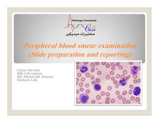
  • 1. Peripheral blood smear examination (Slide preparation and reporting) Caesar Abu Arra MSc Life sciences BSc Medical lab. Sciences Medicare Labs
  • 2. Aim of blood smear  Evaluation of anemia  Infections • bacteria, malaria, microfilaria..etc.  Abnormal cells • blasts, inclusions..etc.  Cells morphology and count Abu Jad Caesar
  • 3. Making blood films  Three basic steps to make blood film:  Preparation of blood smear.  Fixation of blood smear.  Staining of blood smear. Abu Jad Caesar
  • 4. Preparation of blood smear  Different types of blood smears:  The wedge smear slide to slide method.  The spun smear.  Two additional types of blood smears (used for specific purposes): o Buffy coat smear for WBCs < 1.0×10⁹/L. o Thick blood smears for blood parasites . Abu Jad Caesar
  • 5. Wedge blood smear  Smears should be made within 1 hour of blood collection from EDTA specimens stored at room temperature to avoid distortion of cell morphology.  Small drop (10 µl)  Hold the spreader slide at a 30°- 45° angle, and draw it back against the drop of blood.  Air dry at room temperature. Abu Jad Caesar
  • 6. Abu Jad Caesar Wedge blood smear
  • 7. Procedure notes 1. The smear should be made without delay. Any delay results in an abnormal distribution of the white blood cells, with many of the large white cells accumulating at the thin edge of the smear. 2. HCT↑ ↓ The angle of the spreader slide HCT↓ ↑ The angle of the spreader slide Abu Jad Caesar
  • 8. large angle low HCT small angle high HCT Abu Jad Caesar Procedure notes
  • 9. Spin method  Automated method  Placeadrop of blood in thecenter of aglassslide.  Spin atahighspeed in aspecialcentrifugecytospin.  Blood spreads uniformly. Abu Jad Caesar
  • 10. Characteristics of a Good Smear  Thick at one end, thinning out to a smooth rounded feather edge.  Should occupy 2/3 of the total slide area.  Should not touch any edge of the slide. Abu Jad Caesar
  • 11. tail body head Abu Jad Caesar Characteristics of a Good Smear
  • 12. Common causes of a poor blood smear  Drop of blood too large or too small.  Spreader slide pushed across the slide in a jerky manner.  Failure to keep the spreader slide at a 30° angle with the slide.  Failure to push the spreader slide completely across the slide. Abu Jad Caesar
  • 13. Common causes of a poor blood smear  Holes in film: Slide contaminated with fat or grease  Cellular degenerative changes: delay in fixing  Inadequate fixing time Methanol contaminated with water. Abu Jad Caesar
  • 14. Biologic causes of a poor smear  Cold agglutinin - RBCs will clump together. Warm the blood at 37° C for 5 minutes.  Lipemia - holes will appear in the smear. Nothing you can do to correct this.  Rouleaux - RBC’s will form into stacks as coins. Nothing you can do to correct this. Abu Jad Caesar
  • 15. Staining of blood smear  Romanowsky stain: Any Combination of: Eosin Y (Acidic or anionic dye): o Binds to cationic sites (cytoplasm & Hb) Methylene blue (Basic, cationic or Azure B dye): o Binds to anionic sites (nucleus and RNA) Abu Jad Caesar
  • 16. Staining of blood smear  Buffer:  Used to maintain an adequate pH.  0.05M Na2PO4 (pH 6.4) or dH2O (pH 6.4-6.8) Abu Jad Caesar
  • 17. pH of the phosphate buffer  If the pH is too acidic: RBC :Pinker WBC nuclei : very pale staining (very pale purple)  If the pH is too basic: RBC :Grayish blue WBC nuclei :very deeply purple Abu Jad Caesar
  • 18. pH of the phosphate buffer Abu Jad Caesar
  • 19. Staining Troubleshooting  Too Acid Stain: Insufficient staining time Prolonged buffering or washing Old stain  Correction:  Lengthen staining time  Check stain and buffer pH  Shorten buffering or wash time Abu Jad Caesar
  • 20. Staining Troubleshooting  Too Alkaline Stain: Thick blood smear Prolonged staining Insufficient washing Alkaline pH of stain components  Correction :  Check pH  Shorten stain time  Prolong buffering time Abu Jad Caesar
  • 22.  Macroscopic view:  Quality of the smear  The microscopic view:  Progressing from low power to high power: Blood film examination - preliminary Abu Jad Caesar
  • 23. Blood film examination - preliminary Abu Jad Caesar
  • 24.  Low-Power (10x) Scan  Determine the overall staining quality of the blood smear.  Determine if there is a good distribution of the cells. o Discussed later  Find an optimal area for the detailed examination and enumeration of cells Blood film examination - preliminary Abu Jad Caesar
  • 25.  High-Power (40x) Scan  Determine the WBC estimate. o WBCs are counted in 10 fields and averaged X 2000 o The estimate could be reported according to the table below: Blood film examination - preliminary Abu Jad Caesar
  • 26. • High-Power (40x) Scan  Grading scale for WBC count (X103 cell/µl) Blood film examination - preliminary Low WBC High WBC Mild 3-4.5 Mild 10-15 Moderate 2-3 Moderate 15-30 Severe <2 Marked 30-100 Extreme >100 Abu Jad Caesar
  • 27.  Oil Immersion (100x) Examination  Perform a 100 WBC differential count.  Correct WBC count that has greater than 10 NRBCs per 100 WBCs. o Report as: WBC count was corrected due to presence of NRBCs. Blood film examination - preliminary Abu Jad Caesar
  • 28.  Oil Immersion (100x) Examination  to correct a WBC count (/µl): Uncorrected WBC (/ ) X l Blood film examination - preliminary Abu Jad Caesar
  • 29.  Oil Immersion (100x) Examination  Evaluate the RBCs for: o Anisocytosis o Poikilocytosis o Inclusions o Hypochromasia o Polychromasia. Blood film examination - preliminary Abu Jad Caesar
  • 30.  Oil Immersion (100x) Examination  Perform a platelet estimate and evaluate platelet morphology: o Platelets are counted in 10 fields and averaged: • X 15  Automated smear. • X 20  Wedge smear is used. Blood film examination - preliminary Abu Jad Caesar
  • 31.  Oil Immersion (100x) Examination  Grading scale for platelets count (X103 cell/µl) Blood film examination - preliminary Abu Jad Caesar Low Platelets High Platelets Mild 100-150 Mild 500-700 Moderate 50-100 Moderate 700-900 Severe <50 Severe 900-1000 Extreme >1000
  • 32.  Oil Immersion (100x) Examination  The absolute WBC count (/µl) may be determined: Relative count (%) X Total WBC count (/µl) Blood film examination - preliminary Abu Jad Caesar
  • 33.  Relative vs. Absolute value:  Relative value: o Measures the percentage of corresponding WBC type in peripheral blood. Blood film examination - preliminary Abu Jad Caesar
  • 34.  Relative vs. Absolute value:  Absolute value: o Measures the total count of corresponding WBC type /µl in peripheral blood. o Gives more meaningful information than the percentage. o It is the preferred reporting method for the WBC differential. • Reported as absolute cytosis or absolute cytopenia. Blood film examination - preliminary Abu Jad Caesar
  • 35.  Relative vs. Absolute value: Example: leukopenic patient and increased susceptibility to infection and sepsis:  WBC=5000 Relative Neutrophils=50% o Absolute= 2500 cell/µl Normal  WBC=2000 Relative Neutrophils=85% o Absolute= 1700 cell/µl Low Blood film examination - preliminary Abu Jad Caesar
  • 37.  Examination of blood films should include:  RBC Size, Shape, color Hemoglobin distribution Arrangement and distribution Inclusions nucleated RBCs Examination of blood films Abu Jad Caesar
  • 38.  Examination of blood films should include:  WBC  Total counts  Differential counts  Abnormal and immature WBC Examination of blood films Abu Jad Caesar
  • 39.  Examination of blood films should include: Platelets  Counts should be verified by estimation  Size  Clumping Examination of blood films Abu Jad Caesar
  • 40.  Examination of blood films should include: Parasites Examination of blood films Abu Jad Caesar
  • 41.  Pancytopenia Deficiency in all the three cell lines RBC, WBC, and platelets.  Aplastic anemia  When report, recommend bone marrow assessment.  Bicytopenia Deficiency in two of the three cell lines  Viruses and drugs Terms Abu Jad Caesar
  • 42. PARASITES Abu Jad Caesar  Thick film When parasites are scanty  Thin film  Identification of species
  • 43. PARASITES Abu Jad CaesarAbu Jad Caesar Plasmodium falciparum Trypanosoma spp. Microfilaria
  • 45.  1 to 4 μm in diameter and vary in shape.  Reddish-purple granules  Life span 9-12 days  One megakaryocyte can release several thousand platelets (4000).  Report as platelets are adequate with normal-sized ones OR platelets are normal Abu Jad Caesar Platelets
  • 46.  Large and Giant platelets Large platelets (4-7 µm) oITP Abu Jad Caesar Platelets
  • 47.  Large and Giant platelets Giant platelet (7-20 µm) oPlatelets seems to be size of RBC. oBernard Soulier syndrome Abu Jad Caesar Platelets
  • 48.  Large and Giant platelets NOTE: o When estimate, Large platelets and platelet aggregates NOT counted. Report as Occasional, Few, Many Abu Jad Caesar Platelets
  • 49.  Platelet anisocytosis: Small large giant platelets will be seen  Report as: Platelets anisocytosis ranging in size from tiny to large giant platelets were seen. Abu Jad Caesar Platelets
  • 50.  Thrombocytosis  Post infection and Inflammation  Report as: o Thrombocytosis (with grading). o Platelets were estimated and approved to be about ( /µl)  In case of microcytosis (RBC and PLT diameters are similar) report as : o Thrombocytosis due severe microcytosis. Abu Jad Caesar Platelets
  • 51.  Thrombocytopenia: Could be due to: o Decreased production Aplastic anemia o Increased destruction ITP Abu Jad Caesar Platelets
  • 52.  Thrombocytopenia:  In case of no aggregates report as: o Thrombocytopenia (with grading). o No platelet clumps are noted OR Negative for microaggregates. o Platelets were estimated and approved to be about ( /µl) Abu Jad Caesar Platelets
  • 53.  Pseudothrombocytopenia: In vitro EDTA dependent phenomena oPlatelet clumping Abu Jad Caesar Platelets
  • 54.  Pseudothrombocytopenia: In vitro EDTA dependent phenomena oPlatelet satellitism • Platelets adhere to neutrophil surface Abu Jad Caesar Platelets
  • 55.  Pseudothrombocytopenia: Repeat on new samples (EDTA and sodium citrated) Scan for platelet clumping in thin and thick portion of smear Abu Jad Caesar Platelets
  • 56.  Pseudothrombocytopenia: Reporting criteria: Two samples were drawn to rule out EDTA- induced pseudothrombocytopenia: o EDTA smear revealed platelet clumps / Platelet satellitism o Na+ citrated smear revealed normal platelets count and distribution. Conclusion: Platelets are adequate Abu Jad Caesar Platelets
  • 57. Morphology of Normal RBC  Biconcave disc  Diameter 7 ~ 8 μm  Central pallor occupy 3rd of total size  Approx same as nucleus of mature lymphocyte  Stained with eosin component of Romanowsky dyes Abu Jad Caesar RBC
  • 59. RBC Abnormality  Examination of blood films for RBC’s should include:  Anisocytosis  Poikilocytosis  Hemoglobin distribution  Arrangement and Distribution  Inclusions  NRBCs Abu Jad Caesar
  • 61. Arrangement and Distribution  Normal arrangement and distribution:  The thin portion of the smear where RBC’s are slightly separated from one another or at most, barely touching, with no overlap.  The thin area should represent at least 3rd of the entire film. Abu Jad Caesar
  • 62. Arrangement and Distribution  Normal arrangement and distribution:  The reviewer should avoid the thicker portion of the slide where cells are overlapping and the edges of smear where cells may be distorted in size, shape, and color. o An exception is to be made when scanning for platelet clumping Abu Jad Caesar
  • 63. Arrangement and Distribution  Rouleaux  RBC’s appear as stacks of coins (linear)  Due to ↑ Globulins or fibrinogens  seen in: o Multiple myeloma o Macroglobulinemia Abu Jad Caesar
  • 64. Arrangement and Distribution  Agglutination  More irregular and round clumping than linear rouleaux  Seen in: o Cold agglutinin o Anti RBC antibodies o Autoimmune HA o Macroglobulinemia Abu Jad Caesar
  • 65. Inclusions  Basophilic stippling (Punctate basophilia)  Aggregates of Ribrosomes.  Multiple blue black inclusions (coarse or punctate )  Seen in: o Lead and arsenic poisoning o Thalassemias o Alcoholism o Megaloblastic anemias Abu Jad Caesar
  • 66. Inclusions  Howell Jolly bodies  Large round inclusions which are remnant of nuclear chromatin  Appear singly or doubly in an eccentric position on the cell periphery  Deep purple Abu Jad Caesar
  • 67. Inclusions  Howell Jolly bodies  Seen in: o Postsplenectomy o Megalobalstic anaemia o Haemolytic anaemia Abu Jad Caesar
  • 68. Inclusions  Pappenheimer Bodies  Ferric compound complexed with protein  Small dark blue bodies of uniform size. Abu Jad Caesar
  • 69. Inclusions  Pappenheimer Bodies  Unlike basophilic stippling: o Basophilic stippling appears homogeneously over the cell whereas Pappenheimers tend to appear as single or clusters in the cells periphery.  Unlike Howell–Jolly bodies: o Howell–Jolly bodies appear to be larger Abu Jad Caesar
  • 70. Inclusions  Pappenheimer Bodies  Seen in: o Sideroblastic erythropoiesis o Postsplenectomy o Hemolytic anemia o Thalassemia Abu Jad Caesar
  • 71. Inclusions  Heinz bodies  Result of denatured or precipitated hemoglobin  Purple, blue, large, single or multiple inclusions attached to the inner surface of the red blood cell. Abu Jad Caesar
  • 72. Inclusions  Heinz bodies  Stained by supravital stains  Not Stained by Romanowsky stain.  Seen in: o Postsplenectomy o oxidant stress of drugs and chemicals Abu Jad Caesar
  • 73. Inclusions  Cabot rings  Represent a part of the mitotic spindle, remnants of microtubules, or a fragment of the nuclear membrane.  Have no internal structure Abu Jad Caesar
  • 74. Inclusions  Cabot rings  Red or reddish purple  Could be appear in a figure-of-eight conformation Abu Jad Caesar
  • 75. Inclusions  Cabot rings  Seen rarely in o Megaloblastic anemias o Postsplenectomy Abu Jad Caesar
  • 76. Poikilocytosis  Shape variation  Mention the abnormal shapes rather than the term poikilocytosis.  Grading scale: Abu Jad Caesar
  • 77. Poikilocytosis  Spherocytes  Loss of biconcavity o ↓ Surface-to-volume ratio o ↑ Osmotic fragility  Smaller diameter  Dense-staining o No central pallor area Abu Jad Caesar
  • 78. Poikilocytosis  Spherocytes  Seen in: o Hereditary spherocytosis o Immune hemolytic anemia Abu Jad Caesar
  • 79. Poikilocytosis  Target cell  Codocytes  ↑ membrane cholesterol and phospholipid and ↓ cellular hemoglobin.  ↑ surface-to-volume ratio Abu Jad Caesar
  • 80. Poikilocytosis  Target cell  Seen in: o Obstructive liver disease o Iron deficiency anemia o Thalassemia o Post-transfusion Abu Jad Caesar
  • 81. Poikilocytosis  Leptocyte  Synonymous with target cell, BUT: o Red cells with large unstained central area. Abu Jad Caesar
  • 82. Poikilocytosis  Leptocyte  Seen in: o IDA o Thalassemia Abu Jad Caesar
  • 83. Poikilocytosis  Elliptocyte and ovalocyte  Elliptocytes o Pencil, rod, or cigar shaped o Hb appears to be concentrated on both ends of the cell o Seen in IDA Abu Jad Caesar
  • 84. Poikilocytosis  Elliptocyte and ovalocyte  Ovalocyte o More egg-shaped o Have a greater tendency to vary in their Hb content o Seen in megaloblastic anemia Abu Jad Caesar
  • 85. Poikilocytosis  Elliptocyte and ovalocyte Abu Jad Caesar
  • 86. Poikilocytosis  Teardrop cell  Dacrocytes  Physiologic mechanism is unknown  Seen in: o IDA o Pernicious anemia Abu Jad Caesar
  • 87. Poikilocytosis  Stomatocyte  RBC with narrow slit-like area of central pallor  Normal size, but are not biconcave  Seen in: o Acute alcoholism o Hemolytic anemia o Malignancies Abu Jad Caesar
  • 88. Poikilocytosis  Acanthocyte Thorn Cells, Spur Cells 3 to 12 irregular, uneven length spicules distributed along the periphery of the cell membrane Abu Jad Caesar
  • 89. Poikilocytosis  Acanthocyte  Seen in: o Severe hepatic disease o Autoimmune HA o Postsplenectomy o Vitamin E deficiency Abu Jad Caesar
  • 90. Poikilocytosis  Echinocyte  Burr Cells, crenated cells  10 to 30 regular spicules, evenly placed over the surface of RBC  Considered pathologic and should be reported. Abu Jad Caesar
  • 91. Poikilocytosis  Echinocyte  Seen in: o Liver disease o Renal disease o Severe burns Abu Jad Caesar
  • 92. Poikilocytosis  Fragmented cells  Several types 1- Schistocytes 2- Helmet Cells (bite cell) usually 2 projections 3- Keratocytes Abu Jad Caesar
  • 93. Poikilocytosis  Fragmented cells  Sometimes counted as platelets o Estimate platelets and report thrombocytosis due to presence of RBC fragments  Seen in : DIC, TTP, HUS Abu Jad Caesar
  • 94. Poikilocytosis  Fragmented cells  Several types 4- Prekeratocyte (Blister cell) resemble a women’s handbag o RBC’s containing a peripherally located vacuole. o Keratocytes are RBC’s that have been caught on fibrin strands in circulation, and rather than splitting, the cell hangs over the fibrin fusing two sides of the cell together, creating a vacuole. o Seen in:  Oxidative stress G6PD deficiency Abu Jad Caesar
  • 95. Anisocytosis  Size variation  Grading scale: Abu Jad Caesar
  • 96. Anisocytosis  Size variation  Normocytic  Microcytic  Macrocytic Abu Jad Caesar
  • 97. Anisocytosis  Normocyte  MCV 80-100 fl  Diameter 7 to 8 μm Abu Jad Caesar
  • 98. Anisocytosis  Microcytes  MCV <80 fl  Diameter <7 μm  Results from a defect in hemoglobin formation  Seen in: o IDA o Thalassemia Abu Jad Caesar
  • 99. Anisocytosis  Macrocytes  MCV >100 fl  Diameter >9 μm  Result from impaired DNA synthesis.  Seen in:  Megaloblastic anemia (↓ B12 and folic acid).  Aplastic anaemia Abu Jad Caesar
  • 100. Anisocytosis  Anisocytosis is a feature of most anemias. o Report as anisocytosis (with Qual. or Quan. Grading): Abu Jad Caesar RDW: 16-18  Slight RDW: 18-22  Moderate RDW: >22 Marked or severe
  • 101. Anisocytosis  Mixture of large and small with normal MCV and high RDW usually due: Recent blood transfusion Anemia or recovery stages of anemia. o Report as Anisocytosis with dimorphic RBC population (with grading): Abu Jad Caesar RDW: 16-18  Slight RDW: 18-22  Moderate RDW: >22 Marked or severe
  • 102. Hemoglobin Distribution  MCHC used in conjunction with MCV used do describe anemias Normochromia Hypochromia Hyperchromia Polychromasia Anisochromia Abu Jad Caesar
  • 103. Hemoglobin Distribution  Normochromia: MCHC 32-36%  Normal intensity of staining.  The central pallor < 3µm (<3rd of total size) Abu Jad Caesar
  • 104. Hemoglobin Distribution  Hypochromia : MCHC < 32%  Unusually palely RBC (↓Hb content)  The central pallor > 3µm (>1/3 rd of total size)  Seen in: o IDA o Thalassemia Abu Jad Caesar
  • 105. Hemoglobin Distribution  Hypochromia : MCHC < 32%  Grading: Abu Jad Caesar
  • 106. Hemoglobin Distribution  Hyperchromia : MCHC > 36%  Decreased or absent central pallor  Seen in: o Sperocytosis (↓surface-to-volume ratio) o hemolytic anemias (hemolysis caused by burns) Abu Jad Caesar
  • 107. Hemoglobin Distribution  Hyperchromia : MCHC > 36%  Even though true hyperchromia does exist it is not reported as such: o It is reported in terms of the cell abnormalities resulting from the increased volume of hemoglobin and the decreased surface area (e.g spherocytes). Abu Jad Caesar
  • 108. Hemoglobin Distribution  Polychromasia  Premature RBC in circulation called Polychromatophilic cells actually reticulocytes.  Gray-blue in color and usually larger than normal RBC o Result of the residual RNA involved in Hb synthesis. Abu Jad Caesar
  • 109. Hemoglobin Distribution  Polychromasia  Seen in: o Acute and chronic blood loss. o Recovering from anemias Abu Jad Caesar
  • 110. Hemoglobin Distribution  Polychromasia  Grading excellent indicator of therapeutic effectiveness when patient is given iron or vitamin therapy as treatment of anemia Abu Jad Caesar
  • 111. Hemoglobin Distribution  Anisochromia  Hypochromic cells and normochromic cells in the same film  Seen in: o Development or recovering from anemias o Post-transfusion Abu Jad Caesar
  • 112. Hemoglobin Distribution  MCHC used in conjunction with MCV used do describe anemias Abu Jad Caesar
  • 113. White blood cells  WBC categories:  Granulocytes (PMN): o Neutrophils, Eosinophils and basophiles o Granules in their cell cytoplasm. o Multilobed nucleus  Agranulocytes (MNC): o Lymphocytes and monocytes. o Do not have granules (contain non specific azurophilic granules) o Nonlobular nuclei. Abu Jad Caesar
  • 115. Neutrophil  Terms  Shift to the left o Release of younger granulocytes (specifically bands and metamyelocytes from the bone marrow). Abu Jad Caesar
  • 116. Neutrophil  Terms  Shift to the right o An abnormal cell maturation situation that occurs when hypersegmented cells are seen. • It is indicative of vitamin B12 and/or folate deficiency. Abu Jad Caesar
  • 117. Neutrophil  Cytoplasm o Pink  Nucleus o Dark purple blue o Dense chromatin o 3-5 lobes Abu Jad Caesar
  • 118. Neutrophil  Report as: o Mature/normal appearing neutrophils. o In case of leukocytosis neutrophilic leukocytosis o Absolute / relative neutrophilia or neutropenia Abu Jad Caesar
  • 119. Neutrophil  Neutrophilia o Acute bacterial infection. o Many inflammatory processes.  Neutropenia o Typhoid fever o Brucellosis o Viral diseases. Abu Jad Caesar
  • 120. Abnormal neutrophil  Abnormal neutrophil morphology: Band or stab forms Hypersegmentation (≥ 6 lobes) Vacuolization Toxic granulation Dohle bodies Nuclear pyknosis and karyorrhexis Abu Jad Caesar
  • 121. Band or Stab  Cytoplasm o Pink  Nucleus o U shaped nucleus of uniform thickness. o Purplish red o Dense chromatin Abu Jad Caesar
  • 122. Band or Stab  Normally form 5-10% of WBC’s  Increased bands: o Acute infection (usually bacterial) o Non infectious inflammatory disease Abu Jad Caesar
  • 123. Band or Stab  Include it when calculate absolute neutrophil  Report as: o Many/Few neutrophilc bands OR with shift to the left Abu Jad Caesar
  • 124. Hypersegmentation  Neutropils with ≥ 6 lobes  Normally < 3% of WBC’s  Seen in megaloblastic anemia Abu Jad Caesar
  • 125. Hypersegmentation  Report as: o Few/many hypersegmented neutrophils OR with shift to the right. o Further hematological examinations are recommended (B12 & folic acid). Abu Jad Caesar
  • 126. Vacuolization  Vacuoles in neutrophil contain ingested microorganisms.  Seen in: o Severe infection (leukemoid reaction) o Non infectious inflammation Abu Jad Caesar
  • 127. Toxic granulation  ↑ staining density and number of granules  Seen in: o Bacterial infection and leukemoid reaction o Other inflammation Abu Jad Caesar
  • 128. Döhle bodies  Pale blue inclusions at the periphery of the cytoplasm  Strands of RER that have aggregated o Bacterial infection and leukemoid reaction o Inflammation Abu Jad Caesar
  • 129. Nuclear pyknosis and karyorrhexis  Degenerative changes in nucleus of neutrophil :  Pyknosis: Condensation and shrinkage of cells through degeneration.  Karyorrhexis: A necrotic stage with fragmentation of the nucleus, whereby chromatin is distributed irregularly throughout the cytoplasm. Abu Jad Caesar
  • 130. Nuclear pyknosis and karyorrhexis  Due to:  Prolonged time in circulation.  Increased numbers in inflammatory and neoplastic disorders  Old blood samples.  New fresh sample is preferred. Abu Jad Caesar
  • 131. Nuclear pyknosis and karyorrhexis  Could be mistaken with NRBC’s. Abu Jad Caesar
  • 133. Eosinophil  Cytoplasm o Full of orange-red granules  Nucleus o Blue purple o Coarsely granular chromatin o 2 Lobes (like a pair of glass) Abu Jad Caesar
  • 134. Eosinophil  Eosinophilia o Parasitic infection o Allergic Disorders o Eosinophilic leukemia Abu Jad Caesar
  • 136. Basophil  Cytoplasm o Pale blue with coarse violet-blue granules  Nucleus o Blue purple o Coarsely granular chromatin o 2 Lobes Abu Jad Caesar
  • 137. Basophil  Basophilia o Allergic Reactions o Infections Smallpox Chicken pox Influenza Abu Jad Caesar
  • 139. Monocyte  Cytoplasm o Pale gray-blue o Vacuoles and granules may be observed  Nucleus o Blue-purple o large irregularly, folded, kidney shaped, ….etc Abu Jad Caesar
  • 140. Monocyte Often mistaken for a large lymphocyte Abu Jad Caesar
  • 141. Monocyte  Monocytosis o Tuberculosis o Brucellosis o Malaria Abu Jad Caesar
  • 143. Lymphocyte  Cytoplasm o Light sky blue  Nucleus o Dark blue o Dense chromatin o Round Abu Jad Caesar
  • 144. Lymphocyte  Report as: o Mature-appearing lymphocytes with homogenous chromatin pattern (Resting lymphocytes). o In case of leukocytosis Lymphocytic leukocytosis Abu Jad Caesar
  • 145. Lymphocyte  Lymphocytosis o Lymphocytic leukemia o Hepatitis o Mumps o Syphilis  Lymphocytopenia o Severe bacterial infection Abu Jad Caesar
  • 147. Abnormal lymphocyte  Abnormal lymphocyte morphology: Reactive lymphocytes Smudge cells (Basket cell) Turk cell (Transformed lymphocyte or immunoblast) Atypical Lymphocytes (Malignant-appearing cell) All should be reported Abu Jad Caesar
  • 148. Reactive lymphocyte  Normally < 10% of the total lymphocytes  Nucleus: o Slightly large with more open chromatin  Cytoplasm: o Abundant and irregular (↓N:C) Abu Jad Caesar
  • 149. Reactive lymphocyte  Seen in: o Infectious mononucleosis o Viral infections  The term atypical should not be used interchangeably with reactive lymphocyte. Abu Jad Caesar
  • 150. Smudge cell  Normally < 1%  Remnants of cells that lack: o Identifiable cytoplasmic membrane and Nuclear structure.  Associated with abnormally fragile lymphocytes (mostly CLL)  Should be reported. Abu Jad Caesar
  • 151. Turk cell  Reactive lymphocyte with large nucleolus, abundant and deeply basophilic cytoplasm  Seen in bacterial and viral infection  Round nucleus Abu Jad Caesar
  • 152. Atypical Lymphocyte  Malignant-appearing cells  Discussed later Abu Jad Caesar
  • 153. Leukemia  Two major types according to cell maturity:  Acute o The malignant cells are immature (stem cells, blasts, or other immature precursors.  Chronic o The cells are predominantly mature. Abu Jad Caesar
  • 154. Leukemia  Two major types according to cell lineage: Myeloid (Myelogenous) o Granulocytic leukemia o Monocytic leukemia o Megakaryocytic leukemia o Erythrocytic leukemia Lymphoid (Lymphocytic) Abu Jad Caesar
  • 155. Leukemia  So, four major types of leukemias:  Acute myeloid leukemia (AML)  Chronic myeloid leukemia (CML)  Acute lymphoblastic leukemia (ALL)  Chronic lymphocytic leukemia (CLL) Abu Jad Caesar
  • 156. Acute vs. Chronic Abu Jad Caesar
  • 157. WBC Reporting  WBC Reporting should includes: Leukocytosis/Leukocytopenia. Maturation stage Absolute value of major cell. Chromatin (fine, coarse, clumped) Nucleolus ?? Amount of cytoplasm ??. Abu Jad Caesar
  • 158. WBC Reporting  WBC Reporting should includes: For Leukemia, recommend flow cytometry for immunological markers For CML Recommend Philadelphia chromosome (BCR–ABL fusion genepositive >95%) Abu Jad Caesar
  • 167. CML  CML is characterized by three phases Chronic phase Accelerated phase Blast phase Abu Jad Caesar
  • 168. CML  Chronic phase  Neutrophilic leukocytosis o From segmented neutrophils to occasional blasts (≤10%) But mainly mature  Basophilia/eosinophilia Abu Jad Caesar
  • 169. CML  Chronic phase  Thrombocytosis (~50% of cases).  Normocytic anemia  ↑ uric acid , LDH, Vitamin B12 Abu Jad Caesar
  • 170. CML  Accelerated phase  Worsening of anemia  Basophils (>20%).  Shift to the more immature myeloid forms (blasts 10-19%). Abu Jad Caesar
  • 171. CML  Accelerated phase  Persistent thrombocytopenia ( 100 X 109/L)  Persistent thrombocytosis ( 1000 X 109/L) Abu Jad Caesar
  • 172. CML  Blast phase  Blast crisis (≥20%)  Conversion of CML to AML Abu Jad Caesar
  • 174. Leukemoid reaction  Moderate or marked leukocytosis Usually o >30000 cell/µl Absolute neutrophilia Shift to the left o Mostly neutrophilic metamyelocytes and band forms. o Immaturity is similarly observed in the early stages of CML Abu Jad Caesar
  • 175. Leukemoid reaction  Not a result of a leukemic disease Infectious disease o Bacterial (most common) o Toxoplasma and viral (less common) Other inflammatory process o Uremia, acute gout and burns Drug and chemical intoxication Abu Jad Caesar
  • 176. Leukemoid reaction  Reactive changes of neutrophils  Usually appear as follow arrangement 1- Toxic granulation o Nonspecific reactive changes 2- Döhle bodies o Nonspecific reactive changes 3- Cytoplasmic vacuolization o Strongly indicates a serious bacterial infection Abu Jad Caesar
  • 177. Leukemoid reaction  Reactive changes of neutrophils  Usually appear as follow arrangement 1- Toxic granulation o Nonspecific reactive changes 2- Döhle bodies o Nonspecific reactive changes 3- Cytoplasmic vacuolization o Strongly indicates a serious bacterial infection Abu Jad Caesar
  • 179.  Report as: WBC immaturity. Neutrophils reactive changes Features in keeping with marked response to infectious or inflammatory processes (Leukemoid reaction). Abu Jad Caesar Leukemoid reaction
  • 180.  Slight leukocytosis with neutrophilia With or without the shift to the left. Toxic granulation  Report as: Features in keeping with mild response to infection or inflammatory process. Abu Jad Caesar Mild infection
  • 181.  Mild to severe normocytic normochromic anemia  ↓ platelet counts  WBC count is variable (↓ to ↑↑↑)  Auer rods in blasts cytoplasm (for AML)  The blood smear usually reveals blasts or other immature cells  Circulating NRBC are occasionally seen Abu Jad Caesar AML & ALL
  • 184. Abu Jad Caesar For AML AML & ALL
  • 185. Abu Jad Caesar AML & ALL For ALL
  • 197.  Normochromic normocytic anemia  ↑ Indirect serum bilirubin level  Thrombocytopenia is not common. Abu Jad Caesar CLL
  • 198.  Lymphocytic leukocytosis From mature appearing lymphocytes to prolymphocytes (10%) o Small lymphocytes or slightly larger than a normal lymphocyte and have a hypercondensed nuclear chromatin pattern  Smudge cells are common Abu Jad Caesar CLL
  • 200.  WBC Reporting o Leukocytosis/Leukocytopenia. o Maturation stage o Smudge cell o Chromatin (fine, coarse, clumped) o Nucleolus ?? (for prolymphocyte) o Amount of cytoplasm ??. o For definitive diagnosis, recommend flow cytometry for immunological markers. Abu Jad Caesar WBC Reporting for CLL
  • 201.  WBC Reporting o Mostly reported as: Mature-appearing lymphocyte with hypercondensed nuclear chromatin pattern and few/many prolymphocytes Abu Jad Caesar WBC Reporting for CLL
  • 202. Abu Jad Caesar CLL  A lymphoblasts  B lymphocytes  C smudge cells
  • 203. THANK YOU A B U J A D C A E S A R