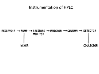
HPLC in Pathology
Malvika Tripathi
Resident of Pathology
Contents
• A physical method of separation in which the
components to be separated are distributed between
two phases: one of which is stationary (stationary
phase), whereas the other (the mobile phase) moves in
a definite direction.
• Chromatogram: A graphical presentation of detector
response, concentration of analyte in the effluent, or
other quantity used as a measure of effluent
concentration versus effluent volume or time.
• It is used in the clinical laboratory for separating and
quantifying a variety of clinically relevant analytes.
Basics of chromatography
• Mobile phase – A gas or liquid which percolates through
or along the stationary bed in a definite direction.
• Stationary phase - The stationary phase is one of the two
phases forming a chromatographic system. It may be a
solid, a gel, or a liquid. If a liquid, it may be distributed on
a solid support. This solid support may or may not
contribute to the separation process.
• During this process, the mobile phase carries the sample
through a bed, layer, or column containing the stationary
phase.
• As the mobile phase flows past the stationary phase, the
solutes may –
1. Reside only on the stationary phase (no migration) –
Solutes with high affinity for stationary phase & migrate
slower,
2. Reside only in the mobile phase (migration with the
mobile phase) – Solutes with less affinity for stationary
phase & migrate faster,
3. distribute between the two phases (differential
migration).
• Thus the lower affinity solutes separate from solutes
having greater affinities for the stationary phase.
• Strongly bound solutes subsequently are displaced from
the stationary phase by changing the physical or
chemical nature of the mobile phase.
Separation mechanisms
• Chromatographic separations are classified by the
chemical or physical mechanisms used to separate
the solutes. These include -
1. Ion-exchange,
2. Partition,
3. Adsorption,
4. Size-exclusion, and
5. Affinity mechanisms.
Ion exchange chromatography
• Ion-exchange chromatography is based on an exchange of
ions between a charged stationary surface and ions of the
opposite charge in the mobile phase.
• Depending on the conditions, solutes are either cations
(positively charged) or anions (negatively charged).
• They are separated depending on the differences in their ionic
charge or the magnitude of their ionic charges.
• It has many clinical applications including separation of - (1)
amino acids, (2) peptides, (3) proteins, (4) nucleotides, (5)
oligonucleotides, and (6) nucleic acids.
Partition chromatography
• The differential distribution of solutes between two
immiscible liquids is the basis for separation by partition
chromatography.
• Operationally, one of the immiscible liquids serves as the
stationary phase.
• Separation is based on differences in the relative
solubility of solute molecules between the stationary and
mobile phases.
Adsorption chromatography
• The basis of separation by adsorption chromatography is
the differences between the adsorption and desorption of
solutes at the surface of a solid particle.
• Electrostatic, hydrogen-bonding, and dispersive
interactions are the physical forces that control this type of
chromatography.
Size exclusion chromatography
• Also known as gel-filtration, gel permeation, steric-
exclusion, molecular-exclusion, or molecular sieve
chromatography.
• Separates solutes on the basis of their molecular sizes.
• A variety of materials are used as stationary phases for size
exclusion chromatography, including -
(1) cross-linked dextran,
(2) polyacrylamide
(3) agarose,
(4) polystyrene-divinyibenzene,
(5) porous glass, and
(6) combinations of the above.
• Beads of these materials are porous with pore sizes that
allow small molecules to be temporarily entrapped.
• Molecules too large to enter the pores remain entirely in
the mobile phase and are rapidly eluted from the column.
• Molecules that are intermediate in size have access to
various fractions of the pore volume and elute between the
large and small molecules.
• In practice, this type of chromatography is used more for
preparative than for analytical purposes.
Affinity chromatography
• In affinity chromatography, the unique and specific biological
interaction of the analyte and ligand is used for the
separation.
• Enzyme-substrate, hormone-receptor, or antigen-antibody
interactions are used in this type of chromatography.
• In the clinical laboratory, affinity chromatography has been
used to separate analytes, such as glycated hemoglobin
(phenyl boronate columns) and low-density and very low-
density lipoproteins (heparin columns).
• It has also been used to prepare larger quantities of proteins
and antibodies for further study.
Planar chromatography
• In planar chromatography, solutes are separated on a
planar surface of the stationary phase.
• Paper and TLC are sub classifications of planar
chromatography.
• In paper chromatography, the stationary phase is a layer of
water or a polar solvent coated onto the paper fibers.
• In TLC a thin layer of sorbent is spread uniformly on a glass
plate or on a plastic or aluminum sheet.
Paper chromatography
Thin layer chromatography
• Sample is added as a small spot or band near the
bottom edge of the plate.
• The plate then is placed in a closed
glass container or tank with the lower edge in, and the sample
band just above, the mobile phase.
• The mobile phase then migrates up the plate by capillary action.
• After the mobile phase travels a desired distance, the plate is
removed from the tank and dried.
• Separated components are located and identified by UV illumination,
fluorescence or spraying with color generating reagents.
Column chromatography
• In column chromatography, the stationary phase is
coated onto, or chemically bonded to, support particles
that are then "packed" into a tube, or the stationary
phase is coated onto the inner surface of the tube.
• Gas chromatography (GC) and Liquid chromatography
(LC) are sub classifications of column chromatography.
Gas chromatography
• Gas chromatography (GC) is useful for compounds that are
naturally volatile or can be easily converted into a volatile
form.
• GC has been a widely used method for decades owing to its
high resolution, low detection limits, accuracy, and short
analytic time.
• Two types of stationary phases commonly used in GC are
solid absorbent (gas–solid chromatography [GSC]) and liquids
coated on solid supports (gas–liquid chromatography [GLC]).
• In GSC, the same material (usually alumina, silica, or
activated carbon) acts as both the stationary phase and the
support phase.
• GLC uses liquid phases such as polymers, hydrocarbons,
fluorocarbons, liquid crystals, and molten organic salts to
coat the solid support material.
• Calcine diatomaceous earth graded into appropriate size
ranges is often used as a stationary phase because it is a
stable inorganic substance.
• Mobile phase – Helium, hydrogen, nitrogen, steam &
supercritical fluids (carbon dioxide, nitrogen oxide,
ammonia).
• Sample injector - syringe pipet or an automated syringe
pipet system.
• Each injection port is heated to very high temperatures.
• Samples are vaporized and swept onto the column.
• If the molecule of interest is not volatile enough for direct
injection, it is necessary to derivatize it into a more volatile
form.
• Derivatization – silylation (most common), alkylation, and
acylation.
• Columns – Packed or Capillary.
• Packed -1–5 m long and 2–4 mm in diameter, and are filled
with a stationary phase.
• Capillary - 5–100 m in length and from 0.1–0.8 mm in
diameter, and have a stationary phase located on their
interior surface.
• Capillary columns are more efficient.
• Detectors - flame ionization detector (most widely used),
thermal conductivity detector, nitrogen–phosphorus
detector, an electron capture detector, flame photometric
detector, and a mass spectrometric detector.
Liquid chromatography
• GC as a separation technique has some restrictions that
make liquid chromatography a suitable alternative.
• Many organic compounds are too unstable or are
insufficiently volatile to be assayed by GC without prior
chemical derivatization.
• Liquid chromatography techniques use lower temperatures
for separation, thereby achieving better separation of
thermolabile compounds.
• These two factors allow liquid chromatography to separate
compounds that cannot be separated by GC.
• High-performance liquid chromatography (HPLC) emerged
in the late 1960s as a viable form of liquid chromatography
that provided advantages over other forms of liquid
chromatography and gas chromatography.
• HPLC uses small, rigid supports and special mechanical
pumps, producing high pressure to pass the mobile phase
through the column.
• The resolution achieved with HPLC columns is superior to
that of other forms of liquid chromatography, analysis
times are usually much shorter, and reproducibility is
greatly improved.
• All of these attributes of HPLC render it a better method of
separation over other forms of liquid chromatography.
Instrumentation of HPLC
Reservoir
• Solvents used as the mobile phase are contained in solvent
reservoirs.
• In their simplest forms, the reservoirs are glass bottles or
flasks into which "feed lines" to the pump are inserted.
• To remove particles from solvents, inline filters are placed
on the inlets of the feed lines.
Pumps
• The most critical feature of a liquid chromatographic
system (apart from the column) is the pump.
• Both constant pressure and constant displacement pumps
are used in liquid chromatographs with the latter used
more widely.
• During its operation, the constant displacement pump
withdraws (aspirates) the mobile phase from the solvent
reservoir and delivers a reproducibly constant flow of it
through the chromatographic system.
• The HPLC pump is operated in either an isocratic or gradient
mode.
• In the isocratic mode, the mobile phase composition remains
constant throughout the chromatographic run.
• This mode is usually used for simpler separations and
separations of those compounds with similar structures
and/or retention times.
• Most HPLC separations are performed under isocratic
conditions.
• Gradient mode is used for more complex separations.
• In this mode, mobile phase composition is changed during
the run in either a stepwise or continuous fashion.
Injector
• The most widely used type of injector is the fixed-loop
injector.
• In the fill position, an aliquot of sample is introduced at
atmospheric pressure into a stainless steel loop.
• In the inject mode, the sample loop is rotated into the
flowing stream of the mobile phase, and the sample flows
into the chromatographic column.
• These injectors are -
(1) precise,
(2) function at high pressures, and
(3) have been programmed for use in automated
systems.
Column
• Both packed and capillary columns are used in HPLC.
• For use in the clinical laboratory, most analytical HPLC
columns are fabricated from tubes made of 316 stainless
steel that have internal diameter ranging from 0.1 mm to 5
mm and lengths from 50 mm to 250 mm.
• In addition, columns (termed "nanobore") are being
developed having IDs ranging from 25 to 100 pm.
• Generally, columns having smaller IDs-
(1) are more efficient,
(2) have lower detection limits, and
(3) require decreased volumes of mobile phase.
• Capillary columns used in LC are constructed by coating the
inner wall of a fused-silica tube with a thin film of liquid
phase.
• These columns vary from 0.1 to 1.0 mm in ID and from 10
to 50 cm in length.
• To prevent an analytical column from irreversibly adsorbing
proteins contained in the sample aliquot, with a
subsequent reduction in both resolution and column life, a
guard column is placed between the injector and analytical
column.
• A guard column is packed with the same or similar
stationary phase as the analytical column.
• It collects particulate matter and any strongly retained
components from the sample and thus conserves the life of
the analytical column.
• Types of column packings –
1. Particulate column packing
2. Monolithic Particulate Column Packings
• Particulate column packing - Particulate packings have
diameters ranging from 1.8 to 10 micrometer.
• In general, the smaller the diameter of the particle, the more
efficient the column.
• Types of particulate packings include bonded, polymeric,
chiral, and restricted access materials.
• In bonded phase packing, the stationary phase is bonded
chemically to the surface of silica particles through a silica
ester or silicone polymeric linkage.
• Bonded phase packings
(1) are mechanically and chemically stable,
(2) have long lifetimes, and
(3) provide excellent chromatographic performance.
• In normal-phase HPLC, the functional groups of the
stationary phase are polar relative to those of the mobile
phase, which usually consists of nonpolar solvents, such as
hexane.
• Reversed phase HPLC requires a nonpolar stationary phase.
• The most popular reversed-phase packing is the C18 type,
in which octadecylsilane molecules are bonded to silica
particles.
Detectors
Sample preparation for HPLC
• Sample preparation is an important step in chromatographic
analysis and includes procedures for sample concentration,
purification and derivatization.
• The direct injection of a crude biological sample onto an
HPLC column can cause major problems and it is usually
essential that some form of sample clean-up be undertaken
prior to analysis.
• Failure to partially purify the sample can allow particulate
matter to enter the HPLC system and also allow soluble
components to irreversibly bind to the stationary phase.
• An initial clean-up and extraction step often involves sample
homogenization, Enzymatic hydrolysis provides a milder but
slower alternative.
• Hydrolysis can be combined with protein denaturation and
precipitation using perchloric acid to remove unwanted
proteins in a biological sample.
• Alternatively, protein can be removed by the addition of
other acids, salts (e.g. ammonium sulphate), solvents or by
ultra filtration through a semi-permeable membrane.
• At this stage, providing that the compounds of interest are
present in sufficient concentration, they can be injected
onto the HPLC column.
• As a final step, the sample should be dissolved in an
appropriate solvent (preferably the HPLC mobile phase)
and either centrifuged or filtered prior to injection onto the
HPLC column.
• If the concentration of the compounds of interest is
insufficient to allow detection the sample may require
derivatization.
Derivatization
• Pre column derivatization or post column derivatization can
be performed.
• The advantages of pre-column derivatization are not only
that sample detection can be enhanced but also that
extraction, purification and chromatography of the
compounds can be modified. In addition, a large choice of
reagents are available.
• However, the disadvantages of pre-column derivatization
are that samples may degrade during the derivatization
procedure and that the chemical reactions may not go to
completion, resulting in spurious peaks in the
chromatogram.
Applications of HPLC
1. Screening of hemoglobinopathies
 Cation: exchange high performance liquid chromatography
(HPLC) has emerged as the method of choice for
quantification of HbA2, HbF and for detection and
quantization of the Hb variants.
 In this method phosphate buffers at different concentrations
(mobile phase), pass under pressure through an ionic
exchange column (stationary phase).
 The stationary phase consists of a temperature controlled
analytical cartridge containing a resin of anionic or cationic
particles (3-5 μm).
• The hemoglobins are separated according to their ionic
interaction with the stationary phase.
• The separated hemoglobins then pass through the flow cell
of the filter photometer, where changes in the absorbance
(415 nm) are measured.
• Each hemoglobin is characterized by a specific retention
time, which is the elapsed time from the sample injection
to the apex of a hemoglobin peak.
• Specific software turns the raw data collected from each
analysis into a report.
• The report presents the percentages of hemoglobins F, A1C,
A and A2 and provides qualitative and quantitative
determination of abnormal hemoglobins.
• The expected normal range for HbA2 is between 1.7% and
3.2% in normal subjects, while in β-thalassaemia carriers
when it is between 4.0% and 7%.
• Since Hb Lepore and HbE are co-eluted with HbA2, their
presence in the sample gives a falsely high percentage
(>10%) of HbA2.
• This amount of HbA2 is almost never present in β-
thalassaemia carriers.
• Therefore samples found to have a level of HbA2 greater
than 10% should be further tested for the possible
presence of a hemoglobin variant running with the
HbA2 peak.
2. Estimation of Glycosylated hemoglobin.
 The measurement of HbA1c in human blood is most
important for the long term control of the glycaemic state in
diabetic patients.
 Because there was no internationally agreed reference
method the IFCC Working Group on HbA1c Standardization
developed a reference method.
 In a first step haemoglobin is cleaved into peptides by the
enzyme endoproteinase Glu-C.
 In a second step the glycated and non-glycated N terminal
hexapeptides of the chain obtained are separated and
quantified by HPLC and electrospray ionization mass
spectrometry or in a two-dimensional approach using HPLC
and capillary electrophoresis with UV-detection.
Other uses
References
1. Tietz fundamentals of clinical chemistry 6th edition
2. Henry's Clinical Diagnosis and Management by Laboratory
Methods 22nd edition
3. Applications of HPLC in biochemistry Volume 17
4. Prevention of Thalassaemias and Other Haemoglobin
Disorders: Volume 2: Laboratory Protocols.2nd edition. Old
J, Harteveld CL, Traeger-Synodinos J, et al. Nicosia,
Cyprus: Thalassaemia International Federation; 2012.
5. Approved IFCC Reference Method for the Measurement of
HbA1c in Human Blood.
Clin Chem Lab Med 2002; 40(1):78–89 © 2002 by Walter de
Gruyter · Berlin · New York
Thank you

HPLC in Pathology

  • 1.
    HPLC in Pathology MalvikaTripathi Resident of Pathology
  • 2.
  • 3.
    • A physicalmethod of separation in which the components to be separated are distributed between two phases: one of which is stationary (stationary phase), whereas the other (the mobile phase) moves in a definite direction. • Chromatogram: A graphical presentation of detector response, concentration of analyte in the effluent, or other quantity used as a measure of effluent concentration versus effluent volume or time. • It is used in the clinical laboratory for separating and quantifying a variety of clinically relevant analytes.
  • 4.
    Basics of chromatography •Mobile phase – A gas or liquid which percolates through or along the stationary bed in a definite direction. • Stationary phase - The stationary phase is one of the two phases forming a chromatographic system. It may be a solid, a gel, or a liquid. If a liquid, it may be distributed on a solid support. This solid support may or may not contribute to the separation process.
  • 5.
    • During thisprocess, the mobile phase carries the sample through a bed, layer, or column containing the stationary phase. • As the mobile phase flows past the stationary phase, the solutes may – 1. Reside only on the stationary phase (no migration) – Solutes with high affinity for stationary phase & migrate slower, 2. Reside only in the mobile phase (migration with the mobile phase) – Solutes with less affinity for stationary phase & migrate faster, 3. distribute between the two phases (differential migration).
  • 6.
    • Thus thelower affinity solutes separate from solutes having greater affinities for the stationary phase. • Strongly bound solutes subsequently are displaced from the stationary phase by changing the physical or chemical nature of the mobile phase.
  • 7.
    Separation mechanisms • Chromatographicseparations are classified by the chemical or physical mechanisms used to separate the solutes. These include - 1. Ion-exchange, 2. Partition, 3. Adsorption, 4. Size-exclusion, and 5. Affinity mechanisms.
  • 8.
    Ion exchange chromatography •Ion-exchange chromatography is based on an exchange of ions between a charged stationary surface and ions of the opposite charge in the mobile phase. • Depending on the conditions, solutes are either cations (positively charged) or anions (negatively charged). • They are separated depending on the differences in their ionic charge or the magnitude of their ionic charges. • It has many clinical applications including separation of - (1) amino acids, (2) peptides, (3) proteins, (4) nucleotides, (5) oligonucleotides, and (6) nucleic acids.
  • 9.
    Partition chromatography • Thedifferential distribution of solutes between two immiscible liquids is the basis for separation by partition chromatography. • Operationally, one of the immiscible liquids serves as the stationary phase. • Separation is based on differences in the relative solubility of solute molecules between the stationary and mobile phases.
  • 10.
    Adsorption chromatography • Thebasis of separation by adsorption chromatography is the differences between the adsorption and desorption of solutes at the surface of a solid particle. • Electrostatic, hydrogen-bonding, and dispersive interactions are the physical forces that control this type of chromatography.
  • 11.
    Size exclusion chromatography •Also known as gel-filtration, gel permeation, steric- exclusion, molecular-exclusion, or molecular sieve chromatography. • Separates solutes on the basis of their molecular sizes. • A variety of materials are used as stationary phases for size exclusion chromatography, including - (1) cross-linked dextran, (2) polyacrylamide (3) agarose, (4) polystyrene-divinyibenzene, (5) porous glass, and (6) combinations of the above.
  • 12.
    • Beads ofthese materials are porous with pore sizes that allow small molecules to be temporarily entrapped. • Molecules too large to enter the pores remain entirely in the mobile phase and are rapidly eluted from the column. • Molecules that are intermediate in size have access to various fractions of the pore volume and elute between the large and small molecules. • In practice, this type of chromatography is used more for preparative than for analytical purposes.
  • 14.
    Affinity chromatography • Inaffinity chromatography, the unique and specific biological interaction of the analyte and ligand is used for the separation. • Enzyme-substrate, hormone-receptor, or antigen-antibody interactions are used in this type of chromatography. • In the clinical laboratory, affinity chromatography has been used to separate analytes, such as glycated hemoglobin (phenyl boronate columns) and low-density and very low- density lipoproteins (heparin columns). • It has also been used to prepare larger quantities of proteins and antibodies for further study.
  • 16.
    Planar chromatography • Inplanar chromatography, solutes are separated on a planar surface of the stationary phase. • Paper and TLC are sub classifications of planar chromatography. • In paper chromatography, the stationary phase is a layer of water or a polar solvent coated onto the paper fibers. • In TLC a thin layer of sorbent is spread uniformly on a glass plate or on a plastic or aluminum sheet.
  • 17.
  • 18.
    Thin layer chromatography •Sample is added as a small spot or band near the bottom edge of the plate. • The plate then is placed in a closed glass container or tank with the lower edge in, and the sample band just above, the mobile phase. • The mobile phase then migrates up the plate by capillary action. • After the mobile phase travels a desired distance, the plate is removed from the tank and dried. • Separated components are located and identified by UV illumination, fluorescence or spraying with color generating reagents.
  • 19.
    Column chromatography • Incolumn chromatography, the stationary phase is coated onto, or chemically bonded to, support particles that are then "packed" into a tube, or the stationary phase is coated onto the inner surface of the tube. • Gas chromatography (GC) and Liquid chromatography (LC) are sub classifications of column chromatography.
  • 20.
    Gas chromatography • Gaschromatography (GC) is useful for compounds that are naturally volatile or can be easily converted into a volatile form. • GC has been a widely used method for decades owing to its high resolution, low detection limits, accuracy, and short analytic time. • Two types of stationary phases commonly used in GC are solid absorbent (gas–solid chromatography [GSC]) and liquids coated on solid supports (gas–liquid chromatography [GLC]).
  • 21.
    • In GSC,the same material (usually alumina, silica, or activated carbon) acts as both the stationary phase and the support phase. • GLC uses liquid phases such as polymers, hydrocarbons, fluorocarbons, liquid crystals, and molten organic salts to coat the solid support material. • Calcine diatomaceous earth graded into appropriate size ranges is often used as a stationary phase because it is a stable inorganic substance.
  • 23.
    • Mobile phase– Helium, hydrogen, nitrogen, steam & supercritical fluids (carbon dioxide, nitrogen oxide, ammonia). • Sample injector - syringe pipet or an automated syringe pipet system. • Each injection port is heated to very high temperatures. • Samples are vaporized and swept onto the column. • If the molecule of interest is not volatile enough for direct injection, it is necessary to derivatize it into a more volatile form. • Derivatization – silylation (most common), alkylation, and acylation.
  • 24.
    • Columns –Packed or Capillary. • Packed -1–5 m long and 2–4 mm in diameter, and are filled with a stationary phase. • Capillary - 5–100 m in length and from 0.1–0.8 mm in diameter, and have a stationary phase located on their interior surface. • Capillary columns are more efficient. • Detectors - flame ionization detector (most widely used), thermal conductivity detector, nitrogen–phosphorus detector, an electron capture detector, flame photometric detector, and a mass spectrometric detector.
  • 25.
    Liquid chromatography • GCas a separation technique has some restrictions that make liquid chromatography a suitable alternative. • Many organic compounds are too unstable or are insufficiently volatile to be assayed by GC without prior chemical derivatization. • Liquid chromatography techniques use lower temperatures for separation, thereby achieving better separation of thermolabile compounds. • These two factors allow liquid chromatography to separate compounds that cannot be separated by GC.
  • 26.
    • High-performance liquidchromatography (HPLC) emerged in the late 1960s as a viable form of liquid chromatography that provided advantages over other forms of liquid chromatography and gas chromatography. • HPLC uses small, rigid supports and special mechanical pumps, producing high pressure to pass the mobile phase through the column. • The resolution achieved with HPLC columns is superior to that of other forms of liquid chromatography, analysis times are usually much shorter, and reproducibility is greatly improved. • All of these attributes of HPLC render it a better method of separation over other forms of liquid chromatography.
  • 27.
  • 28.
    Reservoir • Solvents usedas the mobile phase are contained in solvent reservoirs. • In their simplest forms, the reservoirs are glass bottles or flasks into which "feed lines" to the pump are inserted. • To remove particles from solvents, inline filters are placed on the inlets of the feed lines.
  • 29.
    Pumps • The mostcritical feature of a liquid chromatographic system (apart from the column) is the pump. • Both constant pressure and constant displacement pumps are used in liquid chromatographs with the latter used more widely. • During its operation, the constant displacement pump withdraws (aspirates) the mobile phase from the solvent reservoir and delivers a reproducibly constant flow of it through the chromatographic system.
  • 30.
    • The HPLCpump is operated in either an isocratic or gradient mode. • In the isocratic mode, the mobile phase composition remains constant throughout the chromatographic run. • This mode is usually used for simpler separations and separations of those compounds with similar structures and/or retention times. • Most HPLC separations are performed under isocratic conditions. • Gradient mode is used for more complex separations. • In this mode, mobile phase composition is changed during the run in either a stepwise or continuous fashion.
  • 31.
    Injector • The mostwidely used type of injector is the fixed-loop injector. • In the fill position, an aliquot of sample is introduced at atmospheric pressure into a stainless steel loop. • In the inject mode, the sample loop is rotated into the flowing stream of the mobile phase, and the sample flows into the chromatographic column. • These injectors are - (1) precise, (2) function at high pressures, and (3) have been programmed for use in automated systems.
  • 32.
    Column • Both packedand capillary columns are used in HPLC. • For use in the clinical laboratory, most analytical HPLC columns are fabricated from tubes made of 316 stainless steel that have internal diameter ranging from 0.1 mm to 5 mm and lengths from 50 mm to 250 mm. • In addition, columns (termed "nanobore") are being developed having IDs ranging from 25 to 100 pm.
  • 33.
    • Generally, columnshaving smaller IDs- (1) are more efficient, (2) have lower detection limits, and (3) require decreased volumes of mobile phase. • Capillary columns used in LC are constructed by coating the inner wall of a fused-silica tube with a thin film of liquid phase. • These columns vary from 0.1 to 1.0 mm in ID and from 10 to 50 cm in length.
  • 35.
    • To preventan analytical column from irreversibly adsorbing proteins contained in the sample aliquot, with a subsequent reduction in both resolution and column life, a guard column is placed between the injector and analytical column. • A guard column is packed with the same or similar stationary phase as the analytical column. • It collects particulate matter and any strongly retained components from the sample and thus conserves the life of the analytical column. • Types of column packings – 1. Particulate column packing 2. Monolithic Particulate Column Packings
  • 36.
    • Particulate columnpacking - Particulate packings have diameters ranging from 1.8 to 10 micrometer. • In general, the smaller the diameter of the particle, the more efficient the column. • Types of particulate packings include bonded, polymeric, chiral, and restricted access materials. • In bonded phase packing, the stationary phase is bonded chemically to the surface of silica particles through a silica ester or silicone polymeric linkage. • Bonded phase packings (1) are mechanically and chemically stable, (2) have long lifetimes, and (3) provide excellent chromatographic performance.
  • 37.
    • In normal-phaseHPLC, the functional groups of the stationary phase are polar relative to those of the mobile phase, which usually consists of nonpolar solvents, such as hexane. • Reversed phase HPLC requires a nonpolar stationary phase. • The most popular reversed-phase packing is the C18 type, in which octadecylsilane molecules are bonded to silica particles.
  • 38.
  • 39.
    Sample preparation forHPLC • Sample preparation is an important step in chromatographic analysis and includes procedures for sample concentration, purification and derivatization. • The direct injection of a crude biological sample onto an HPLC column can cause major problems and it is usually essential that some form of sample clean-up be undertaken prior to analysis. • Failure to partially purify the sample can allow particulate matter to enter the HPLC system and also allow soluble components to irreversibly bind to the stationary phase.
  • 40.
    • An initialclean-up and extraction step often involves sample homogenization, Enzymatic hydrolysis provides a milder but slower alternative. • Hydrolysis can be combined with protein denaturation and precipitation using perchloric acid to remove unwanted proteins in a biological sample. • Alternatively, protein can be removed by the addition of other acids, salts (e.g. ammonium sulphate), solvents or by ultra filtration through a semi-permeable membrane.
  • 41.
    • At thisstage, providing that the compounds of interest are present in sufficient concentration, they can be injected onto the HPLC column. • As a final step, the sample should be dissolved in an appropriate solvent (preferably the HPLC mobile phase) and either centrifuged or filtered prior to injection onto the HPLC column. • If the concentration of the compounds of interest is insufficient to allow detection the sample may require derivatization.
  • 42.
    Derivatization • Pre columnderivatization or post column derivatization can be performed. • The advantages of pre-column derivatization are not only that sample detection can be enhanced but also that extraction, purification and chromatography of the compounds can be modified. In addition, a large choice of reagents are available. • However, the disadvantages of pre-column derivatization are that samples may degrade during the derivatization procedure and that the chemical reactions may not go to completion, resulting in spurious peaks in the chromatogram.
  • 45.
    Applications of HPLC 1.Screening of hemoglobinopathies  Cation: exchange high performance liquid chromatography (HPLC) has emerged as the method of choice for quantification of HbA2, HbF and for detection and quantization of the Hb variants.  In this method phosphate buffers at different concentrations (mobile phase), pass under pressure through an ionic exchange column (stationary phase).  The stationary phase consists of a temperature controlled analytical cartridge containing a resin of anionic or cationic particles (3-5 μm).
  • 46.
    • The hemoglobinsare separated according to their ionic interaction with the stationary phase. • The separated hemoglobins then pass through the flow cell of the filter photometer, where changes in the absorbance (415 nm) are measured. • Each hemoglobin is characterized by a specific retention time, which is the elapsed time from the sample injection to the apex of a hemoglobin peak. • Specific software turns the raw data collected from each analysis into a report. • The report presents the percentages of hemoglobins F, A1C, A and A2 and provides qualitative and quantitative determination of abnormal hemoglobins.
  • 47.
    • The expectednormal range for HbA2 is between 1.7% and 3.2% in normal subjects, while in β-thalassaemia carriers when it is between 4.0% and 7%. • Since Hb Lepore and HbE are co-eluted with HbA2, their presence in the sample gives a falsely high percentage (>10%) of HbA2. • This amount of HbA2 is almost never present in β- thalassaemia carriers. • Therefore samples found to have a level of HbA2 greater than 10% should be further tested for the possible presence of a hemoglobin variant running with the HbA2 peak.
  • 48.
    2. Estimation ofGlycosylated hemoglobin.  The measurement of HbA1c in human blood is most important for the long term control of the glycaemic state in diabetic patients.  Because there was no internationally agreed reference method the IFCC Working Group on HbA1c Standardization developed a reference method.  In a first step haemoglobin is cleaved into peptides by the enzyme endoproteinase Glu-C.  In a second step the glycated and non-glycated N terminal hexapeptides of the chain obtained are separated and quantified by HPLC and electrospray ionization mass spectrometry or in a two-dimensional approach using HPLC and capillary electrophoresis with UV-detection.
  • 49.
  • 50.
    References 1. Tietz fundamentalsof clinical chemistry 6th edition 2. Henry's Clinical Diagnosis and Management by Laboratory Methods 22nd edition 3. Applications of HPLC in biochemistry Volume 17 4. Prevention of Thalassaemias and Other Haemoglobin Disorders: Volume 2: Laboratory Protocols.2nd edition. Old J, Harteveld CL, Traeger-Synodinos J, et al. Nicosia, Cyprus: Thalassaemia International Federation; 2012. 5. Approved IFCC Reference Method for the Measurement of HbA1c in Human Blood. Clin Chem Lab Med 2002; 40(1):78–89 © 2002 by Walter de Gruyter · Berlin · New York
  • 51.