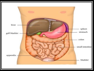
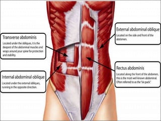
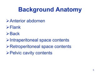
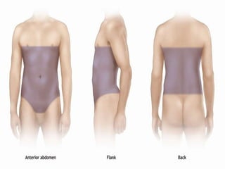
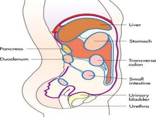
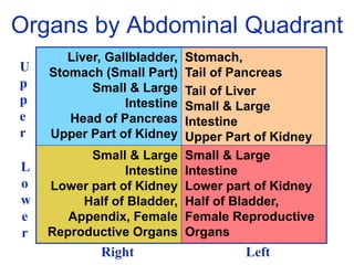
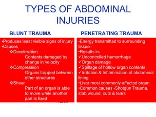
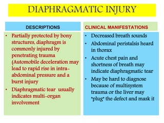
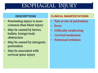
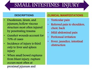
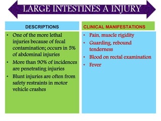
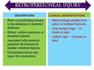
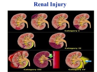
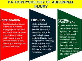
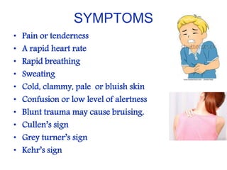
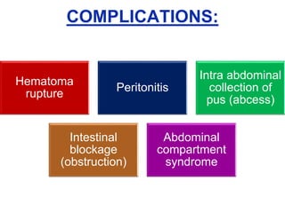
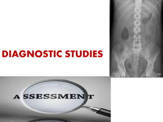
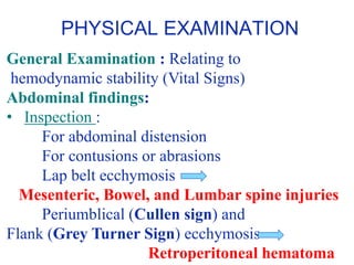
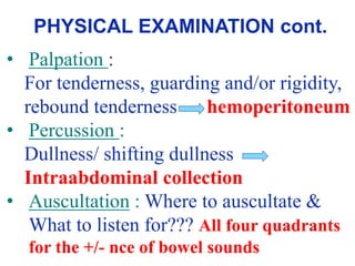
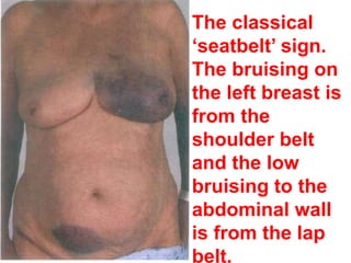
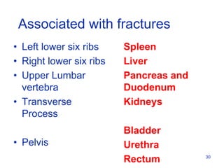
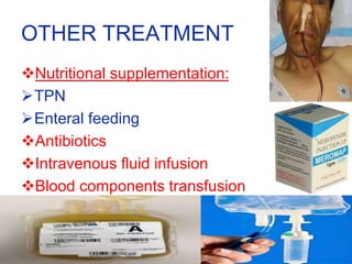
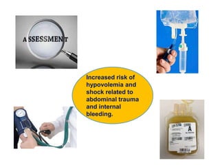
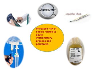
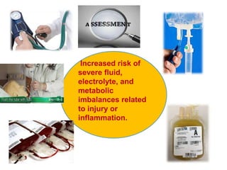
Content will be helpful for B.Sc. and M.Sc. nursing students as it describes causes, signs and symptoms, diagnosis,emergency mangement , medical and nursing management.
Approach to Management of Upper Gastrointestinal (GI) BleedingArun Vasireddy
Upper gastrointestinal bleeding is gastrointestinal bleeding in the upper gastrointestinal tract, commonly defined as bleeding arising from the esophagus, stomach, or duodenum. Blood may be observed in vomit (hematemesis) or in altered form in the stool (melena). Depending on the severity of the blood loss, there may be symptoms of insufficient circulating blood volume and shock. As a result, upper gastrointestinal bleeding is considered a medical emergency and typically requires hospital care for urgent diagnosis and treatment. Upper gastrointestinal bleeding can be caused by peptic ulcers, gastric erosions, esophageal varices, and some rarer causes such as gastric cancer.
The initial assessment includes measurement of the blood pressure and heart rate, as well as blood tests to determine hemoglobin concentration. In significant bleeding, fluid replacement is often required, as well as blood transfusion, before the source of bleeding can be determined by endoscopy of the upper digestive tract with an esophagogastroduodenoscopy. Depending on the source, endoscopic therapy can be applied to reduce rebleeding risk. Specific medical treatments (such as proton pump inhibitors for peptic ulcer disease) or procedures (such as TIPS for variceal hemorrhage) may be used. Recurrent or refractory bleeding may lead to need for surgery, although this has become uncommon as a result of improved endoscopic and medical treatment.
CHEST INJURY- BLUNT/ Trauma Surgery
Dear viewers,
Greetings from “Surgical Educator”
Today I have uploaded a video on CHEST INJURY- BLUNT- an important topic in trauma. Even the blunt chest trauma can turn into penetrating one because of jagged edges of the broken ribs. I haven’t talked elaborately but have included the essential minimum an undergraduate medical student should know. I have talked about pathophysiology, clinical approach, symptoms, signs, investigations, different individual types of Chest injuries and management of all the varieties of Chest injuries. My aim is, after watching this video all of you should be able to arrive at a correct working diagnosis of the type of chest injury and should also be able to institute immediate lifesaving treatment to the patients if there is a need. You can watch the video in the following links:
surgicaleducator.blogspot.com
youtube.com/c/surgicaleducator
Thank you for watching the video.
Lecture on abdominal trauma during Basic Life Support 2018 course in Sibu Hospital. Encompasses blunt and penetrating trauma, principles and tips of management
This is a lucid presentation on the management of abdominal trauma/injury in children of paediatric age. Even though it focused on paediatric trauma, the information is also relevant to adult trauma. It went ahead to discuss the definition, causes, and initial management of abdominal trauma. It further went to highlight the management algorithms and outcomes of management. Finally, the presentation ended with management of abdominal compartment syndrome
This is a content of PERSONALITY, FACTORS AFFECTING PERSONALITY & HUMAN BEHAVIOR. This content also explains important theories of personality in brief. I have prepared it for my Advance Nursing Practice presentation. Hope it will be helpful for Msc. nursing students.
Defecation
Normal defecation begins with movement in the left colon, moving stool toward the anus. When stool reaches the rectum, the distention causes relaxation of the internal sphincter and an awareness of the need to defecate. At the time of defecation, the external sphincter relaxes, and abdominal muscles contract, increasing intrarectal pressure and forcing the stool out
The Valsalva maneuver exerts pressure to expel faeces through a voluntary contraction of the abdominal muscles while maintaining forced expiration against a closed airway. Patients with cardiovascular disease, glaucoma, increased intracranial pressure, or a new surgical wound are at greater risk for cardiac dysrhythmias and elevated blood pressure with the Valsalva maneuver and need to avoid straining to pass the stool.
Normal defecation is painless, resulting in passage of soft, formed stool
CONSTIPATION
Constipation is a symptom, not a disease. Improper diet, reduced fluid intake, lack of exercise, and certain medications can cause constipation. For example, patients receiving opiates for pain after surgery often require a stool softener or laxative to prevent constipation. The signs of constipation include infrequent bowel movements (less than every 3 days), difficulty passing stools, excessive straining, inability to defecate at will, and hard feaces
IMPACTION
Fecal impaction results from unrelieved constipation. It is a collection of hardened feces wedged in the rectum that a person cannot expel. In cases of severe impaction the mass extends up into the sigmoid colon.
DIARRHEA
Diarrhea is an increase in the number of stools and the passage of liquid, unformed feces. It is associated with disorders affecting digestion, absorption, and secretion in the GI tract. Intestinal contents pass through the small and large intestine too quickly to allow for the usual absorption of fluid and nutrients. Irritation within the colon results in increased mucus secretion. As a result, feces become watery, and the patient is unable to control the urge to defecate. Normally an anal bag is safe and effective in long-term treatment of patients with fecal incontinence at home, in hospice, or in the hospital. Fecal incontinence is expensive and a potentially dangerous condition in terms of contamination and risk of skin ulceration
HEMORRHOIDS
Hemorrhoids are dilated, engorged veins in the lining of the rectum. They are either external or internal.
FLATULENCE
As gas accumulates in the lumen of the intestines, the bowel wall stretches and distends (flatulence). It is a common cause of abdominal fullness, pain, and cramping. Normally intestinal gas escapes through the mouth (belching) or the anus (passing of flatus)
FECAL INCONTINENCE
Fecal incontinence is the inability to control passage of feces and gas from the anus. Incontinence harms a patient’s body image
PREPARATION AND GIVING OF LAXATIVESACCORDING TO POTTER AND PERRY,
An enema is the instillation of a solution into the rectum and sig
India Clinical Trials Market: Industry Size and Growth Trends [2030] Analyzed...Kumar Satyam
According to TechSci Research report, "India Clinical Trials Market- By Region, Competition, Forecast & Opportunities, 2030F," the India Clinical Trials Market was valued at USD 2.05 billion in 2024 and is projected to grow at a compound annual growth rate (CAGR) of 8.64% through 2030. The market is driven by a variety of factors, making India an attractive destination for pharmaceutical companies and researchers. India's vast and diverse patient population, cost-effective operational environment, and a large pool of skilled medical professionals contribute significantly to the market's growth. Additionally, increasing government support in streamlining regulations and the growing prevalence of lifestyle diseases further propel the clinical trials market.
Growing Prevalence of Lifestyle Diseases
The rising incidence of lifestyle diseases such as diabetes, cardiovascular diseases, and cancer is a major trend driving the clinical trials market in India. These conditions necessitate the development and testing of new treatment methods, creating a robust demand for clinical trials. The increasing burden of these diseases highlights the need for innovative therapies and underscores the importance of India as a key player in global clinical research.
R3 Stem Cells and Kidney Repair A New Horizon in Nephrology.pptxR3 Stem Cell
R3 Stem Cells and Kidney Repair: A New Horizon in Nephrology" explores groundbreaking advancements in the use of R3 stem cells for kidney disease treatment. This insightful piece delves into the potential of these cells to regenerate damaged kidney tissue, offering new hope for patients and reshaping the future of nephrology.
CHAPTER 1 SEMESTER V - ROLE OF PEADIATRIC NURSE.pdfSachin Sharma
Pediatric nurses play a vital role in the health and well-being of children. Their responsibilities are wide-ranging, and their objectives can be categorized into several key areas:
1. Direct Patient Care:
Objective: Provide comprehensive and compassionate care to infants, children, and adolescents in various healthcare settings (hospitals, clinics, etc.).
This includes tasks like:
Monitoring vital signs and physical condition.
Administering medications and treatments.
Performing procedures as directed by doctors.
Assisting with daily living activities (bathing, feeding).
Providing emotional support and pain management.
2. Health Promotion and Education:
Objective: Promote healthy behaviors and educate children, families, and communities about preventive healthcare.
This includes tasks like:
Administering vaccinations.
Providing education on nutrition, hygiene, and development.
Offering breastfeeding and childbirth support.
Counseling families on safety and injury prevention.
3. Collaboration and Advocacy:
Objective: Collaborate effectively with doctors, social workers, therapists, and other healthcare professionals to ensure coordinated care for children.
Objective: Advocate for the rights and best interests of their patients, especially when children cannot speak for themselves.
This includes tasks like:
Communicating effectively with healthcare teams.
Identifying and addressing potential risks to child welfare.
Educating families about their child's condition and treatment options.
4. Professional Development and Research:
Objective: Stay up-to-date on the latest advancements in pediatric healthcare through continuing education and research.
Objective: Contribute to improving the quality of care for children by participating in research initiatives.
This includes tasks like:
Attending workshops and conferences on pediatric nursing.
Participating in clinical trials related to child health.
Implementing evidence-based practices into their daily routines.
By fulfilling these objectives, pediatric nurses play a crucial role in ensuring the optimal health and well-being of children throughout all stages of their development.
Medical Technology Tackles New Health Care Demand - Research Report - March 2...pchutichetpong
M Capital Group (“MCG”) predicts that with, against, despite, and even without the global pandemic, the medical technology (MedTech) industry shows signs of continuous healthy growth, driven by smaller, faster, and cheaper devices, growing demand for home-based applications, technological innovation, strategic acquisitions, investments, and SPAC listings. MCG predicts that this should reflects itself in annual growth of over 6%, well beyond 2028.
According to Chris Mouchabhani, Managing Partner at M Capital Group, “Despite all economic scenarios that one may consider, beyond overall economic shocks, medical technology should remain one of the most promising and robust sectors over the short to medium term and well beyond 2028.”
There is a movement towards home-based care for the elderly, next generation scanning and MRI devices, wearable technology, artificial intelligence incorporation, and online connectivity. Experts also see a focus on predictive, preventive, personalized, participatory, and precision medicine, with rising levels of integration of home care and technological innovation.
The average cost of treatment has been rising across the board, creating additional financial burdens to governments, healthcare providers and insurance companies. According to MCG, cost-per-inpatient-stay in the United States alone rose on average annually by over 13% between 2014 to 2021, leading MedTech to focus research efforts on optimized medical equipment at lower price points, whilst emphasizing portability and ease of use. Namely, 46% of the 1,008 medical technology companies in the 2021 MedTech Innovator (“MTI”) database are focusing on prevention, wellness, detection, or diagnosis, signaling a clear push for preventive care to also tackle costs.
In addition, there has also been a lasting impact on consumer and medical demand for home care, supported by the pandemic. Lockdowns, closure of care facilities, and healthcare systems subjected to capacity pressure, accelerated demand away from traditional inpatient care. Now, outpatient care solutions are driving industry production, with nearly 70% of recent diagnostics start-up companies producing products in areas such as ambulatory clinics, at-home care, and self-administered diagnostics.
Deep Leg Vein Thrombosis (DVT): Meaning, Causes, Symptoms, Treatment, and Mor...The Lifesciences Magazine
Deep Leg Vein Thrombosis occurs when a blood clot forms in one or more of the deep veins in the legs. These clots can impede blood flow, leading to severe complications.
Health Education on prevention of hypertensionRadhika kulvi
Hypertension is a chronic condition of concern due to its role in the causation of coronary heart diseases. Hypertension is a worldwide epidemic and important risk factor for coronary artery disease, stroke and renal diseases. Blood pressure is the force exerted by the blood against the walls of the blood vessels and is sufficient to maintain tissue perfusion during activity and rest. Hypertension is sustained elevation of BP. In adults, HTN exists when systolic blood pressure is equal to or greater than 140mmHg or diastolic BP is equal to or greater than 90mmHg. The
6. • Anterior abdomen:
Trans-nipple line, Anterior axillary lines,
Inguinal ligaments and Symphysis pubis.
• Flank:
Anterior and posterior axillary line;
Sixth intercostal to iliac crest.
• Back:
Posterior axillary line; Tip of scapula to
Iliac crest.
7. • Upper Peritoneal cavity
Covered by lower aspect of bony thorax. Includes Diaphragm, Liver,
Spleen, Stomach, Transverse colon.
• Lower Peritoneal cavity:
Small bowel Ascending and Descending colon, Sigmoid colon
• Retroperitoneal space:
A Potential space Behind “true” abdominal cavity
Abdominal Aorta, Inferior vena cava, Parts of Duodenum, Pancreas,
kidneys, Ureters and posterior aspects of Ascending and Descending
colons
• Pelvic cavity:
Rectum, Bladder, iliac vessels and Internal genitalia in women.
8. Organs by Abdominal Quadrant
Small & Large
Intestine
Lower part of Kidney
Half of Bladder,
Female Reproductive
Organs
Small & Large
Intestine
Lower part of Kidney
Half of Bladder,
Appendix, Female
Reproductive Organs
Stomach,
Tail of Pancreas
Tail of Liver
Small & Large
Intestine
Upper Part of Kidney
Liver, Gallbladder,
Stomach (Small Part)
Small & Large
Intestine
Head of Pancreas
Upper Part of Kidney
U
p
p
e
r
L
o
w
e
r
Right Left
10. TYPES OF ABDOMINAL
INJURIES
BLUNT TRAUMA PENETRATING TRAUMA
•Energy transmitted to surrounding
tissue
•Results in-
Uncontrolled hemorrhage
Organ damage
Spillage of hollow organ contents
Irritation & Inflammation of abdominal
lining
•Liver most commonly affected organ
•Common causes -Shotgun Trauma,
stab wound, cuts & tears
•Produces least visible signs of injury
•Causes
Deceleration
Contents damaged by
change in velocity
Compression
Organs trapped between
other structures
Shear
Part of an organ is able
to move while another
part is fixed
12. DIAPHRAGMATIC INJURY
DESCRIPTIONS
• Partially protected by bony
structures, diaphragm is
commonly injured by
penetrating trauma
(Automobile deceleration may
lead to rapid rise in intra-
abdominal pressure and a
burst injury
• Diaphragmatic tear usually
indicates multi-organ
involvement
CLINICAL MANIFESTATIONS
• Decreased breath sounds
• Abdominal peristalsis heard
in thorax
• Acute chest pain and
shortness of breath may
indicate diaphragmatic tear
• May be hard to diagnose
because of multisystem
trauma or the liver may
"plug" the defect and mask it
13. ESOPHAGEAL INJURY
DESCRIPTIONS
• Penetrating injury is more
common than blunt injury
• May be caused by knives,
bullets, foreign body
obstruction
• May be caused by iatrogenic
perforation
• May be associated with
cervical spine injury
CLINICAL MANIFESTATIONS
• Pain at site of perforation
• Fever
• Difficulty swallowing
• Cervical tenderness
• Peritoneal irritation
14. STOMACH INJURY
DESCRIPTIONS
• Penetrating injury is
more common than blunt
injury; in one-third of
patients, both the
anterior and the posterior
walls are penetrated
• May occur as a
complication from
cardiopulmonary
resuscitation or from
gastric dilation
CLINICAL MANIFESTATIONS
• Epigastric pain
• Epigastric tenderness
• Signs of peritonitis
• Bloody gastric
drainage
15. LIVER INJURY
DESCRIPTIONS
• Most commonly injured
organ; blunt injuries (70% of
total) usually occur from
motor vehicle crashes and
steering wheel trauma
• Highest mortality from blunt
injury and gunshot wound
• Hemorrhage is most common
cause of death from liver
injury; overall mortality
10%–15%
CLINICAL MANIFESTATIONS
• Persistent hypotension
despite adequate fluid
resuscitation
• Guarding over right upper or
lower quadrant; rebound
abdominal tenderness
• Dullness to percussion
• Abdominal distention and
peritoneal irritation
• Persistent thoracic bleed
16. SPLEEN INJURY
DESCRIPTIONS
• Most commonly injured
organ with blunt abdominal
trauma
• Injured in penetrating
trauma of the left upper
quadrant
CLINICAL MANIFESTATIONS
• Hypotension, tachycardia,
shortness of breath
• Peritoneal irritation
• Abdominal wall tenderness
• Left upper quadrant pain
• Fixed dullness to percussion
in left flank; dullness to
percussion in right flank that
disappears with change of
position
17. PANCREAS INJURY
DESCRIPTIONS
• Most often penetrating injury
(gunshot wounds at close
range)
• Blunt injury from
deceleration; injury from
steering wheel
• Often associated (40%) with
other organ damage (liver,
spleen, vessels)
CLINICAL MANIFESTATIONS
• Pain over pancreas
• Paralytic ileus
• Symptoms may occur late
(after 24 hr); epigastric pain
radiating to back; nausea,
vomiting
• Tenderness to deep palpation
18. SMALL INTESTINES INJURY
DESCRIPTIONS
• Duodenum, ileum, and
jejunum; hollow viscous
structure most often injured
by penetrating trauma
• Gunshot wounds account for
70% of cases
• Incidence of injury is third
only to liver and spleen
injury
• When small bowel ruptures
from blunt injury, rupture
occurs most often at
proximal jejunum and
CLINICAL MANIFESTATIONS
• Testicular pain
• Referred pain to shoulders,
chest, back
• Mild abdominal pain
• Peritoneal irritation
• Fever, jaundice, intestinal
obstruction
19. LARGE INTESTINES A INJURY
DESCRIPTIONS
• One of the more lethal
injuries because of fecal
contamination; occurs in 5%
of abdominal injuries
• More than 90% of incidences
are penetrating injuries
• Blunt injuries are often from
safety restraints in motor
vehicle crashes
CLINICAL MANIFESTATIONS
• Pain, muscle rigidity
• Guarding, rebound
tenderness
• Blood on rectal examination
• Fever
20. RETROPERITONEAL INJURY
DESCRIPTIONS
• Blunt or penetrating trauma
to the abdomen or posterior
abdomen.
• Kidney, ureters, pancreas, or
duodenal injuries
• Associated with posterior
posterior rib fractures &
lumbar vertebral injuries.
• Deceleration forces may
injure the renal artery
CLINICAL MANIFESTATIONS
• Haemorrhage usually from
pelvic or lumbar fractures:
• Gray turner’s sign – 12
hours or later
• cullen’s sign – 12 hours or
later
21. Renal Injury
.
Classification of Injury
• Grade I : Contusion or Subcapsular
Hematoma
• Grade II: Non Expanding Hematoma, <1
cm deep ,no extravasation
• Grade III: Laceration >1cm with urinary
Extravasation
• Grade IV: Parenchymal Laceration
• Grade V: Renovascular injury
22. PATHOPHYSIOLOGY OF ABDOMINAL
INJURY
DECELERATION
• Rapid decelaration causes
differential movement
among adjacent structures.
As a result, shear forces are
created & cause hollow,
solid, visceral organs &
vascular pedicles to tear,
especially at relatively fixed
points of attachment.
CRUSHING
• . Intra abdominal
contents are crushed
between the anterior
abdominal wall & the
vertebral column or
posterior thoracic cage.
This produces a crushing
effect, to which solid
viscera (eg. spleen, liver,
kidneys) are especially
vulnerable.
EXTERNAL
COMPRESSION
• Direct blows or from
external compression
against a fixed object
(eg. lap belt, spinal
column). External
compressive forces
result in a sudden &
dramatic rise in
intraabdominal pressure
& culminate in rupture
of a hollow organ .
23. SYMPTOMS
• Pain or tenderness
• A rapid heart rate
• Rapid breathing
• Sweating
• Cold, clammy, pale or bluish skin
• Confusion or low level of alertness
• Blunt trauma may cause bruising.
• Cullen’s sign
• Grey turner’s sign
• Kehr’s sign
26. HISTORY TAKING
AMPLE History
• A: Allergy
• M: Medications
• P: Past medical history
• L: Last meal
• E: Event - What happened
27. General Examination : Relating to
hemodynamic stability (Vital Signs)
Abdominal findings:
• Inspection :
For abdominal distension
For contusions or abrasions
Lap belt ecchymosis
Mesenteric, Bowel, and Lumbar spine injuries
Periumblical (Cullen sign) and
Flank (Grey Turner Sign) ecchymosis –
Retroperitoneal hematoma
PHYSICAL EXAMINATION
28. • Palpation :
For tenderness, guarding and/or rigidity,
rebound tenderness – hemoperitoneum
• Percussion :
Dullness/ shifting dullness
Intraabdominal collection
• Auscultation : Where to auscultate &
What to listen for??? All four quadrants
for the +/- nce of bowel sounds
PHYSICAL EXAMINATION cont.
29. The classical
‘seatbelt’ sign.
The bruising on
the left breast is
from the
shoulder belt
and the low
bruising to the
abdominal wall
is from the lap
belt.
30. • Left lower six ribs
• Right lower six ribs
• Upper Lumbar
vertebra
• Transverse
Process
• Pelvis
Spleen
Liver
Pancreas and
Duodenum
Kidneys
Bladder
Urethra
Rectum 30
Associated with fractures
32. Diagnostic studies
• Drug & alcohol screens
• Rigid sigmoidoscopy: is indicated for
patients presenting with injuries in the
pelvis or if blood is found on rectal
examination.
• magnetic resonance
cholangiopancreatography (MRCP) for
the diagnosis of bile duct injuries
• chest, and cervical spine radiographs
• Arteriographs
34. Initial Assessment and Resuscitation
Primary survey
Identification & treatment of life threatening
conditions
• Airway , with cervical spine precautions
• Breathing
• Circulation
• Disability
• Exposure
35. Emergency Care
• I V fluids
• Control external bleeding
• Dressing of wounds
• Protect eviscerated organs with a sterile
dressing
• Stabilize an impaled object in place
• Give high flow oxygen
• Immobilize the patient with a fractured pelvis
• Keep the patient warm
• Analgesics
36. MANGEMENT BASED ON ORGANS
• DIAPHRAGMATIC TEARS :
repaired surgically to prevent visceral
herniation in later years.
• ESOPHAGEAL INJURY:
gastric decompression with a nasogastric
tube, antibiotic therapy
surgical repair of the esophageal tear.
• GASTRIC INJURY:
partial gastrectomy may be needed if
extensive injury has occurred.
37. MANGEMENT BASED ON ORGANS
• LIVER INJURY
managed nonoperatively or operatively, depending on
the degree of injury and the amount of bleeding.
Albumin transfusion
Blood glucose regulation
• SPLEEN INJURY
• splenectomy is the treatment of choice when the
patient is markedly hemodynamically unstable, or
when the spleen is totally macerated.
38. MANGEMENT BASED ON ORGANS
• PANCREATIC INJURY :
depends on the degree of pancreatic
damage, but drainage of the area is usually
necessary to prevent pancreatic fistula
formation and surrounding tissue damage
from pancreatic enzymes.
• SMALL AND LARGE BOWEL :
Perforation or lacerations are managed by
surgical exploration and repair.
Colostomy