(1) Perform a primary survey and assess for ABCDE issues.
(2) Consider a seatbelt sign and evaluate for abdominal tenderness or rigidity which suggest occult injury.
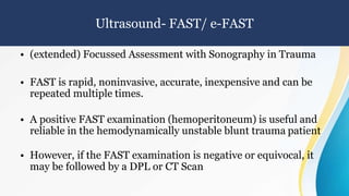
(3) Perform a FAST exam to check for hemoperitoneum which, if positive, indicates need for surgical consultation given the mechanism of injury.
(4) If the patient is stable, further evaluation with CT scan would be most accurate to diagnose potential solid organ or retroperitoneal injuries from the handlebar impact.