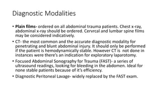
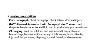
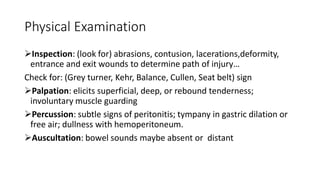
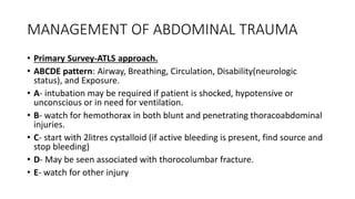
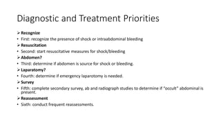
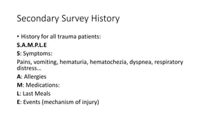
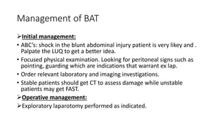
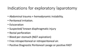
ABDOMINAL TRAUMA
GROUP B TOPIC PRESENTATION
Introduction
• Abdominal trauma can be defined as the adverse effect of a physical
force upon a person resulting in injury.
• “Abdomen” for trauma purposes is everything below the nipple line,
inferiorly to the public line.
• This includes:
• Peritoneum- liver, spleen, stomach, last ¼ of duodenum, small bowel,
transverse colon, sigmoid colon.
• Retroperitoneum- first ¼ of the duodenum, pancreas, kidneys,
ureters, ascending/descending colon, major vessels.
Types of Injury
Blunt Abdominal Trauma
• Greater mortality than Penetrating Abdominal Trauma because it’s more
difficult to diagnose and is commonly associated with trauma to multiple
organs/system.
• Most commonly injured organs are: spleen (most common), liver, intestine
which is most likely a hollow viscus.
• Most common causes are: motor vehicle accident (50-75% of cases)
followed by blows to the abdomen(15%) and falls (6-9%)
Penetrating Abdominal Trauma
• Stabbing is most common followed by firearm wounds.
• Most commonly injured organs are: liver, small intestines and colon.
GENERAL CLINICAL FEATURES
• Features of shock—pallor, tachycardia, hypotension, cold
• periphery, sweating, oliguria.
• Abdominal distension.
• Pain, tenderness, rebound tenderness, guarding and rigidity, dullness
in the flank on percussion.
• Respiratory distress, cyanosis depending on the amount of blood
• loss.
• Bruising over the skin of the abdominal wall.
• Features specific of individual organ injuries.
Physical Examination
Grey Turners sign: bruising discoloration of lower flanks, lower back;
associated with retroperitoneal bleeding of pancreas, kidney or pelvic
fracture.
Seat belt sign: Diagonal and lower abdominal abrasion, secondary to
restraining belt. Consider fracture of lumbar spine (chance fracture) and
bladder and/or bowel perforation.
Cullen sign: Bluish discoloration around umbilicus indicates peritoneal
bleeding, often pancreatic hemorrhage.
Kehr sign: shoulder pain while supine caused by diaphragmatic irritation
(splenic injury, free air, intra-abdominal bleeding)
Balance sign: Dull percussion in LUQ sign of splenic injury; blood
accumulation in subcapsular or extracapsular spleen.
Penetrating Abdominal Injury
• Most commonly due to gun shot wound and stab wounds. Gun shot wounds
always penetrate the peritoneum/ retroperitoneum. However, severity of stab
wounds vary based on depth.
Initial management:
• ABCs- primarily concerned with blood loss. Fluid replacement to maintain systolic
pressure > 90mmHg.
• Focused physical examination.
• Order essential laboratory and imaging investigation for the trauma patient.
• Stable patients should get CT scan to assess damage, in urgent situations Focused
Assessment with Sonography for Trauma is prefared.
• Any violation of the peritoneum/ retroperitoneum requires prophylactic
antibiotics (cover GI flora) and tetanus booster.
Surgery Management:
• All gun shot wounds to the abdomen are management by exploratory
laparotomy.
• Stab wounds may be treated conservatively or require exploratory
laparotomy.
Pathophysiology of injury
Blunt Abdominal Trauma
• Rupture or burst injury of a hollow organ by sudden rises in intra-
abdominal pressures.
• Crushing effect due to impact.
• Acceleration and deceleration results in shear injury.
• Seat beat injury commonly seen as “seat belt sign” which is indicative
of intraperitoneal injury.
INVESTIGATIONS
Laboratory Tests
• Hematocrit- below 30% increases the likelihood of intraabdominal injury.
• Leukocyte count- In Blunt Abdominal Trauma (BAT), the white blood cell (WBC)
count is nonspecific and of little value. Catecholamine release due to trauma can
cause demargination and may elevate the WBC to 12,000 to 20,000/mm3 with a
moderate left shift. Solid or hollow viscus injury can cause comparable elevations
• Pancreatic enzymes- Normal serum amylase and lipase concentrations cannot
exclude significant pancreatic injury. And while elevated concentrated raise the
possibility of pancreatic injury.
• Liver function tests- hepatic injury is associated with elevations in liver
transaminase concentrations.
• Urinalysis- Gross hematuria suggests serious renal injury and mandates further
investigation.
Diagnostic Modalities
• Plain films- ordered on all abdominal trauma patients. Chest x-ray,
abdominal x-ray should be ordered. Cervrcal and lumbar spine films
may be considered indicatively.
• CT- the most common and the accurate diagnostic modality for
penetrating and blunt abdominal injury. It should only be performed
if the patient is hemodynamically stable. However CT is not done in
instances were there’s an indication for exploratory laparotomy.
• Focused Abdominal Sonography for Trauma (FAST)- a series of
ultrasound readings, looking for bleeding in the abdomen. Ideal for
none stable patients because of it’s efficiency.
• Diagnostic Peritoneal Lavage- widely replaced by the FAST exam.
Imaging Investigations
• Plain radiograph: chest radiograph check intraabdominal injury.
• (FAST) Focused Assessment with Sonography for Trauma: used to
diagnose free intraperitoneal fluid and to evaluate organ hematoma.
• CT imaging: used for solid visceral lesions and intraperitoneal
hemorrhage because of its accuracy. It is however, insensitive for
injury of the pancreas, diaphragm, small bowel, and mesentery.
Physical Examination
Inspection: (look for) abrasions, contusion, lacerations,deformity,
entrance and exit wounds to determine path of injury…
Check for: (Grey turner, Kehr, Balance, Cullen, Seat belt) sign
Palpation: elicits superficial, deep, or rebound tenderness;
involuntary muscle guarding
Percussion: subtle signs of peritonitis; tympany in gastric dilation or
free air; dullness with hemoperitoneum.
Auscultation: bowel sounds maybe absent or distant
MANAGEMENT OF ABDOMINAL TRAUMA
• Primary Survey-ATLS approach.
• ABCDE pattern: Airway, Breathing, Circulation, Disability(neurologic
status), and Exposure.
• A- intubation may be required if patient is shocked, hypotensive or
unconscious or in need for ventilation.
• B- watch for hemothorax in both blunt and penetrating thoracoabdominal
injuries.
• C- start with 2litres cystalloid (if active bleeding is present, find source and
stop bleeding)
• D- May be seen associated with thorocolumbar fracture.
• E- watch for other injury
Diagnostic and Treatment Priorities
Recognize
• First: recognize the presence of shock or intraabdominal bleeding
Resuscitation
• Second: start resuscitative measures for shock/bleeding
Abdomen?
• Third: determine if abdomen is source for shock or bleeding.
Laparatomy?
• Fourth: determine if emergency laparotomy is needed.
Survey
• Fifth: complete secondary survey, ab and radiograph studies to determine if “occult” abdominal is
present.
Reassessment
• Sixth: conduct frequent reassessments.
Secondary Survey History
• History for all trauma patients:
S.A.M.P.L.E
S: Symptoms:
Pains, vomiting, hematuria, hematochezia, dyspnea, respiratory
distress…
A: Allergies
M: Medications:
L: Last Meals
E: Events (mechanism of injury)
Management of BAT
Initial management:
• ABC’s: shock in the blunt abdominal injury patient is very likey and .
Palpate the LUQ to get a better idea.
• Focused physical examination. Looking for peritoneal signs such as
pointing, guarding which are indications that warrant ex lap.
• Order relevant laboratory and imaging investigations.
• Stable patients should get CT to assess damage while unstable
patients may get FAST.
Operative management:
Exploratory laparotomy performed as indicated.
Indications for exploratory laparotomy
• Abdominal trauma + hemodynamic instability.
• Peritoneal irritation.
• Evisceration
• Suspected/ known diaphragmatic injury
• Rectal perforation
• Bleed per stomach (NGT aspiration)
• Free intraperitoneal or retroperitoneal air.
• Positive Diagnostic Peritoneal Lavage or positive FAST
Specific notes: Diaphragm
• Signs of injury: chest pain, dyspnea, respiratory distress, decreased
breath sounds, signs of chest trauma, abdominal pain/ tenderness.
• Left hemidiaphragm more frequently injured.
• On chest x-ray about 50% of patients shows abdominal viscera in
hemithorax, elevated hemidiaphragm.
Management:
• Laparotomy/ laparoscopy for repair
Complications:
• Henia, strangulation…
Specific notes: Liver
In general:
• Penetrating trauma involving the liver will get an exploratory laparotomy.
• Blunt trauma involving the liver can be managed with observation if:
-Patient is hemodynamically stable, AND
-There are no peritoneal signs, AND
-There are no associated injuries requiring laparoscopy, AND
-There is no need for excessive transfusions
• A repeat CT should be attained at 2-3 days.
Technically, operative management is required for an AAST (American
Association for Surgery of Trauma) liver injury grade III or higher.
Specific notes: Spleen
• This is the most injured organ in blunt abdominal injury, also seen and
should be exposed when there is injury to the left lower rib cage (7th, 8th,
9th, 10th, rib fracture).
• 30% 0f splenic injury patients present with hypotensive shock.
• Immediate laparotomy is required if:
-Ongoing hemodynamic instability
-Expanding hematoma
-Patient has a coagulopathy
-AAST spleen injury grade III or greater ( hematoma > 50% of surface area or
laceration >3 cm)
-In case of splenectomy, patients needs vaccination against capsulated
bacteria such as pneumococcus and meningococcus.
• Non-operative management may be undertaken in stable patients
who don’t require laparotomy.
-Admission + strict bed rest for 2-3 days.
- Null Per Oral
-NG decompression; keep prepped up for emergency surgery incase
condition deteriorates.
-Serial Hematocrit to see if bleeding as stopped.
-At 3 days reassess: follow up CT, may resume diet and light activity.
-Light activity for 3 months.
Specific notes: other organs
• Stomach: bloody aspirate from NG tube suggests damage to stomach.
Subphragmatic free air on chest x-ray, free abdominal fluid on CT,
start antibiotics to cover gut flora then repair.
• Bowel: shows up on CT as hollow viscus injury. Subdiaphragmatic free
air on chest x-ray, free abdominal fluid seen on CT. Start antibiotics to
cover gut flora and repair.
• Pancreas: uncommon, difficult to diagnose. May present weeks/
months later as a pancreatic pseudocyst.
Complications of Abdominal Trauma
• Complications of blunt abdominal trauma include: peritonitis,
haemorrhagic shock, death.
• Common injuries are divided into two categories: solid organ
(e.g., liver, spleen, pancreas, kidneys) and hollow organ (e.g.,
stomach, large and small bowel, gall bladder, urinary bladder)
injuries. Solid organ injuries range from minor injuries such as
small, haemodynamically insignificant liver, spleen, or kidney
lacerations
• Other complications include: abdominal compartment
syndrome, pancreatic pseudo cyst, operative coagulopathy and
post operative complications.
References
• 1 Brouwers M, Kho ME, Browman GP, Burgers JS, Cluzeau F, Feder G, Fervers B,
Graham ID, Grimshaw J, Hanna S, Littlejohns P, Makarski J, Zitzelsberger L for the
AGREE Next Steps Consortium. AGREE II: Advancing guideline development, reporting
and evaluation in healthcare. Can Med Assoc J. 2010. Available online July 5,
2010. doi:10.1503/cmaj.090449
2 Australian Trauma Quality Improvement program. (2012) Caring for the severely
injured in Australia – Inaugural report of the Australian trauma registry 2010 -2012
3 Wilkes, G. (2014). Abdominal Trauma. In C. P, J. G, K. AM, M. Little, & B. A, Textbook
of Adult Emergency Medicine, 3rd edition (pp. 99-103). Sydney: Elsevier.
4 Diercks, D. C. (2016, 12 18). Initial evaluation and management of blunt abdominal
trauma in adults. Retrieved from Up to date: http://www.uptodate.com/contents/initial-
evaluation-and-management-of-blunt-abdominal-trauma-in-adults

GROUP B- TOPIC PRESENTATION ON ABDOMINAL TRAUMA_060447.pdf

  • 1.
    ABDOMINAL TRAUMA GROUP BTOPIC PRESENTATION
  • 2.
    Introduction • Abdominal traumacan be defined as the adverse effect of a physical force upon a person resulting in injury. • “Abdomen” for trauma purposes is everything below the nipple line, inferiorly to the public line. • This includes: • Peritoneum- liver, spleen, stomach, last ¼ of duodenum, small bowel, transverse colon, sigmoid colon. • Retroperitoneum- first ¼ of the duodenum, pancreas, kidneys, ureters, ascending/descending colon, major vessels.
  • 3.
    Types of Injury BluntAbdominal Trauma • Greater mortality than Penetrating Abdominal Trauma because it’s more difficult to diagnose and is commonly associated with trauma to multiple organs/system. • Most commonly injured organs are: spleen (most common), liver, intestine which is most likely a hollow viscus. • Most common causes are: motor vehicle accident (50-75% of cases) followed by blows to the abdomen(15%) and falls (6-9%) Penetrating Abdominal Trauma • Stabbing is most common followed by firearm wounds. • Most commonly injured organs are: liver, small intestines and colon.
  • 4.
    GENERAL CLINICAL FEATURES •Features of shock—pallor, tachycardia, hypotension, cold • periphery, sweating, oliguria. • Abdominal distension. • Pain, tenderness, rebound tenderness, guarding and rigidity, dullness in the flank on percussion. • Respiratory distress, cyanosis depending on the amount of blood • loss. • Bruising over the skin of the abdominal wall. • Features specific of individual organ injuries.
  • 5.
    Physical Examination Grey Turnerssign: bruising discoloration of lower flanks, lower back; associated with retroperitoneal bleeding of pancreas, kidney or pelvic fracture. Seat belt sign: Diagonal and lower abdominal abrasion, secondary to restraining belt. Consider fracture of lumbar spine (chance fracture) and bladder and/or bowel perforation. Cullen sign: Bluish discoloration around umbilicus indicates peritoneal bleeding, often pancreatic hemorrhage. Kehr sign: shoulder pain while supine caused by diaphragmatic irritation (splenic injury, free air, intra-abdominal bleeding) Balance sign: Dull percussion in LUQ sign of splenic injury; blood accumulation in subcapsular or extracapsular spleen.
  • 6.
    Penetrating Abdominal Injury •Most commonly due to gun shot wound and stab wounds. Gun shot wounds always penetrate the peritoneum/ retroperitoneum. However, severity of stab wounds vary based on depth. Initial management: • ABCs- primarily concerned with blood loss. Fluid replacement to maintain systolic pressure > 90mmHg. • Focused physical examination. • Order essential laboratory and imaging investigation for the trauma patient. • Stable patients should get CT scan to assess damage, in urgent situations Focused Assessment with Sonography for Trauma is prefared. • Any violation of the peritoneum/ retroperitoneum requires prophylactic antibiotics (cover GI flora) and tetanus booster.
  • 7.
    Surgery Management: • Allgun shot wounds to the abdomen are management by exploratory laparotomy. • Stab wounds may be treated conservatively or require exploratory laparotomy.
  • 8.
    Pathophysiology of injury BluntAbdominal Trauma • Rupture or burst injury of a hollow organ by sudden rises in intra- abdominal pressures. • Crushing effect due to impact. • Acceleration and deceleration results in shear injury. • Seat beat injury commonly seen as “seat belt sign” which is indicative of intraperitoneal injury.
  • 9.
    INVESTIGATIONS Laboratory Tests • Hematocrit-below 30% increases the likelihood of intraabdominal injury. • Leukocyte count- In Blunt Abdominal Trauma (BAT), the white blood cell (WBC) count is nonspecific and of little value. Catecholamine release due to trauma can cause demargination and may elevate the WBC to 12,000 to 20,000/mm3 with a moderate left shift. Solid or hollow viscus injury can cause comparable elevations • Pancreatic enzymes- Normal serum amylase and lipase concentrations cannot exclude significant pancreatic injury. And while elevated concentrated raise the possibility of pancreatic injury. • Liver function tests- hepatic injury is associated with elevations in liver transaminase concentrations. • Urinalysis- Gross hematuria suggests serious renal injury and mandates further investigation.
  • 10.
    Diagnostic Modalities • Plainfilms- ordered on all abdominal trauma patients. Chest x-ray, abdominal x-ray should be ordered. Cervrcal and lumbar spine films may be considered indicatively. • CT- the most common and the accurate diagnostic modality for penetrating and blunt abdominal injury. It should only be performed if the patient is hemodynamically stable. However CT is not done in instances were there’s an indication for exploratory laparotomy. • Focused Abdominal Sonography for Trauma (FAST)- a series of ultrasound readings, looking for bleeding in the abdomen. Ideal for none stable patients because of it’s efficiency. • Diagnostic Peritoneal Lavage- widely replaced by the FAST exam.
  • 11.
    Imaging Investigations • Plainradiograph: chest radiograph check intraabdominal injury. • (FAST) Focused Assessment with Sonography for Trauma: used to diagnose free intraperitoneal fluid and to evaluate organ hematoma. • CT imaging: used for solid visceral lesions and intraperitoneal hemorrhage because of its accuracy. It is however, insensitive for injury of the pancreas, diaphragm, small bowel, and mesentery.
  • 12.
    Physical Examination Inspection: (lookfor) abrasions, contusion, lacerations,deformity, entrance and exit wounds to determine path of injury… Check for: (Grey turner, Kehr, Balance, Cullen, Seat belt) sign Palpation: elicits superficial, deep, or rebound tenderness; involuntary muscle guarding Percussion: subtle signs of peritonitis; tympany in gastric dilation or free air; dullness with hemoperitoneum. Auscultation: bowel sounds maybe absent or distant
  • 13.
    MANAGEMENT OF ABDOMINALTRAUMA • Primary Survey-ATLS approach. • ABCDE pattern: Airway, Breathing, Circulation, Disability(neurologic status), and Exposure. • A- intubation may be required if patient is shocked, hypotensive or unconscious or in need for ventilation. • B- watch for hemothorax in both blunt and penetrating thoracoabdominal injuries. • C- start with 2litres cystalloid (if active bleeding is present, find source and stop bleeding) • D- May be seen associated with thorocolumbar fracture. • E- watch for other injury
  • 14.
    Diagnostic and TreatmentPriorities Recognize • First: recognize the presence of shock or intraabdominal bleeding Resuscitation • Second: start resuscitative measures for shock/bleeding Abdomen? • Third: determine if abdomen is source for shock or bleeding. Laparatomy? • Fourth: determine if emergency laparotomy is needed. Survey • Fifth: complete secondary survey, ab and radiograph studies to determine if “occult” abdominal is present. Reassessment • Sixth: conduct frequent reassessments.
  • 15.
    Secondary Survey History •History for all trauma patients: S.A.M.P.L.E S: Symptoms: Pains, vomiting, hematuria, hematochezia, dyspnea, respiratory distress… A: Allergies M: Medications: L: Last Meals E: Events (mechanism of injury)
  • 16.
    Management of BAT Initialmanagement: • ABC’s: shock in the blunt abdominal injury patient is very likey and . Palpate the LUQ to get a better idea. • Focused physical examination. Looking for peritoneal signs such as pointing, guarding which are indications that warrant ex lap. • Order relevant laboratory and imaging investigations. • Stable patients should get CT to assess damage while unstable patients may get FAST. Operative management: Exploratory laparotomy performed as indicated.
  • 17.
    Indications for exploratorylaparotomy • Abdominal trauma + hemodynamic instability. • Peritoneal irritation. • Evisceration • Suspected/ known diaphragmatic injury • Rectal perforation • Bleed per stomach (NGT aspiration) • Free intraperitoneal or retroperitoneal air. • Positive Diagnostic Peritoneal Lavage or positive FAST
  • 18.
    Specific notes: Diaphragm •Signs of injury: chest pain, dyspnea, respiratory distress, decreased breath sounds, signs of chest trauma, abdominal pain/ tenderness. • Left hemidiaphragm more frequently injured. • On chest x-ray about 50% of patients shows abdominal viscera in hemithorax, elevated hemidiaphragm. Management: • Laparotomy/ laparoscopy for repair Complications: • Henia, strangulation…
  • 19.
    Specific notes: Liver Ingeneral: • Penetrating trauma involving the liver will get an exploratory laparotomy. • Blunt trauma involving the liver can be managed with observation if: -Patient is hemodynamically stable, AND -There are no peritoneal signs, AND -There are no associated injuries requiring laparoscopy, AND -There is no need for excessive transfusions • A repeat CT should be attained at 2-3 days. Technically, operative management is required for an AAST (American Association for Surgery of Trauma) liver injury grade III or higher.
  • 20.
    Specific notes: Spleen •This is the most injured organ in blunt abdominal injury, also seen and should be exposed when there is injury to the left lower rib cage (7th, 8th, 9th, 10th, rib fracture). • 30% 0f splenic injury patients present with hypotensive shock. • Immediate laparotomy is required if: -Ongoing hemodynamic instability -Expanding hematoma -Patient has a coagulopathy -AAST spleen injury grade III or greater ( hematoma > 50% of surface area or laceration >3 cm) -In case of splenectomy, patients needs vaccination against capsulated bacteria such as pneumococcus and meningococcus.
  • 21.
    • Non-operative managementmay be undertaken in stable patients who don’t require laparotomy. -Admission + strict bed rest for 2-3 days. - Null Per Oral -NG decompression; keep prepped up for emergency surgery incase condition deteriorates. -Serial Hematocrit to see if bleeding as stopped. -At 3 days reassess: follow up CT, may resume diet and light activity. -Light activity for 3 months.
  • 22.
    Specific notes: otherorgans • Stomach: bloody aspirate from NG tube suggests damage to stomach. Subphragmatic free air on chest x-ray, free abdominal fluid on CT, start antibiotics to cover gut flora then repair. • Bowel: shows up on CT as hollow viscus injury. Subdiaphragmatic free air on chest x-ray, free abdominal fluid seen on CT. Start antibiotics to cover gut flora and repair. • Pancreas: uncommon, difficult to diagnose. May present weeks/ months later as a pancreatic pseudocyst.
  • 23.
    Complications of AbdominalTrauma • Complications of blunt abdominal trauma include: peritonitis, haemorrhagic shock, death. • Common injuries are divided into two categories: solid organ (e.g., liver, spleen, pancreas, kidneys) and hollow organ (e.g., stomach, large and small bowel, gall bladder, urinary bladder) injuries. Solid organ injuries range from minor injuries such as small, haemodynamically insignificant liver, spleen, or kidney lacerations • Other complications include: abdominal compartment syndrome, pancreatic pseudo cyst, operative coagulopathy and post operative complications.
  • 24.
    References • 1 BrouwersM, Kho ME, Browman GP, Burgers JS, Cluzeau F, Feder G, Fervers B, Graham ID, Grimshaw J, Hanna S, Littlejohns P, Makarski J, Zitzelsberger L for the AGREE Next Steps Consortium. AGREE II: Advancing guideline development, reporting and evaluation in healthcare. Can Med Assoc J. 2010. Available online July 5, 2010. doi:10.1503/cmaj.090449 2 Australian Trauma Quality Improvement program. (2012) Caring for the severely injured in Australia – Inaugural report of the Australian trauma registry 2010 -2012 3 Wilkes, G. (2014). Abdominal Trauma. In C. P, J. G, K. AM, M. Little, & B. A, Textbook of Adult Emergency Medicine, 3rd edition (pp. 99-103). Sydney: Elsevier. 4 Diercks, D. C. (2016, 12 18). Initial evaluation and management of blunt abdominal trauma in adults. Retrieved from Up to date: http://www.uptodate.com/contents/initial- evaluation-and-management-of-blunt-abdominal-trauma-in-adults