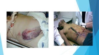
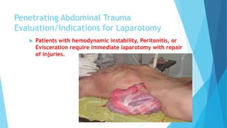
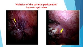
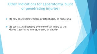
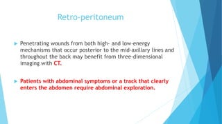
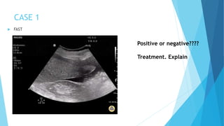
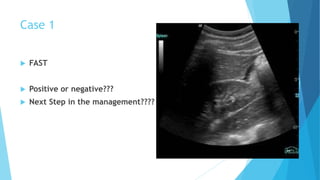
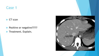
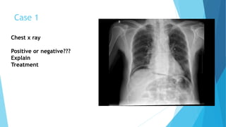
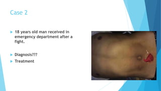
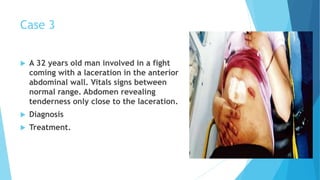
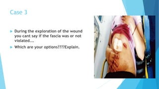
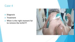
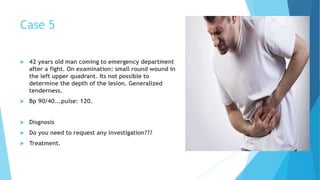
ABDOMINAL INJURIES
Annia Ramos Perez
Consultant General Surgeon.
Introduction
 Abdominal injuries are frequently encountered in the management of
trauma patients.
 Of all patients in the 2017 NTDB, 13% sustained abdominal injuries,
associated with an overall mortality rate of 7.7%.
 During the evaluation of the injured patient, the abdomen is of high
priority because the vital nature of the contained organs and
structures.
Introduction
 Blunt trauma can result in the laceration of solid organs usually
causing bleeding, which in its most severe form manifests as
hemorrhagic shock or as visceral perforation of the GI tract.
 Penetrating trauma to the abdomen can result in laceration of
solid organs and perforation of hollow organs, which must be
discovered and repaired at the time of laparotomy.
 Penetrating abdominal injury:
 Any penetrating injury that could have
entered the peritoneal cavity or retro-
peritoneum inflicting damage on the
abdominal contents. In general, the
entry wounds for an abdominal injury
extend from the fifth intercostal space
to the perineum.
CLASIFICATION
Abdominal injury
Open
Loss of continuity of
the abdominal wall
1-Penetrating
Fascia
Peritoneum
2-Not
Penetrating
Blunt
(closed)
No loss of
continuity of the
abdominal wall
INITIAL EVALUATION/General Principles
 The doctor evaluating the abdomen should answer two questions:
 (a) Is there an intra-abdominal injury
 (b) Does this injury require operative repair?
 While addressing these issues, two principles should not be violated:
 (a) the ABCs should be adequately assessed before focusing on the abdomen
(b) clinical examination should be the most important element of the
evaluation.
INITIAL EVALUATION/General Principles
 Clinical examination can determine the need for emergent
exploration following abdominal trauma by the presence
of one or both of two signs:
 (a) peritonitis
 (b) hemodynamic instability.
In the absence of these two signs, there is time for more
detailed investigations.
Clinical Examination
 Despite the technological advances, which undoubtedly add greatly to
the ability to evaluate the abdomen, clinical examination remains of
paramount importance.
 Hemodynamic stability (or instability) is a term, which is widely used
but insufficiently understood.
 At the initial stage, the hemodynamic status is usually assessed by
crude methods, such as blood pressure, heart rate, and urine output
monitoring, serum hemoglobin measurement, and evaluation of the skin
and capillary refill.
Clinical Examination
 Although such information is very important, one needs to
remember the limitations of using these measurements to
assess hemodynamic stability.
 Hypotension may occur in the presence of spinal cord injury
without blood loss.
 Hypertension may occur even in the presence of blood loss due
to increased intracranial pressure.
 A blood pressure of 110 mm Hg may be normal for a 25-year-old
man, abnormally high for a six-month old baby, and profusely low
for a 70-year-old hypertensive woman.
Clinical Examination
 The heart rate can be high for causes unrelated to bleeding, such
as pain or anxiety.
 On the other hand, a normal or low heart rate cannot exclude
bleeding, particularly in patients with high spinal cord injuries,
chronic cocaine intoxication, or beta-blocker medication.
 Paradoxical bradycardia occurs in up to 29% of hypotensive
trauma patients.
Clinical Examination
 For these reasons a more precise description of the hemodynamic status of
the patient is highly desirable.
 Now, noninvasive technology allows continuous monitoring of the cardiac
output, stroke volume, and transcutaneous oxygen tension. These portable
devices can be connected to the patient by simple self-adhesive patches,
like EKG leads, and provide hemodynamic and oxygen tissue perfusion
monitoring even during the very early posttraumatic stages.
Clinical Examination
 In summary, hemodynamic instability is a valid reason for
laparotomy as long as the physician recognizes the limitations
of vital signs and incorporates additional elements in the
decision-making to increase its accuracy.
Clinical Examination
 Diffuse abdominal tenderness is the other major indicator of the
need for laparotomy.
 It is important to distinguish between superficial soft tissue
tenderness, such as caused by a stab wound or a seatbelt, and deep
tenderness caused by a true abdominal organ injury.
 In the presence of penetrating injuries, it is the tenderness on deep
palpation away from the wound site that denotes internal injuries.
 A significant part of the trauma population is simply non-evaluable
because of associated head injuries, spinal cord injuries, or
intoxication.
 Such patients receive the most benefit from additional studies.
Ultrasonography
 The focused abdominal sonography for trauma (FAST) emerged
recently as an important tool in the initial diagnosis of intra-
abdominal injuries.
 Performed by surgeons or emergency room physicians, FAST has
shown an excellent sensitivity in identifying intra-abdominal fluid.
 Because of its ease of use, repeatability, and avoidance of radiation
exposure it has rapidly become an essential part of the initial
evaluation.
 In the presence of multiple Intraperitoneal injuries, blood is most
often found by FAST in the right upper quadrant.
Ultrasonography
 At this point, it seems that the most significant contribution of the
FAST examination is in the detection of intra-abdominal fluid in the
hemodynamically unstable and clinically unevaluable blunt trauma
victim.
 These patients should be immediately taken to the operating room for
abdominal exploration.
 Negative FAST exams do not preclude the need for further evaluation of
the abdomen by other imaging modalities, most commonly computed
tomography (CT).
 https://youtu.be/paJXt-YG2qg
Diagnostic Peritoneal Lavage
https://youtu.be/aRw3qQGjTzI
Diagnostic Peritoneal Lavage
 Catheter is inserted with a direction toward the pelvis and the
guidewire is removed.
 If gross aspiration of the catheter yields no gross blood, bilious
content, fecal material or particles of food; one liter of warm
normal saline (in children 10-15 mL/kg) is infused into the
peritoneal cavity.
 The fluid is then siphoned back into the empty container which is
lowered to below the level of the patient.
 A specimen of the recovered fluid is then examined
macroscopically, microscopically, and biochemically.
Diagnostic Peritoneal Lavage/POSITIVE
 More than 100,000 red blood cells/mm3
 More than 500 white blood cells/mm3
 Amylase of more than 19 IU/L
 The major advantage of DPL is a sensitivity rate higher than 95%
for identifying Intraperitoneal hemorrhage.
 However, because the technique is invasive and fails to identify
the source of bleeding and the need for operative repair, DPL is
used with decreasing frequency over the years.
Abdominal Computed Tomography
 Abdominal CT is becoming the test of preference for evaluating
the abdomen of patients with blunt abdominal trauma who are
hemodynamically stable and complain of abdominal tenderness or
are unevaluable.
 It allows estimation of the amount of intra-abdominal fluid and
accurate imaging of solid parenchymal injuries in most patients.
 Abdominal CT plays a major role in the decision to manage the
injured spleen, liver, or kidney nonoperatively
Abdominal Computed Tomography
 Abdominal CT has become the mainstay of imaging for the stable
blunt trauma patient and has led to the emergence of
Nonoperative management of many solid abdominal organ
injuries.
 Abdominal CT is typically performed with IV contrast timed to
capture the portal venous phase, which best demonstrates the
vasculature and visceral perfusion of the solid abdominal organs.
Abdominal Computed Tomography
 CT provides excellent visualization of the solid organs, allowing
the characterization of injury severity (injury grade) and the
recognition of active bleeding, which appears as contrast
extravasation.
 The retroperitoneal structures are also well visualized on CT,
identifying injuries that are difficult to evaluate with FAST or
DPL
Abdominal Computed Tomography
 As the use of abdominal CT in blunt trauma is increasing, it is
important to recognize its LIMITATIONS
 CT has limitations in detecting hollow visceral and
mesenteric injuries
 This diagnosis is usually suspected on the basis of indirect
signs:
1. Free fluid in the absence of solid visceral injury
2. Pneumoperitoneum
3. Mesenteric fat streaking
4. Bowel wall thickening
5. Extravasation of luminal contrast.
Abdominal Computed Tomography
CT scan of the abdomen following a gunshot wound. The
bullet tract (arrows) is outside the peritoneal cavity. This
patient does not need laparotomy.
Rigid Sigmoidoscopy
 The value of rigid Sigmoidoscopy is significant when it comes to
evaluating the extra-peritoneal rectum.
 Injuries to this part of the intestinal tract may not produce
symptoms initially and escape diagnosis until septic complications.
 Classically, gunshot wounds to the gluteal regions, particularly if the
trajectory crosses the midline, have the potential of producing
initially asymptomatic extra-peritoneal rectal injuries.
 Rectal examination may be helpful but is often unreliable.
 Rigid Sigmoidoscopy reveals intraluminal blood or the exact
injury.
Diagnostic Laparoscopy
 The major limitations of diagnostic laparoscopy in trauma are
related to:
1. The inability to “run” the bowel
2. Diagnose retroperitoneal injuries
3. Expose adequately deep-lying organs
4. Estimate accurately the quantity of hemoperitoneum.
 Studies have shown that close to half of the existing injuries can
be missed by laparoscopy.
Diagnostic Laparoscopy
 On the other hand, diagnostic laparoscopy has an excellent
sensitivity and specificity (>95%) when used as a screening tool
to establish the presence of peritoneal violation,
hemoperitoneum, or enteric content spillage.
Immediate Management
 Immediate management of abdominal injuries consists of
resuscitation and evaluation.
 Patients in shock require initiation of resuscitation with crystalloid
solutions and blood products, as well as a rapid assessment for the
source of bleeding.
 Retained foreign bodies traversing the abdominal wall should be
maintained throughout the initial evaluation and protected from
excessive movement. These should then be removed only after
defining a definitive plan, which almost always includes abdominal
operation.
Blunt Abdominal Trauma Evaluation
 Patients who present with blunt versus penetrating
mechanisms of injury frequently require varying
approaches to evaluation.
Indications for Emergency Laparotomy
 Blunt trauma patients who are unstable and have intra-
abdominal fluid identified on FAST require an emergent
laparotomy to manage bleeding.
 If FAST is unavailable, aspiration of 10 mL or more of gross
blood on DPL also suggests an intra-abdominal source of
hemorrhage requiring emergent operation
 Patients with peritonitis require abdominal exploration to
evaluate for hollow visceral injury.
 Other patients will undergo further workup of the abdomen to
evaluate for intra-abdominal injury.
Algorithm for the
evaluation and
management of
blunt abdominal
trauma PATIENTS.
Penetrating Abdominal Trauma Evaluation
 Penetrating abdominal trauma is typically evaluated differently
than blunt mechanisms.
 Because of the high rate of intra-abdominal injury, patients
sustaining anterior abdominal gunshot wounds are frequently
transferred quickly to the operating room for laparotomy.
 Depending on the location of the penetrating wound, the chest
may require evaluation for mediastinal, pleural, or pulmonary
injuries.
Penetrating Abdominal Trauma Evaluation
 It may be valuable to attempt to determine the trajectory of the
missiles while preparing for surgery because this may assist in
directing the exploration.
 Penetrating wounds of the skin should be identified with
radiopaque markers and plain radiographs obtained to determine
their location and relation to missile position.
 This evaluation should be brief and not delay operation,
especially in the hemodynamically unstable patient.
Penetrating Abdominal
Trauma Evaluation
Algorithm for the
evaluation and
management of
anterior abdominal stab
wounds
Penetrating Abdominal Trauma
Evaluation/Indications for Laparotomy
 Patients with hemodynamic instability, Peritonitis, or
Evisceration require immediate laparotomy with repair
of injuries.
Penetrating Abdominal Trauma
Evaluation/Indications for Laparotomy
 Others can have the penetrating wound explored locally to
determine whether the anterior or posterior abdominal fascia
was violated.
 Those without fascial penetration can be discharged to home.
 In the setting of a positive or equivocal local wound exploration,
patients should be monitored with serial abdominal
examinations and determinations of hemoglobin levels every 8
hours.
Penetrating Abdominal Trauma
Evaluation/Indications for Laparotomy
 Throughout this evaluation, the development of peritonitis,
hemodynamic instability, significant decreases in hemoglobin
level, or development of leukocytosis should prompt further
evaluation, usually with laparotomy.
 Patients without clinical change after 24 hours can have a diet
instituted and be discharged to home.
Penetrating Abdominal Trauma
Evaluation/Indications for Laparotomy
 An additional tool that has been used more recently is
laparoscopy, mainly to establish or exclude the presence of
peritoneal penetration.
 It remains fairly well accepted that laparoscopy in most hands is
not sufficient to explore the entire abdomen but it can be used
to identify violation of the parietal peritoneum, which can
then prompt laparotomy.
Violation of the parietal peritoneum/
Laparoscopic view
Other indications for Laparotomy( blunt
or penetrating injuries)
 (1) new onset hematemesis, proctorrhagia, or hematuria
 (2) contrast radiography evidence of an injury to the
kidney (significant injury), ureter, or bladder.
Retro-peritoneum
 Penetrating wounds from both high- and low-energy
mechanisms that occur posterior to the mid-axillary lines and
throughout the back may benefit from three-dimensional
imaging with CT.
 Patients with abdominal symptoms or a track that clearly
enters the abdomen require abdominal exploration.
Exploratory Laparotomy
 Patients who require laparotomy should undergo a
systematic exploration so that all areas of the abdomen
are assessed and injuries are not missed.
 This approach may require abbreviation in the setting of
deteriorating physiologic condition.
 As a standard technique, the abdomen is opened from the
xiphoid process to pubic symphysis to provide adequate
exposure of all abdominal structures.
Exploratory Laparotomy
 The Falciform ligament is divided, separating the liver from the
abdominal wall to improve retraction and perihepatic packing.
 Using a hand-held retractor, blood is quickly evacuated from all four
quadrants of the abdomen and laparotomy sponges are placed to
provide temporary hemostasis.
 The entire GI tract is carefully evaluated, from the gastro-
esophageal junction to the rectum at the peritoneal reflection.
Exploratory Laparotomy
 This includes entering the lesser sac to evaluate the posterior
stomach and the pancreas.
 Areas stained with blood that are of concern for injury should be
explored further with careful dissection
Exploratory Laparotomy
 Developing physiologic compromise should be promptly recognized
this requires open lines of communication with anesthesia
providers throughout the operation.
 In this setting, the operation should be abbreviated, with the
only goals becoming hemorrhage and contamination control
with temporary abdominal closure( CONTROL DAMAGE SURGERY)
 Otherwise, the abdominal fascia can be closed in a single layer
and the subcutaneous wound addressed as dictated by the level of
intra-abdominal contamination.
DAMAGE CONTROL SURGERY
ANTIBIOTIC PROPHYLAXIS
 Antibiotic prophylaxis should be given preferably before
the incision is made.
 Ampicillin/ sulbactam was associated with a significantly
lower incidence of wound sepsis than cephalosporin.
Other point in the treatment
 Analgesia
 Tetanus vaccine( metallic objects).
Bibliography
 Sabiston Text Book Of Surgery. 19 Edition.
 Swartz Text Book of Surgery. 11 Edition.
 Oxford Text Book Of Surgery. 2nd Edition.
 Feliciano, Trauma, Text Book Of Surgery.6th Edition.
 ATLS. 8 Edition
QUESTIONS?????
 USE YOUR INDIVIDUAL CHATS AND SHARE YOUR QUESTIONS
INTERACTIVE SESSION
 I WILL SHARE SOME CLINICAL PICTURES
 I WILL SELECT DIRECTLY WHO WILL ANSWER THE QUESTION
 YOU WILL HAVE ONE MINUTE TO THINK IN YOUR ANSWER.
CASE 1
 A 47 years old man, involved in RTA 7 hours ago, arrived
to the A&E Department.
 1-IS THERE ANY SIGN OF ABDOMINAL INJURY?
 2-NAME OF THE SIGN
 3-MEANING OF THE SIGN
CASE 1 Abdominal examination: distended abdomen,
absence of bowel sounds, no guarding or
tenderness, no abdominal pain. Normal pulse
and blood pressure.
 4.What is the next step in the treatment?
 LAPAROTOMY OR REQUEST INVESTIGATIONS???
 5-Which investigations will you need in order
to determine the cause of the abdominal
distension.
CASE 1
 FAST
Positive or negative????
Treatment. Explain
Case 1
 FAST
 Positive or negative???
 Next Step in the management????
Case 1
 CT scan
 Positive or negative?????
 Treatment. Explain.
Case 1
Chest x ray
Positive or negative???
Explain
Treatment
Case 2
 18 years old man received in
emergency department after a
fight.
 Diagnosis???
 Treatment
Case 3
 A 32 years old man involved in a fight
coming with a laceration in the anterior
abdominal wall. Vitals signs between
normal range. Abdomen revealing
tenderness only close to the laceration.
 Diagnosis
 Treatment.
Case 3
 During the exploration of the wound
you cant say if the fascia was or not
violated….
 Which are your options????Explain.
Case 4
 Diagnosis
 Treatment
 When is the right moment for
to remove the knife???
Case 5
 42 years old man coming to emergency department
after a fight. On examination: small round wound in
the left upper quadrant. Its not possible to
determine the depth of the lesion. Generalized
tenderness.
 Bp 90/40….pulse: 120.
 Disgnosis
 Do you need to request any investigation???
 Treatment.
THANKS

Abdominal injuries, lecture

  • 1.
    ABDOMINAL INJURIES Annia RamosPerez Consultant General Surgeon.
  • 2.
    Introduction  Abdominal injuriesare frequently encountered in the management of trauma patients.  Of all patients in the 2017 NTDB, 13% sustained abdominal injuries, associated with an overall mortality rate of 7.7%.  During the evaluation of the injured patient, the abdomen is of high priority because the vital nature of the contained organs and structures.
  • 4.
    Introduction  Blunt traumacan result in the laceration of solid organs usually causing bleeding, which in its most severe form manifests as hemorrhagic shock or as visceral perforation of the GI tract.  Penetrating trauma to the abdomen can result in laceration of solid organs and perforation of hollow organs, which must be discovered and repaired at the time of laparotomy.
  • 6.
     Penetrating abdominalinjury:  Any penetrating injury that could have entered the peritoneal cavity or retro- peritoneum inflicting damage on the abdominal contents. In general, the entry wounds for an abdominal injury extend from the fifth intercostal space to the perineum.
  • 7.
    CLASIFICATION Abdominal injury Open Loss ofcontinuity of the abdominal wall 1-Penetrating Fascia Peritoneum 2-Not Penetrating Blunt (closed) No loss of continuity of the abdominal wall
  • 9.
    INITIAL EVALUATION/General Principles The doctor evaluating the abdomen should answer two questions:  (a) Is there an intra-abdominal injury  (b) Does this injury require operative repair?  While addressing these issues, two principles should not be violated:  (a) the ABCs should be adequately assessed before focusing on the abdomen (b) clinical examination should be the most important element of the evaluation.
  • 11.
    INITIAL EVALUATION/General Principles Clinical examination can determine the need for emergent exploration following abdominal trauma by the presence of one or both of two signs:  (a) peritonitis  (b) hemodynamic instability. In the absence of these two signs, there is time for more detailed investigations.
  • 12.
    Clinical Examination  Despitethe technological advances, which undoubtedly add greatly to the ability to evaluate the abdomen, clinical examination remains of paramount importance.  Hemodynamic stability (or instability) is a term, which is widely used but insufficiently understood.  At the initial stage, the hemodynamic status is usually assessed by crude methods, such as blood pressure, heart rate, and urine output monitoring, serum hemoglobin measurement, and evaluation of the skin and capillary refill.
  • 13.
    Clinical Examination  Althoughsuch information is very important, one needs to remember the limitations of using these measurements to assess hemodynamic stability.  Hypotension may occur in the presence of spinal cord injury without blood loss.  Hypertension may occur even in the presence of blood loss due to increased intracranial pressure.  A blood pressure of 110 mm Hg may be normal for a 25-year-old man, abnormally high for a six-month old baby, and profusely low for a 70-year-old hypertensive woman.
  • 14.
    Clinical Examination  Theheart rate can be high for causes unrelated to bleeding, such as pain or anxiety.  On the other hand, a normal or low heart rate cannot exclude bleeding, particularly in patients with high spinal cord injuries, chronic cocaine intoxication, or beta-blocker medication.  Paradoxical bradycardia occurs in up to 29% of hypotensive trauma patients.
  • 15.
    Clinical Examination  Forthese reasons a more precise description of the hemodynamic status of the patient is highly desirable.  Now, noninvasive technology allows continuous monitoring of the cardiac output, stroke volume, and transcutaneous oxygen tension. These portable devices can be connected to the patient by simple self-adhesive patches, like EKG leads, and provide hemodynamic and oxygen tissue perfusion monitoring even during the very early posttraumatic stages.
  • 16.
    Clinical Examination  Insummary, hemodynamic instability is a valid reason for laparotomy as long as the physician recognizes the limitations of vital signs and incorporates additional elements in the decision-making to increase its accuracy.
  • 17.
    Clinical Examination  Diffuseabdominal tenderness is the other major indicator of the need for laparotomy.  It is important to distinguish between superficial soft tissue tenderness, such as caused by a stab wound or a seatbelt, and deep tenderness caused by a true abdominal organ injury.  In the presence of penetrating injuries, it is the tenderness on deep palpation away from the wound site that denotes internal injuries.  A significant part of the trauma population is simply non-evaluable because of associated head injuries, spinal cord injuries, or intoxication.  Such patients receive the most benefit from additional studies.
  • 19.
    Ultrasonography  The focusedabdominal sonography for trauma (FAST) emerged recently as an important tool in the initial diagnosis of intra- abdominal injuries.  Performed by surgeons or emergency room physicians, FAST has shown an excellent sensitivity in identifying intra-abdominal fluid.  Because of its ease of use, repeatability, and avoidance of radiation exposure it has rapidly become an essential part of the initial evaluation.  In the presence of multiple Intraperitoneal injuries, blood is most often found by FAST in the right upper quadrant.
  • 21.
    Ultrasonography  At thispoint, it seems that the most significant contribution of the FAST examination is in the detection of intra-abdominal fluid in the hemodynamically unstable and clinically unevaluable blunt trauma victim.  These patients should be immediately taken to the operating room for abdominal exploration.  Negative FAST exams do not preclude the need for further evaluation of the abdomen by other imaging modalities, most commonly computed tomography (CT).  https://youtu.be/paJXt-YG2qg
  • 22.
  • 23.
    Diagnostic Peritoneal Lavage Catheter is inserted with a direction toward the pelvis and the guidewire is removed.  If gross aspiration of the catheter yields no gross blood, bilious content, fecal material or particles of food; one liter of warm normal saline (in children 10-15 mL/kg) is infused into the peritoneal cavity.  The fluid is then siphoned back into the empty container which is lowered to below the level of the patient.  A specimen of the recovered fluid is then examined macroscopically, microscopically, and biochemically.
  • 24.
    Diagnostic Peritoneal Lavage/POSITIVE More than 100,000 red blood cells/mm3  More than 500 white blood cells/mm3  Amylase of more than 19 IU/L  The major advantage of DPL is a sensitivity rate higher than 95% for identifying Intraperitoneal hemorrhage.  However, because the technique is invasive and fails to identify the source of bleeding and the need for operative repair, DPL is used with decreasing frequency over the years.
  • 25.
    Abdominal Computed Tomography Abdominal CT is becoming the test of preference for evaluating the abdomen of patients with blunt abdominal trauma who are hemodynamically stable and complain of abdominal tenderness or are unevaluable.  It allows estimation of the amount of intra-abdominal fluid and accurate imaging of solid parenchymal injuries in most patients.  Abdominal CT plays a major role in the decision to manage the injured spleen, liver, or kidney nonoperatively
  • 27.
    Abdominal Computed Tomography Abdominal CT has become the mainstay of imaging for the stable blunt trauma patient and has led to the emergence of Nonoperative management of many solid abdominal organ injuries.  Abdominal CT is typically performed with IV contrast timed to capture the portal venous phase, which best demonstrates the vasculature and visceral perfusion of the solid abdominal organs.
  • 28.
    Abdominal Computed Tomography CT provides excellent visualization of the solid organs, allowing the characterization of injury severity (injury grade) and the recognition of active bleeding, which appears as contrast extravasation.  The retroperitoneal structures are also well visualized on CT, identifying injuries that are difficult to evaluate with FAST or DPL
  • 29.
    Abdominal Computed Tomography As the use of abdominal CT in blunt trauma is increasing, it is important to recognize its LIMITATIONS  CT has limitations in detecting hollow visceral and mesenteric injuries  This diagnosis is usually suspected on the basis of indirect signs: 1. Free fluid in the absence of solid visceral injury 2. Pneumoperitoneum 3. Mesenteric fat streaking 4. Bowel wall thickening 5. Extravasation of luminal contrast.
  • 30.
    Abdominal Computed Tomography CTscan of the abdomen following a gunshot wound. The bullet tract (arrows) is outside the peritoneal cavity. This patient does not need laparotomy.
  • 31.
    Rigid Sigmoidoscopy  Thevalue of rigid Sigmoidoscopy is significant when it comes to evaluating the extra-peritoneal rectum.  Injuries to this part of the intestinal tract may not produce symptoms initially and escape diagnosis until septic complications.  Classically, gunshot wounds to the gluteal regions, particularly if the trajectory crosses the midline, have the potential of producing initially asymptomatic extra-peritoneal rectal injuries.  Rectal examination may be helpful but is often unreliable.  Rigid Sigmoidoscopy reveals intraluminal blood or the exact injury.
  • 33.
    Diagnostic Laparoscopy  Themajor limitations of diagnostic laparoscopy in trauma are related to: 1. The inability to “run” the bowel 2. Diagnose retroperitoneal injuries 3. Expose adequately deep-lying organs 4. Estimate accurately the quantity of hemoperitoneum.  Studies have shown that close to half of the existing injuries can be missed by laparoscopy.
  • 35.
    Diagnostic Laparoscopy  Onthe other hand, diagnostic laparoscopy has an excellent sensitivity and specificity (>95%) when used as a screening tool to establish the presence of peritoneal violation, hemoperitoneum, or enteric content spillage.
  • 36.
    Immediate Management  Immediatemanagement of abdominal injuries consists of resuscitation and evaluation.  Patients in shock require initiation of resuscitation with crystalloid solutions and blood products, as well as a rapid assessment for the source of bleeding.  Retained foreign bodies traversing the abdominal wall should be maintained throughout the initial evaluation and protected from excessive movement. These should then be removed only after defining a definitive plan, which almost always includes abdominal operation.
  • 38.
    Blunt Abdominal TraumaEvaluation  Patients who present with blunt versus penetrating mechanisms of injury frequently require varying approaches to evaluation.
  • 39.
    Indications for EmergencyLaparotomy  Blunt trauma patients who are unstable and have intra- abdominal fluid identified on FAST require an emergent laparotomy to manage bleeding.  If FAST is unavailable, aspiration of 10 mL or more of gross blood on DPL also suggests an intra-abdominal source of hemorrhage requiring emergent operation  Patients with peritonitis require abdominal exploration to evaluate for hollow visceral injury.  Other patients will undergo further workup of the abdomen to evaluate for intra-abdominal injury.
  • 40.
    Algorithm for the evaluationand management of blunt abdominal trauma PATIENTS.
  • 41.
    Penetrating Abdominal TraumaEvaluation  Penetrating abdominal trauma is typically evaluated differently than blunt mechanisms.  Because of the high rate of intra-abdominal injury, patients sustaining anterior abdominal gunshot wounds are frequently transferred quickly to the operating room for laparotomy.  Depending on the location of the penetrating wound, the chest may require evaluation for mediastinal, pleural, or pulmonary injuries.
  • 42.
    Penetrating Abdominal TraumaEvaluation  It may be valuable to attempt to determine the trajectory of the missiles while preparing for surgery because this may assist in directing the exploration.  Penetrating wounds of the skin should be identified with radiopaque markers and plain radiographs obtained to determine their location and relation to missile position.  This evaluation should be brief and not delay operation, especially in the hemodynamically unstable patient.
  • 44.
    Penetrating Abdominal Trauma Evaluation Algorithmfor the evaluation and management of anterior abdominal stab wounds
  • 45.
    Penetrating Abdominal Trauma Evaluation/Indicationsfor Laparotomy  Patients with hemodynamic instability, Peritonitis, or Evisceration require immediate laparotomy with repair of injuries.
  • 46.
    Penetrating Abdominal Trauma Evaluation/Indicationsfor Laparotomy  Others can have the penetrating wound explored locally to determine whether the anterior or posterior abdominal fascia was violated.  Those without fascial penetration can be discharged to home.  In the setting of a positive or equivocal local wound exploration, patients should be monitored with serial abdominal examinations and determinations of hemoglobin levels every 8 hours.
  • 47.
    Penetrating Abdominal Trauma Evaluation/Indicationsfor Laparotomy  Throughout this evaluation, the development of peritonitis, hemodynamic instability, significant decreases in hemoglobin level, or development of leukocytosis should prompt further evaluation, usually with laparotomy.  Patients without clinical change after 24 hours can have a diet instituted and be discharged to home.
  • 48.
    Penetrating Abdominal Trauma Evaluation/Indicationsfor Laparotomy  An additional tool that has been used more recently is laparoscopy, mainly to establish or exclude the presence of peritoneal penetration.  It remains fairly well accepted that laparoscopy in most hands is not sufficient to explore the entire abdomen but it can be used to identify violation of the parietal peritoneum, which can then prompt laparotomy.
  • 49.
    Violation of theparietal peritoneum/ Laparoscopic view
  • 50.
    Other indications forLaparotomy( blunt or penetrating injuries)  (1) new onset hematemesis, proctorrhagia, or hematuria  (2) contrast radiography evidence of an injury to the kidney (significant injury), ureter, or bladder.
  • 51.
    Retro-peritoneum  Penetrating woundsfrom both high- and low-energy mechanisms that occur posterior to the mid-axillary lines and throughout the back may benefit from three-dimensional imaging with CT.  Patients with abdominal symptoms or a track that clearly enters the abdomen require abdominal exploration.
  • 53.
    Exploratory Laparotomy  Patientswho require laparotomy should undergo a systematic exploration so that all areas of the abdomen are assessed and injuries are not missed.  This approach may require abbreviation in the setting of deteriorating physiologic condition.  As a standard technique, the abdomen is opened from the xiphoid process to pubic symphysis to provide adequate exposure of all abdominal structures.
  • 55.
    Exploratory Laparotomy  TheFalciform ligament is divided, separating the liver from the abdominal wall to improve retraction and perihepatic packing.  Using a hand-held retractor, blood is quickly evacuated from all four quadrants of the abdomen and laparotomy sponges are placed to provide temporary hemostasis.  The entire GI tract is carefully evaluated, from the gastro- esophageal junction to the rectum at the peritoneal reflection.
  • 56.
    Exploratory Laparotomy  Thisincludes entering the lesser sac to evaluate the posterior stomach and the pancreas.  Areas stained with blood that are of concern for injury should be explored further with careful dissection
  • 57.
    Exploratory Laparotomy  Developingphysiologic compromise should be promptly recognized this requires open lines of communication with anesthesia providers throughout the operation.  In this setting, the operation should be abbreviated, with the only goals becoming hemorrhage and contamination control with temporary abdominal closure( CONTROL DAMAGE SURGERY)  Otherwise, the abdominal fascia can be closed in a single layer and the subcutaneous wound addressed as dictated by the level of intra-abdominal contamination.
  • 58.
  • 59.
    ANTIBIOTIC PROPHYLAXIS  Antibioticprophylaxis should be given preferably before the incision is made.  Ampicillin/ sulbactam was associated with a significantly lower incidence of wound sepsis than cephalosporin.
  • 60.
    Other point inthe treatment  Analgesia  Tetanus vaccine( metallic objects).
  • 61.
    Bibliography  Sabiston TextBook Of Surgery. 19 Edition.  Swartz Text Book of Surgery. 11 Edition.  Oxford Text Book Of Surgery. 2nd Edition.  Feliciano, Trauma, Text Book Of Surgery.6th Edition.  ATLS. 8 Edition
  • 62.
    QUESTIONS?????  USE YOURINDIVIDUAL CHATS AND SHARE YOUR QUESTIONS
  • 63.
    INTERACTIVE SESSION  IWILL SHARE SOME CLINICAL PICTURES  I WILL SELECT DIRECTLY WHO WILL ANSWER THE QUESTION  YOU WILL HAVE ONE MINUTE TO THINK IN YOUR ANSWER.
  • 64.
    CASE 1  A47 years old man, involved in RTA 7 hours ago, arrived to the A&E Department.  1-IS THERE ANY SIGN OF ABDOMINAL INJURY?  2-NAME OF THE SIGN  3-MEANING OF THE SIGN
  • 65.
    CASE 1 Abdominalexamination: distended abdomen, absence of bowel sounds, no guarding or tenderness, no abdominal pain. Normal pulse and blood pressure.  4.What is the next step in the treatment?  LAPAROTOMY OR REQUEST INVESTIGATIONS???  5-Which investigations will you need in order to determine the cause of the abdominal distension.
  • 66.
    CASE 1  FAST Positiveor negative???? Treatment. Explain
  • 67.
    Case 1  FAST Positive or negative???  Next Step in the management????
  • 68.
    Case 1  CTscan  Positive or negative?????  Treatment. Explain.
  • 69.
    Case 1 Chest xray Positive or negative??? Explain Treatment
  • 70.
    Case 2  18years old man received in emergency department after a fight.  Diagnosis???  Treatment
  • 71.
    Case 3  A32 years old man involved in a fight coming with a laceration in the anterior abdominal wall. Vitals signs between normal range. Abdomen revealing tenderness only close to the laceration.  Diagnosis  Treatment.
  • 72.
    Case 3  Duringthe exploration of the wound you cant say if the fascia was or not violated….  Which are your options????Explain.
  • 73.
    Case 4  Diagnosis Treatment  When is the right moment for to remove the knife???
  • 74.
    Case 5  42years old man coming to emergency department after a fight. On examination: small round wound in the left upper quadrant. Its not possible to determine the depth of the lesion. Generalized tenderness.  Bp 90/40….pulse: 120.  Disgnosis  Do you need to request any investigation???  Treatment.
  • 75.