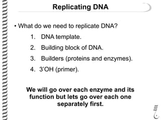
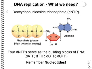
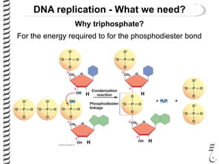
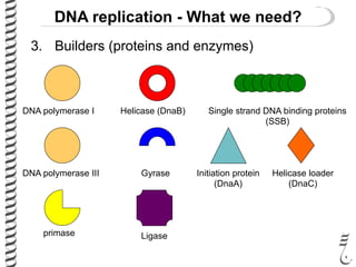
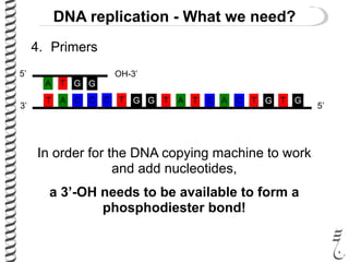
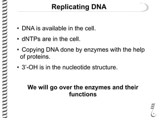
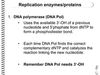
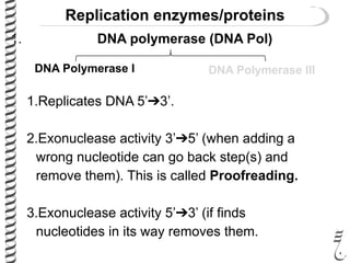
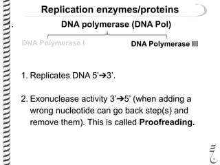
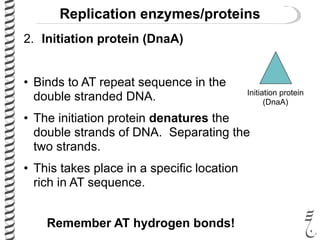
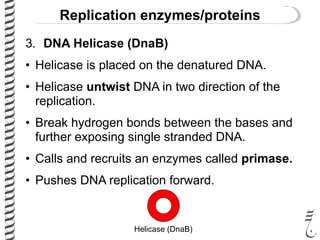
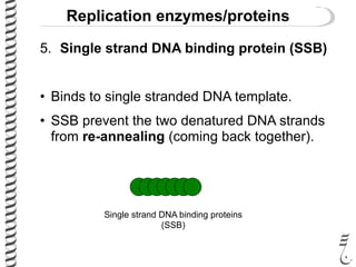
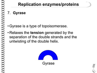
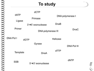
This document introduces the key elements needed for DNA replication in prokaryotes. It discusses the DNA template, building blocks (dNTPs), and proteins/enzymes involved. The main proteins/enzymes that are introduced are: DNA polymerase, which adds new nucleotides; helicase, which unwinds the DNA; single-stranded DNA binding proteins, which prevent rewinding; primase, which adds RNA primers for initiation; and ligase, which seals nicks in the newly synthesized strand. The goal is to explain the chemistry and functions of each component to introduce how prokaryotic DNA replication occurs.
DNA is maintained in a compressed, supercoiled state.
But basis of replication is the formation of strands based on specific bases pairing with their complementary bases
Prokaryotic DNA replication : These slides contains basics of the prokaryotic DNA replication for S.Y.B.Sc and T.Y.B.Sc students of Microbiology and biotechnology
It covers topics like Enzymes used in replication, Semiconservative replication, Meselson and Stahl experiment, Termination of replication, modes of replication: theta and rolling circle, basic rules of replication
It is a powerpoint presentation that discusses about the lesson or topic: DNA Replication. It also talks about the definition, and the concepts about DNA Replication.
Toward the expansion of the genetic alphabet of DNA, several artificial third base pairs (unnatural base pairs) have been created. Organisms are defined by the information encoded in their genomes, and since the origin of life this information has been encoded using a two-base-pair genetic alphabet (A–T and G–C). In vitro, the alphabet have been expanded to include several unnatural base pairs (UBPs). A class of UBPs formed between nucleotides bearing hydrophobic nucleobases, exemplified by the pair formed between d5SICS and dNaM (d5SICS–dNaM) was developed, which is efficiently PCR-amplified and transcribed in vitro, and whose unique mechanism of replication has been characterized. However, expansion of an organism’s genetic alphabet presents new and unprecedented challenges: the unnatural nucleoside triphosphates must be available inside the cell; endogenous polymerases must be able to use the unnatural triphosphates to faithfully replicate DNA containing the UBP within the complex cellular milieu; and finally, the UBP must be stable in the presence of pathways that maintain the integrity of DNA. In a major breakthrough, it was reported that an exogenously expressed algal nucleotide triphosphate transporter efficiently imports the triphosphates of both d5SICS and dNaM (d5SICSTP and dNaMTP) into Escherichia coli, and that the endogenous replication machinery uses them to accurately replicate a plasmid containing d5SICS–dNaM was already reported. Neither the presence of the unnatural triphosphates nor the replication of the UBP introduces a notable growth burden. Thus, the resulting bacterium is the first semi-synthetic organism to propagate stably an expanded genetic alphabet. The unnatural base pair systems now have high potential to open the door to next generation biotechnology.
The material of a talk that I prepared to give in the online camel conference of Oman. Unfortunately, I had a death in the family the day before the conference and the material was presented by my friend Dr. Mohammed Alabri from Oman. The material is in Arabic and focused for camel breeders.
The material of a two days workshop that I gave at Sultan Qaboos University in Oman about the importance of livestock biobanks and how to establish an organized one. The workshop was given in Arabic.
A presentation as a webinar for the Winn Feline Foundation that focuses on recent findings related to the signatures of selection in the domestic cat genome
This was my presentation at the Plant and Animal Genome Conference 2019 in San Diego. My talk was a presentation of the thesis project of my student Mona Abdi. The focus of the presentation and project was the genomic signatures of selection in the domestic cat breeds.
This is a my lecture about camels title "Journey around the camel". The lecture was in Arabic and is related to a book under preparation with the same title. This part of the journey around the camel introduces the major camel breeds in the Arabian Peninsula and their external phenotypes and groupings.
This lecture covers some nice stories about the origins of the words "genome" and the derived word "genomics". the lecture also introduces viral, bacterial, and eukaryotic genomes.
This lecture covers key findings to the development of genomics as a field. This first part covers briefly Mendel to knowing that DNA is the genetic material by Hershey and Chase
Slide 1: Title Slide
Extrachromosomal Inheritance
Slide 2: Introduction to Extrachromosomal Inheritance
Definition: Extrachromosomal inheritance refers to the transmission of genetic material that is not found within the nucleus.
Key Components: Involves genes located in mitochondria, chloroplasts, and plasmids.
Slide 3: Mitochondrial Inheritance
Mitochondria: Organelles responsible for energy production.
Mitochondrial DNA (mtDNA): Circular DNA molecule found in mitochondria.
Inheritance Pattern: Maternally inherited, meaning it is passed from mothers to all their offspring.
Diseases: Examples include Leber’s hereditary optic neuropathy (LHON) and mitochondrial myopathy.
Slide 4: Chloroplast Inheritance
Chloroplasts: Organelles responsible for photosynthesis in plants.
Chloroplast DNA (cpDNA): Circular DNA molecule found in chloroplasts.
Inheritance Pattern: Often maternally inherited in most plants, but can vary in some species.
Examples: Variegation in plants, where leaf color patterns are determined by chloroplast DNA.
Slide 5: Plasmid Inheritance
Plasmids: Small, circular DNA molecules found in bacteria and some eukaryotes.
Features: Can carry antibiotic resistance genes and can be transferred between cells through processes like conjugation.
Significance: Important in biotechnology for gene cloning and genetic engineering.
Slide 6: Mechanisms of Extrachromosomal Inheritance
Non-Mendelian Patterns: Do not follow Mendel’s laws of inheritance.
Cytoplasmic Segregation: During cell division, organelles like mitochondria and chloroplasts are randomly distributed to daughter cells.
Heteroplasmy: Presence of more than one type of organellar genome within a cell, leading to variation in expression.
Slide 7: Examples of Extrachromosomal Inheritance
Four O’clock Plant (Mirabilis jalapa): Shows variegated leaves due to different cpDNA in leaf cells.
Petite Mutants in Yeast: Result from mutations in mitochondrial DNA affecting respiration.
Slide 8: Importance of Extrachromosomal Inheritance
Evolution: Provides insight into the evolution of eukaryotic cells.
Medicine: Understanding mitochondrial inheritance helps in diagnosing and treating mitochondrial diseases.
Agriculture: Chloroplast inheritance can be used in plant breeding and genetic modification.
Slide 9: Recent Research and Advances
Gene Editing: Techniques like CRISPR-Cas9 are being used to edit mitochondrial and chloroplast DNA.
Therapies: Development of mitochondrial replacement therapy (MRT) for preventing mitochondrial diseases.
Slide 10: Conclusion
Summary: Extrachromosomal inheritance involves the transmission of genetic material outside the nucleus and plays a crucial role in genetics, medicine, and biotechnology.
Future Directions: Continued research and technological advancements hold promise for new treatments and applications.
Slide 11: Questions and Discussion
Invite Audience: Open the floor for any questions or further discussion on the topic.
This pdf is about the Schizophrenia.
For more details visit on YouTube; @SELF-EXPLANATORY;
https://www.youtube.com/channel/UCAiarMZDNhe1A3Rnpr_WkzA/videos
Thanks...!
A brief information about the SCOP protein database used in bioinformatics.
The Structural Classification of Proteins (SCOP) database is a comprehensive and authoritative resource for the structural and evolutionary relationships of proteins. It provides a detailed and curated classification of protein structures, grouping them into families, superfamilies, and folds based on their structural and sequence similarities.
Comparing Evolved Extractive Text Summary Scores of Bidirectional Encoder Rep...University of Maribor
Slides from:
11th International Conference on Electrical, Electronics and Computer Engineering (IcETRAN), Niš, 3-6 June 2024
Track: Artificial Intelligence
https://www.etran.rs/2024/en/home-english/
Observation of Io’s Resurfacing via Plume Deposition Using Ground-based Adapt...Sérgio Sacani
Since volcanic activity was first discovered on Io from Voyager images in 1979, changes
on Io’s surface have been monitored from both spacecraft and ground-based telescopes.
Here, we present the highest spatial resolution images of Io ever obtained from a groundbased telescope. These images, acquired by the SHARK-VIS instrument on the Large
Binocular Telescope, show evidence of a major resurfacing event on Io’s trailing hemisphere. When compared to the most recent spacecraft images, the SHARK-VIS images
show that a plume deposit from a powerful eruption at Pillan Patera has covered part
of the long-lived Pele plume deposit. Although this type of resurfacing event may be common on Io, few have been detected due to the rarity of spacecraft visits and the previously low spatial resolution available from Earth-based telescopes. The SHARK-VIS instrument ushers in a new era of high resolution imaging of Io’s surface using adaptive
optics at visible wavelengths.
Cancer cell metabolism: special Reference to Lactate PathwayAADYARAJPANDEY1
Normal Cell Metabolism:
Cellular respiration describes the series of steps that cells use to break down sugar and other chemicals to get the energy we need to function.
Energy is stored in the bonds of glucose and when glucose is broken down, much of that energy is released.
Cell utilize energy in the form of ATP.
The first step of respiration is called glycolysis. In a series of steps, glycolysis breaks glucose into two smaller molecules - a chemical called pyruvate. A small amount of ATP is formed during this process.
Most healthy cells continue the breakdown in a second process, called the Kreb's cycle. The Kreb's cycle allows cells to “burn” the pyruvates made in glycolysis to get more ATP.
The last step in the breakdown of glucose is called oxidative phosphorylation (Ox-Phos).
It takes place in specialized cell structures called mitochondria. This process produces a large amount of ATP. Importantly, cells need oxygen to complete oxidative phosphorylation.
If a cell completes only glycolysis, only 2 molecules of ATP are made per glucose. However, if the cell completes the entire respiration process (glycolysis - Kreb's - oxidative phosphorylation), about 36 molecules of ATP are created, giving it much more energy to use.
IN CANCER CELL:
Unlike healthy cells that "burn" the entire molecule of sugar to capture a large amount of energy as ATP, cancer cells are wasteful.
Cancer cells only partially break down sugar molecules. They overuse the first step of respiration, glycolysis. They frequently do not complete the second step, oxidative phosphorylation.
This results in only 2 molecules of ATP per each glucose molecule instead of the 36 or so ATPs healthy cells gain. As a result, cancer cells need to use a lot more sugar molecules to get enough energy to survive.
introduction to WARBERG PHENOMENA:
WARBURG EFFECT Usually, cancer cells are highly glycolytic (glucose addiction) and take up more glucose than do normal cells from outside.
Otto Heinrich Warburg (; 8 October 1883 – 1 August 1970) In 1931 was awarded the Nobel Prize in Physiology for his "discovery of the nature and mode of action of the respiratory enzyme.
WARNBURG EFFECT : cancer cells under aerobic (well-oxygenated) conditions to metabolize glucose to lactate (aerobic glycolysis) is known as the Warburg effect. Warburg made the observation that tumor slices consume glucose and secrete lactate at a higher rate than normal tissues.
(May 29th, 2024) Advancements in Intravital Microscopy- Insights for Preclini...Scintica Instrumentation
Intravital microscopy (IVM) is a powerful tool utilized to study cellular behavior over time and space in vivo. Much of our understanding of cell biology has been accomplished using various in vitro and ex vivo methods; however, these studies do not necessarily reflect the natural dynamics of biological processes. Unlike traditional cell culture or fixed tissue imaging, IVM allows for the ultra-fast high-resolution imaging of cellular processes over time and space and were studied in its natural environment. Real-time visualization of biological processes in the context of an intact organism helps maintain physiological relevance and provide insights into the progression of disease, response to treatments or developmental processes.
In this webinar we give an overview of advanced applications of the IVM system in preclinical research. IVIM technology is a provider of all-in-one intravital microscopy systems and solutions optimized for in vivo imaging of live animal models at sub-micron resolution. The system’s unique features and user-friendly software enables researchers to probe fast dynamic biological processes such as immune cell tracking, cell-cell interaction as well as vascularization and tumor metastasis with exceptional detail. This webinar will also give an overview of IVM being utilized in drug development, offering a view into the intricate interaction between drugs/nanoparticles and tissues in vivo and allows for the evaluation of therapeutic intervention in a variety of tissues and organs. This interdisciplinary collaboration continues to drive the advancements of novel therapeutic strategies.
Seminar of U.V. Spectroscopy by SAMIR PANDASAMIR PANDA
Spectroscopy is a branch of science dealing the study of interaction of electromagnetic radiation with matter.
Ultraviolet-visible spectroscopy refers to absorption spectroscopy or reflect spectroscopy in the UV-VIS spectral region.
Ultraviolet-visible spectroscopy is an analytical method that can measure the amount of light received by the analyte.
3. AIMS
• Introduce the elements needed to replicate
DNA.
• Introduce the chemistry of adding new DNA
building block and form a new strand of DNA.
• Introduce the proteins and enzymes involved
in the replication of DNA.
• Introduce the function of each enzyme and
protein in the replication of DNA.
4. Replicating DNA
• What do we need to replicate DNA?
1. DNA template.
2. Building block of DNA.
3. Builders (proteins and enzymes).
4. 3’OH (primer).
We will go over each enzyme and its
function but lets go over each one
separately first.
5. DNA replication - What we need?
1. DNA template
G A
C T
A
T
T
A
G
C
G
C
A
T
C
G
C
G
G
C
T
A
A
T
T
A
G
C
A
T
C
G
A
T
C
G
5’
5’
3’
3’
C TT A C C TG G CA T A C T G T G
5’3’
G AA T G G AC C GT A T G A C A C
5’ 3’
Each strand serves as a template for replication.
Remember complementary base-pairing!
6. 2. Deoxyribonucleoside triphosphate (dNTP)
DNA replication - What we need?
H
Four dNTPs serve as the building blocks of DNA
(dATP, dTTP, dGTP, dCTP)
Remember Nucleotides!
7. DNA replication - What we need?
Why triphosphate?
For the energy required to for the phosphodiester bond
+
H H
H H
8. 3. Builders (proteins and enzymes)
DNA replication - What we need?
DNA polymerase I
DNA polymerase III
Helicase (DnaB)
Gyrase
Single strand DNA binding proteins
(SSB)
primase Ligase
Initiation protein
(DnaA)
Helicase loader
(DnaC)
9. 4. Primers
DNA replication - What we need?
G
C T
A
T
T
A
G
C C TG G CA T A C T G T G
5’
5’
OH-3’
3’
In order for the DNA copying machine to work
and add nucleotides,
a 3’-OH needs to be available to form a
phosphodiester bond!
11. Replicating DNA
• DNA is available in the cell.
• dNTPs are in the cell.
• Copying DNA done by enzymes with the help
of proteins.
• 3’-OH is in the nucleotide structure.
We will go over the enzymes and their
functions
12. Replication enzymes/proteins
1. DNA polymerase (DNA Pol)
• It is the DNA copier.
• Uses the dNTPs (DNA building blocks) to
make a complementary strand to the
template.
13. Replication enzymes/proteins
1. DNA polymerase (DNA Pol)
• Uses the available 3’-OH of a previous
nucleotide and 5’phsphate from dNTP to
form a phosphodiester bond.
• Each time DNA Pol finds the correct
complementary dNTP and catalyzes the
reaction linking the new nucleotide.
• Remember DNA Pol needs 3’-OH
14. DNA polymerase (DNA Pol)
DNA Polymerase I DNA Polymerase III
Replication enzymes/proteins
15. 1. DNA polymerase (DNA Pol)
DNA Polymerase I DNA Polymerase III
1.Replicates DNA 5’➔3’.
2.Exonuclease activity 3’➔5’ (when adding a
wrong nucleotide can go back step(s) and
remove them). This is called Proofreading.
3.Exonuclease activity 5’➔3’ (if finds
nucleotides in its way removes them.
Replication enzymes/proteins
16. 1. DNA polymerase (DNA Pol)
DNA Polymerase I DNA Polymerase III
1. Replicates DNA 5’➔3’.
2. Exonuclease activity 3’➔5’ (when adding a
wrong nucleotide can go back step(s) and
remove them). This is called Proofreading.
Replication enzymes/proteins
17. Replication enzymes/proteins
2. Initiation protein (DnaA)
• Binds to AT repeat sequence in the
double stranded DNA.
• The initiation protein denatures the
double strands of DNA. Separating the
two strands.
• This takes place in a specific location
rich in AT sequence.
Remember AT hydrogen bonds!
Initiation protein
(DnaA)
18. Replication enzymes/proteins
3. DNA Helicase (DnaB)
• Helicase is placed on the denatured DNA.
• Helicase untwist DNA in two direction of the
replication.
• Break hydrogen bonds between the bases and
further exposing single stranded DNA.
• Calls and recruits an enzymes called primase.
• Pushes DNA replication forward.
Helicase (DnaB)
19. Replication enzymes/proteins
4. DNA Helicase loader (DnaC)
• As the names suggests, this protein loads and
places the DNA helicase on the denatured
DNA.
Helicase loader
(DnaC)
20. Replication enzymes/proteins
5. Single strand DNA binding protein (SSB)
• Binds to single stranded DNA template.
• SSB prevent the two denatured DNA strands
from re-annealing (coming back together).
Single strand DNA binding proteins
(SSB)
21. Replication enzymes/proteins
6. Primase
• Primase adds a block of nucleotides (primer) to
provide the polymerase with a 3’-OH needed
for the synthesis of DNA.
• The block added is complementary to the
template.
• Primase adds a single primer on one template.
• Primase adds multiple primers on the second
template.
primase
22. Replication enzymes/proteins
7. Gyrase
• Gyrase is a type of topoisomerase.
• Relaxes the tension generated by the
separation of the double strands and the
untwisting of the double helix.
Gyrase
24. Quiz
Which of the following enzymes is
responsible for untwisting the DNA during
replication
a) DNA Pol I
b) Lygase
c) Primase
d) Helicase
e) Gyrase
25. To study
Ligase
DNA polymerase I
DNA polymerase III
DNA Pol I
DNA Pol III
Gyrase
Primase
SSB
DnaA
Helicase
DnaB
DnaCPrimer
Template
dNTP
dATP
dCTP
dGTP
dTTP
5’➔3’ exonuclease
3’➔5’ exonuclease
26. Expectations
• You know what is needed for DNA replication
and synthesis to take place.
• You know the building blocks of DNA.
• You know the enzymes/proteins in the process
of replication.
• You know the function of each enzyme/protein.
Next lecture we will go over the process of
DNA replication and connect it with what we
learned today