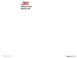
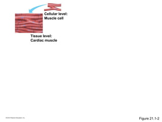
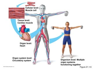
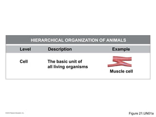
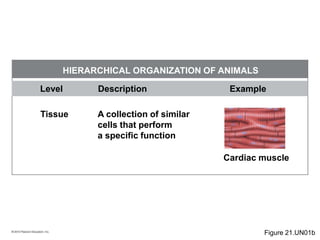
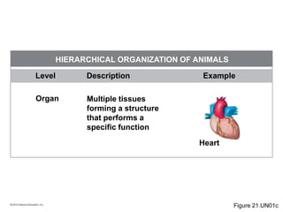
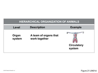
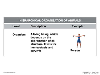
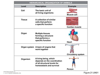
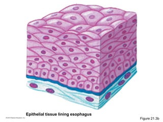
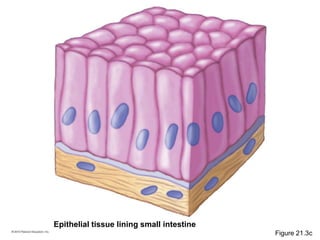
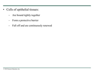
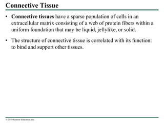
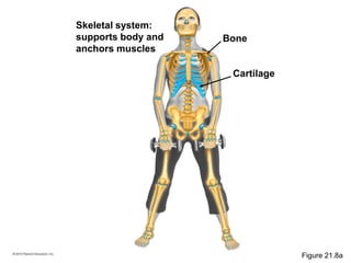
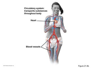
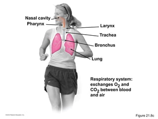
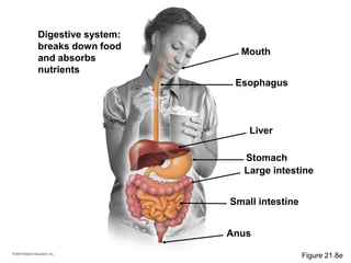
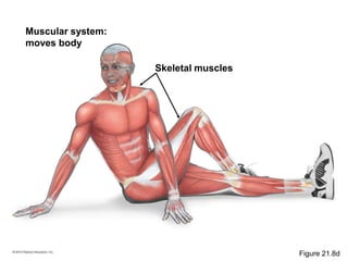
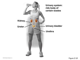
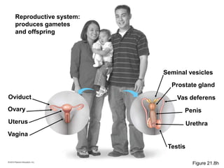
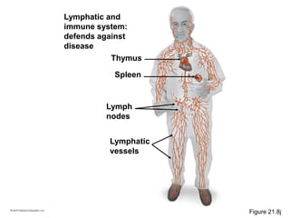
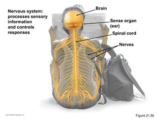
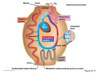
© 2010 Pearson Education, Inc.
THE STRUCTURAL ORGANIZATION OF
ANIMALS
• Life is characterized by a hierarchy of organization.
• In animals:
– Individual cells are grouped into tissues
– Tissues combine to form organs
– Organs are organized into organ systems
– Organ systems make up the entire organism
Cellular level:
Muscle cell
Figure 21.1-1
Cellular level:
Muscle cell
Tissue level:
Cardiac muscle
Figure 21.1-2
Cellular level:
Muscle cell
Tissue level:
Cardiac muscle
Organ level:
Heart
Figure 21.1-3
Cellular level:
Muscle cell
Tissue level:
Cardiac muscle
Organ level:
Heart
Organ system level:
Circulatory system Organism level: Multiple
organ systems
functioning together
Figure 21.1-5
© 2010 Pearson Education, Inc.
Cell
Level Description
The basic unit of
all living organisms
Muscle cell
Example
HIERARCHICAL ORGANIZATION OF ANIMALS
Figure 21.UN01a
© 2010 Pearson Education, Inc.
Level Description Example
HIERARCHICAL ORGANIZATION OF ANIMALS
Tissue A collection of similar
cells that perform
a specific function
Cardiac muscle
Figure 21.UN01b
© 2010 Pearson Education, Inc.
Level Description Example
HIERARCHICAL ORGANIZATION OF ANIMALS
Organ Multiple tissues
forming a structure
that performs a
specific function
Heart
Figure 21.UN01c
© 2010 Pearson Education, Inc.
Level Description Example
HIERARCHICAL ORGANIZATION OF ANIMALS
Organ
system
A team of organs that
work together
Circulatory
system
Figure 21.UN01d
© 2010 Pearson Education, Inc.
Level Description Example
HIERARCHICAL ORGANIZATION OF ANIMALS
Organism A living being, which
depends on the
coordination of all
structural levels for
homeostasis and
survival Person
Figure 21.UN01e
© 2010 Pearson Education, Inc.
Cell
Level
Tissue
Organ
Organ system
Organism
Description
The basic unit of
all living organisms
A collection of similar
cells that perform
a specific function
Multiple tissues
forming a structure
that performs a
specific function
A team of organs that
work together
A living being, which
depends on the coordination
of all structural levels for
homeostasis and survival
Muscle cell
Example
Cardiac muscle
Heart
Circulatory system
Person
HIERARCHICAL ORGANIZATION OF ANIMALS
Figure 21.UN01
© 2010 Pearson Education, Inc.
Form Fits Function
• Analyzing a biological structure gives us clues about:
– What it does
– How it works
© 2010 Pearson Education, Inc.
• Biologists distinguish anatomy from physiology.
– Anatomy is the study of the structure of an organism.
– Physiology is the study of the function of an organism’s structural
equipment.
© 2010 Pearson Education, Inc.
Tissues
• In most multicellular animals, cells are grouped into tissues.
– A tissue is an integrated group of similar cells that perform a specific
function.
– Animals have four main categories of tissue.
© 2010 Pearson Education, Inc.
Epithelial Tissue
• Epithelial tissue, also known as epithelium:
– Covers the surface of the body
– Lines organs and cavities within the body
Some examples of organs
lined with epithelial tissue:
Heart
Lung
Stomach
Small intestine
Large intestine
Urinary bladder
Figure 21.3a
Epithelial tissue lining esophagus
Figure 21.3b
Epithelial tissue lining small intestine
Figure 21.3c
© 2010 Pearson Education, Inc.
• Cells of epithelial tissues:
– Are bound tightly together
– Form a protective barrier
– Fall off and are continuously renewed
© 2010 Pearson Education, Inc.
Connective Tissue
• Connective tissues have a sparse population of cells in an
extracellular matrix consisting of a web of protein fibers within a
uniform foundation that may be liquid, jellylike, or solid.
• The structure of connective tissue is correlated with its function:
to bind and support other tissues.
(a) Loose connective tissue
(under the skin)
(b) Adipose tissue
(c) Blood
(d) Fibrous connective tissue
(forming a tendon)
(e) Cartilage (at the end of a bone)
(f) Bone
Cell
Cells
Matrix
Plasma
Matrix
Cells
Collagen
fiber
Fat
droplets
White blood
cells
Red blood
cell
Cell
nucleus
Collagen
fibers
Figure 21.4
© 2010 Pearson Education, Inc.
• Loose connective tissue:
– Is the most widespread connective tissue
– Binds epithelia to underlying tissues
– Holds organs in place
(a) Loose connective tissue
(under the skin)
Cell
Collagen fiber
Figure 21.4a
© 2010 Pearson Education, Inc.
• Adipose tissue:
– Stores fat
– Stockpiles energy
– Pads and insulates the body
(b) Adipose tissue
Fat
droplets
Figure 21.4b
© 2010 Pearson Education, Inc.
• Blood:
– Is a connective tissue with a matrix of liquid
– Contains red and white blood cells suspended in plasma
(c) Blood
Plasma
White blood cells
Red blood cell
Figure 21.4c
© 2010 Pearson Education, Inc.
• Fibrous connective tissue:
– Has a dense matrix of collagen
– Forms tendons and ligaments
(d) Fibrous connective tissue
(forming a tendon)
Cell nucleus
Collagen fibers
Figure 21.4d
© 2010 Pearson Education, Inc.
• Cartilage:
– Has a strong but rubbery matrix
– Functions as a flexible, boneless skeleton
– Forms the shock-absorbing pads that cushion the vertebrae of the spinal
column
(e) Cartilage (at the end of a bone)
Matrix
Cells
Figure 21.4e
© 2010 Pearson Education, Inc.
• Bone:
– Is a rigid connective tissue
– Has a matrix of collagen fibers hardened with deposits of calcium salts
(f) Bone
Cells
Matrix
Figure 21.4f
© 2010 Pearson Education, Inc.
Muscle Tissue
• Muscle tissue:
– Consists of bundles of long, thin, cylindrical cells called muscle fibers
– Has specialized proteins that contract when stimulated by a nerve
(a) Skeletal muscle
(short segments of
several muscle fibers)
(b) Cardiac muscle
Unit of
muscle
contraction
Muscle
fiber
(cell)
Muscle
fiber
Junction between
two cells
(c) Smooth muscle
Nucleus
Nucleus
Muscle fiber
Nuclei
Figure 21.5
(a) Skeletal muscle
(short segments of
several muscle fibers)
Unit of
muscle
contraction
Muscle fiber
(cell)
Nuclei
Figure 21.5a
(b) Cardiac muscle
Muscle
fiber
Junction between two cells
Nucleus
Figure 21.5b
(c) Smooth muscle
Muscle fiber
Nucleus
Figure 21.5c
© 2010 Pearson Education, Inc.
• Skeletal muscle is:
– Attached to bones by tendons
– Responsible for voluntary movements
– Striated because the contractile apparatus forms a banded pattern in each
cell or fiber
© 2010 Pearson Education, Inc.
• Cardiac muscle is:
– Composed of cells that are branched and striated
– Found only in heart tissue
– Responsible for the contraction of the heart
© 2010 Pearson Education, Inc.
• Smooth muscle is:
– Named for its lack of obvious striations
– Found in the walls of various organs
– Involuntary
© 2010 Pearson Education, Inc.
Nervous Tissue
• Nervous tissue:
– Makes communication of sensory information possible
– Is found in the brain and spinal cord
– Consists of a network of neurons
Brain
Nerve
Spinal cord
Figure 21.6a
Signal-receiving
extensions Cell body
Signal-
transmitting
extensions
LM
Figure 21.6b
© 2010 Pearson Education, Inc.
Organs and Organ Systems
• An organ consists of two or more tissues packaged into one
working unit that performs a specific function.
• Examples include the heart, liver, stomach, brain, and lungs.
© 2010 Pearson Education, Inc.
• Organ systems:
– Are teams of organs that work together
– Perform vital body functions
Blast Animation: Anatomy of the Kidney
Skeletal system:
supports body and
anchors muscles
Cartilage
Bone
Figure 21.8a
Circulatory system:
transports substances
throughout body
Blood vessels
Heart
Figure 21.8b
Respiratory system:
exchanges O2 and
CO2 between blood
and air
Nasal cavity
Bronchus
Pharynx
Trachea
Larynx
Lung
Figure 21.8c
Digestive system:
breaks down food
and absorbs
nutrients
Large intestine
Small intestine
Esophagus
Stomach
Liver
Mouth
Anus
Figure 21.8e
Muscular system:
moves body
Skeletal muscles
Figure 21.8d
Urinary system:
rids body of
certain wastes
Urinary bladder
Urethra
Ureter
Kidney
Figure 21.8f
Reproductive system:
produces gametes
and offspring
Seminal vesicles
Prostate gland
Vas deferens
Urethra
Oviduct
Vagina
Uterus
Ovary Penis
Testis
Figure 21.8h
Lymphatic and
immune system:
defends against
disease
Lymphatic
vessels
Lymph
nodes
Thymus
Spleen
Figure 21.8j
Sense organ
(ear)
Nervous system:
processes sensory
information
and controls
responses Spinal cord
Nerves
Brain
Figure 21.8k
© 2010 Pearson Education, Inc.
EXCHANGES WITH THE EXTERNAL
ENVIRONMENT
• Every organism is an open system, continuously exchanging
chemicals and energy with its surroundings to survive.
© 2010 Pearson Education, Inc.
• An animal’s size and shape affect how it exchanges energy and
materials with its surroundings.
• All living cells must be bathed in a watery solution so that
exchange of materials can occur.
© 2010 Pearson Education, Inc.
• Animals use three organ systems to exchange materials with the
external environment:
– Digestive
– Respiratory
– Urinary
• The circulatory system transports materials inside the body from
these exchange surfaces.
Unabsorbed matter (feces) Metabolic waste products (such as urine)
External environment
Respiratory
systemDigestive
system
Urinary
system
Circulatory
system
Interstitial
fluid
Nutrients
Body cells
Animal
Mouth Food
Anus
Heart
CO2
O2
Figure 21.11
© 2010 Pearson Education, Inc.
REGULATING THE INTERNAL
ENVIRONMENT
• Every living organism has the ability to respond to its
environment.
© 2010 Pearson Education, Inc.
Homeostasis
• Homeostasis is the body’s tendency to maintain relatively steady
conditions in the internal environment when the external
environment changes.
External
environment
Large external
changes
Animal’s internal
environment
Homeostatic
mechanisms
Small internal
changes
Figure 21.12
© 2010 Pearson Education, Inc.
Negative and Positive Feedback
• Most mechanisms of homeostasis depend on a common principle
called negative feedback, in which the results of a process inhibit
that same process, such as a thermostat that turns off a heater
when room temperature rises to the set point.
Animation: Positive Feedback
Animation: Negative Feedback
Thermostat
(control center)
turns heater off
Set point:
Room temperature
20C (68F)
Room
temperature
drops
Response:
Heating
stops
Stimulus:
Room temperature
is above set point
Figure 21.13a
Thermostat
(control center)
turns heater on
Set point:
Room temperature
20C (68F)
Room
temperature
rises
Response:
Heating
starts
Stimulus:
Room temperature
is below set point
Figure 21.13b
© 2010 Pearson Education, Inc.
• Less common is positive feedback in which the results of a
process intensify that same process, such as uterine contractions
during childbirth.
© 2010 Pearson Education, Inc.
• Humans have homeostatic mechanisms that aid in
thermoregulation, which:
– Cool or
– Heat the body
Blast Animation: Negative Feedback: Body Temperature
Body
temperature
drops
Set point:
Body temperature near 37C (98.6F)
Stimulus:
Body temperature
is above set point
Control center
in brain activates
cooling mechanisms
Response:
1. Blood
vessels
dilate
2. Sweat is
produced
Sweat
gland
Skin
Figure 21.14a
Set point:
Body temperature near 37C (98.6F)
Body
temperature
rises
Stimulus:
Body temperature
is below set point
Control center
in brain activates
warming mechanisms
Response:
1. Blood vessels
constrict
2. Muscles shiver
3. Metabolic rate
increases
Skin
Figure 21.14b
© 2010 Pearson Education, Inc.
• Fever:
– Is an abnormally high internal body temperature
– Usually indicates an ongoing fight against infection
© 2010 Pearson Education, Inc.
Osmoregulation
• Living cells depend on a precise balance of:
– Water
– Solutes
• Osmoregulation is the control of the gain or loss of:
– Water
– Dissolved solutes, such as salt
© 2010 Pearson Education, Inc.
Homeostasis in the Urinary System
• The urinary system:
– Plays a central role in homeostasis
– Forms and excretes urine
– Regulates the amount of water and solutes in body fluids
© 2010 Pearson Education, Inc.
• In humans, the two kidneys:
– Are the main processing centers
– Contain many fine tubes called tubules
– Include an intricate network of capillaries
© 2010 Pearson Education, Inc.
• As blood circulates through the kidneys:
– A fraction of it is filtered
– Plasma enters the kidney tubules, forming filtrate
• Filtrate contains:
– Valuable substances that need to be reclaimed (such as water and
glucose)
– Substances to be eliminated, such as urea
© 2010 Pearson Education, Inc.
• The human urinary system includes:
– The kidneys
– Nephrons, the functional units of the kidney
– The urinary bladder, where urine is stored
Animation: Nephron Introduction
Renal artery (red) and
renal vein (blue)
(a) Urinary system
Kidney
Ureter
Urinary
bladder
Urethra
Figure 21.17a
(b) Kidney
Ureter
Nephron
Figure 21.17b
© 2010 Pearson Education, Inc.
• Nephrons:
– Carry out the functions of the urinary system
– Consist of a tubule and its associated vessels
– Number more than a million in a kidney
(c) A nephron and collecting duct
Branch of
renal artery
Branch of
renal vein Collecting
duct
Filter Tubule
To ureter
Figure 21.17c
© 2010 Pearson Education, Inc.
• Nephrons perform four key functions:
– Filtration, forcing water and other small molecules from the blood to
form filtrate
– Reabsorption of water and valuable solutes back into the blood
– Secretion of certain substances, such as ions and drugs, into the filtrate
– Excretion of urine from the kidneys
Filtration Reabsorption Secretion
Renal artery
Renal vein
Capillaries
Tubule
Urine
Filtrate
Excretion
Figure 21.18
© 2010 Pearson Education, Inc.
• Hormonal control of the nephrons allows the body to control its
internal concentration of:
– Water
– Dissolved molecules
© 2010 Pearson Education, Inc.
• Kidney failure can be caused by:
– Injury
– Illness
– Prolonged use of pain relievers, alcohol, or other drugs
• One option for treatment of kidney failure is dialysis, filtration of
blood by a machine.
Used dialyzing
solution (with
urea and
excess salts)
Line from
apparatus
to vein
Fresh dialyzing
solution
Dialyzing
solution
Line from artery
to apparatus
Tubing made of a
selectively permeable
membrane
Pump
Figure 21.19

21 lecture presentation

  • 1.
    © 2010 PearsonEducation, Inc. THE STRUCTURAL ORGANIZATION OF ANIMALS • Life is characterized by a hierarchy of organization. • In animals: – Individual cells are grouped into tissues – Tissues combine to form organs – Organs are organized into organ systems – Organ systems make up the entire organism
  • 2.
  • 3.
    Cellular level: Muscle cell Tissuelevel: Cardiac muscle Figure 21.1-2
  • 4.
    Cellular level: Muscle cell Tissuelevel: Cardiac muscle Organ level: Heart Figure 21.1-3
  • 5.
    Cellular level: Muscle cell Tissuelevel: Cardiac muscle Organ level: Heart Organ system level: Circulatory system Organism level: Multiple organ systems functioning together Figure 21.1-5
  • 6.
    © 2010 PearsonEducation, Inc. Cell Level Description The basic unit of all living organisms Muscle cell Example HIERARCHICAL ORGANIZATION OF ANIMALS Figure 21.UN01a
  • 7.
    © 2010 PearsonEducation, Inc. Level Description Example HIERARCHICAL ORGANIZATION OF ANIMALS Tissue A collection of similar cells that perform a specific function Cardiac muscle Figure 21.UN01b
  • 8.
    © 2010 PearsonEducation, Inc. Level Description Example HIERARCHICAL ORGANIZATION OF ANIMALS Organ Multiple tissues forming a structure that performs a specific function Heart Figure 21.UN01c
  • 9.
    © 2010 PearsonEducation, Inc. Level Description Example HIERARCHICAL ORGANIZATION OF ANIMALS Organ system A team of organs that work together Circulatory system Figure 21.UN01d
  • 10.
    © 2010 PearsonEducation, Inc. Level Description Example HIERARCHICAL ORGANIZATION OF ANIMALS Organism A living being, which depends on the coordination of all structural levels for homeostasis and survival Person Figure 21.UN01e
  • 11.
    © 2010 PearsonEducation, Inc. Cell Level Tissue Organ Organ system Organism Description The basic unit of all living organisms A collection of similar cells that perform a specific function Multiple tissues forming a structure that performs a specific function A team of organs that work together A living being, which depends on the coordination of all structural levels for homeostasis and survival Muscle cell Example Cardiac muscle Heart Circulatory system Person HIERARCHICAL ORGANIZATION OF ANIMALS Figure 21.UN01
  • 12.
    © 2010 PearsonEducation, Inc. Form Fits Function • Analyzing a biological structure gives us clues about: – What it does – How it works
  • 13.
    © 2010 PearsonEducation, Inc. • Biologists distinguish anatomy from physiology. – Anatomy is the study of the structure of an organism. – Physiology is the study of the function of an organism’s structural equipment.
  • 14.
    © 2010 PearsonEducation, Inc. Tissues • In most multicellular animals, cells are grouped into tissues. – A tissue is an integrated group of similar cells that perform a specific function. – Animals have four main categories of tissue.
  • 15.
    © 2010 PearsonEducation, Inc. Epithelial Tissue • Epithelial tissue, also known as epithelium: – Covers the surface of the body – Lines organs and cavities within the body
  • 16.
    Some examples oforgans lined with epithelial tissue: Heart Lung Stomach Small intestine Large intestine Urinary bladder Figure 21.3a
  • 17.
    Epithelial tissue liningesophagus Figure 21.3b
  • 18.
    Epithelial tissue liningsmall intestine Figure 21.3c
  • 19.
    © 2010 PearsonEducation, Inc. • Cells of epithelial tissues: – Are bound tightly together – Form a protective barrier – Fall off and are continuously renewed
  • 20.
    © 2010 PearsonEducation, Inc. Connective Tissue • Connective tissues have a sparse population of cells in an extracellular matrix consisting of a web of protein fibers within a uniform foundation that may be liquid, jellylike, or solid. • The structure of connective tissue is correlated with its function: to bind and support other tissues.
  • 21.
    (a) Loose connectivetissue (under the skin) (b) Adipose tissue (c) Blood (d) Fibrous connective tissue (forming a tendon) (e) Cartilage (at the end of a bone) (f) Bone Cell Cells Matrix Plasma Matrix Cells Collagen fiber Fat droplets White blood cells Red blood cell Cell nucleus Collagen fibers Figure 21.4
  • 22.
    © 2010 PearsonEducation, Inc. • Loose connective tissue: – Is the most widespread connective tissue – Binds epithelia to underlying tissues – Holds organs in place
  • 23.
    (a) Loose connectivetissue (under the skin) Cell Collagen fiber Figure 21.4a
  • 24.
    © 2010 PearsonEducation, Inc. • Adipose tissue: – Stores fat – Stockpiles energy – Pads and insulates the body
  • 25.
  • 26.
    © 2010 PearsonEducation, Inc. • Blood: – Is a connective tissue with a matrix of liquid – Contains red and white blood cells suspended in plasma
  • 27.
    (c) Blood Plasma White bloodcells Red blood cell Figure 21.4c
  • 28.
    © 2010 PearsonEducation, Inc. • Fibrous connective tissue: – Has a dense matrix of collagen – Forms tendons and ligaments
  • 29.
    (d) Fibrous connectivetissue (forming a tendon) Cell nucleus Collagen fibers Figure 21.4d
  • 30.
    © 2010 PearsonEducation, Inc. • Cartilage: – Has a strong but rubbery matrix – Functions as a flexible, boneless skeleton – Forms the shock-absorbing pads that cushion the vertebrae of the spinal column
  • 31.
    (e) Cartilage (atthe end of a bone) Matrix Cells Figure 21.4e
  • 32.
    © 2010 PearsonEducation, Inc. • Bone: – Is a rigid connective tissue – Has a matrix of collagen fibers hardened with deposits of calcium salts
  • 33.
  • 34.
    © 2010 PearsonEducation, Inc. Muscle Tissue • Muscle tissue: – Consists of bundles of long, thin, cylindrical cells called muscle fibers – Has specialized proteins that contract when stimulated by a nerve
  • 35.
    (a) Skeletal muscle (shortsegments of several muscle fibers) (b) Cardiac muscle Unit of muscle contraction Muscle fiber (cell) Muscle fiber Junction between two cells (c) Smooth muscle Nucleus Nucleus Muscle fiber Nuclei Figure 21.5
  • 36.
    (a) Skeletal muscle (shortsegments of several muscle fibers) Unit of muscle contraction Muscle fiber (cell) Nuclei Figure 21.5a
  • 37.
    (b) Cardiac muscle Muscle fiber Junctionbetween two cells Nucleus Figure 21.5b
  • 38.
    (c) Smooth muscle Musclefiber Nucleus Figure 21.5c
  • 39.
    © 2010 PearsonEducation, Inc. • Skeletal muscle is: – Attached to bones by tendons – Responsible for voluntary movements – Striated because the contractile apparatus forms a banded pattern in each cell or fiber
  • 40.
    © 2010 PearsonEducation, Inc. • Cardiac muscle is: – Composed of cells that are branched and striated – Found only in heart tissue – Responsible for the contraction of the heart
  • 41.
    © 2010 PearsonEducation, Inc. • Smooth muscle is: – Named for its lack of obvious striations – Found in the walls of various organs – Involuntary
  • 42.
    © 2010 PearsonEducation, Inc. Nervous Tissue • Nervous tissue: – Makes communication of sensory information possible – Is found in the brain and spinal cord – Consists of a network of neurons
  • 43.
  • 44.
  • 45.
    © 2010 PearsonEducation, Inc. Organs and Organ Systems • An organ consists of two or more tissues packaged into one working unit that performs a specific function. • Examples include the heart, liver, stomach, brain, and lungs.
  • 46.
    © 2010 PearsonEducation, Inc. • Organ systems: – Are teams of organs that work together – Perform vital body functions Blast Animation: Anatomy of the Kidney
  • 47.
    Skeletal system: supports bodyand anchors muscles Cartilage Bone Figure 21.8a
  • 48.
    Circulatory system: transports substances throughoutbody Blood vessels Heart Figure 21.8b
  • 49.
    Respiratory system: exchanges O2and CO2 between blood and air Nasal cavity Bronchus Pharynx Trachea Larynx Lung Figure 21.8c
  • 50.
    Digestive system: breaks downfood and absorbs nutrients Large intestine Small intestine Esophagus Stomach Liver Mouth Anus Figure 21.8e
  • 51.
  • 52.
    Urinary system: rids bodyof certain wastes Urinary bladder Urethra Ureter Kidney Figure 21.8f
  • 53.
    Reproductive system: produces gametes andoffspring Seminal vesicles Prostate gland Vas deferens Urethra Oviduct Vagina Uterus Ovary Penis Testis Figure 21.8h
  • 54.
    Lymphatic and immune system: defendsagainst disease Lymphatic vessels Lymph nodes Thymus Spleen Figure 21.8j
  • 55.
    Sense organ (ear) Nervous system: processessensory information and controls responses Spinal cord Nerves Brain Figure 21.8k
  • 56.
    © 2010 PearsonEducation, Inc. EXCHANGES WITH THE EXTERNAL ENVIRONMENT • Every organism is an open system, continuously exchanging chemicals and energy with its surroundings to survive.
  • 57.
    © 2010 PearsonEducation, Inc. • An animal’s size and shape affect how it exchanges energy and materials with its surroundings. • All living cells must be bathed in a watery solution so that exchange of materials can occur.
  • 58.
    © 2010 PearsonEducation, Inc. • Animals use three organ systems to exchange materials with the external environment: – Digestive – Respiratory – Urinary • The circulatory system transports materials inside the body from these exchange surfaces.
  • 59.
    Unabsorbed matter (feces)Metabolic waste products (such as urine) External environment Respiratory systemDigestive system Urinary system Circulatory system Interstitial fluid Nutrients Body cells Animal Mouth Food Anus Heart CO2 O2 Figure 21.11
  • 60.
    © 2010 PearsonEducation, Inc. REGULATING THE INTERNAL ENVIRONMENT • Every living organism has the ability to respond to its environment.
  • 61.
    © 2010 PearsonEducation, Inc. Homeostasis • Homeostasis is the body’s tendency to maintain relatively steady conditions in the internal environment when the external environment changes.
  • 62.
  • 63.
    © 2010 PearsonEducation, Inc. Negative and Positive Feedback • Most mechanisms of homeostasis depend on a common principle called negative feedback, in which the results of a process inhibit that same process, such as a thermostat that turns off a heater when room temperature rises to the set point. Animation: Positive Feedback Animation: Negative Feedback
  • 64.
    Thermostat (control center) turns heateroff Set point: Room temperature 20C (68F) Room temperature drops Response: Heating stops Stimulus: Room temperature is above set point Figure 21.13a
  • 65.
    Thermostat (control center) turns heateron Set point: Room temperature 20C (68F) Room temperature rises Response: Heating starts Stimulus: Room temperature is below set point Figure 21.13b
  • 66.
    © 2010 PearsonEducation, Inc. • Less common is positive feedback in which the results of a process intensify that same process, such as uterine contractions during childbirth.
  • 67.
    © 2010 PearsonEducation, Inc. • Humans have homeostatic mechanisms that aid in thermoregulation, which: – Cool or – Heat the body Blast Animation: Negative Feedback: Body Temperature
  • 68.
    Body temperature drops Set point: Body temperaturenear 37C (98.6F) Stimulus: Body temperature is above set point Control center in brain activates cooling mechanisms Response: 1. Blood vessels dilate 2. Sweat is produced Sweat gland Skin Figure 21.14a
  • 69.
    Set point: Body temperaturenear 37C (98.6F) Body temperature rises Stimulus: Body temperature is below set point Control center in brain activates warming mechanisms Response: 1. Blood vessels constrict 2. Muscles shiver 3. Metabolic rate increases Skin Figure 21.14b
  • 70.
    © 2010 PearsonEducation, Inc. • Fever: – Is an abnormally high internal body temperature – Usually indicates an ongoing fight against infection
  • 71.
    © 2010 PearsonEducation, Inc. Osmoregulation • Living cells depend on a precise balance of: – Water – Solutes • Osmoregulation is the control of the gain or loss of: – Water – Dissolved solutes, such as salt
  • 72.
    © 2010 PearsonEducation, Inc. Homeostasis in the Urinary System • The urinary system: – Plays a central role in homeostasis – Forms and excretes urine – Regulates the amount of water and solutes in body fluids
  • 73.
    © 2010 PearsonEducation, Inc. • In humans, the two kidneys: – Are the main processing centers – Contain many fine tubes called tubules – Include an intricate network of capillaries
  • 74.
    © 2010 PearsonEducation, Inc. • As blood circulates through the kidneys: – A fraction of it is filtered – Plasma enters the kidney tubules, forming filtrate • Filtrate contains: – Valuable substances that need to be reclaimed (such as water and glucose) – Substances to be eliminated, such as urea
  • 75.
    © 2010 PearsonEducation, Inc. • The human urinary system includes: – The kidneys – Nephrons, the functional units of the kidney – The urinary bladder, where urine is stored Animation: Nephron Introduction
  • 76.
    Renal artery (red)and renal vein (blue) (a) Urinary system Kidney Ureter Urinary bladder Urethra Figure 21.17a
  • 77.
  • 78.
    © 2010 PearsonEducation, Inc. • Nephrons: – Carry out the functions of the urinary system – Consist of a tubule and its associated vessels – Number more than a million in a kidney
  • 79.
    (c) A nephronand collecting duct Branch of renal artery Branch of renal vein Collecting duct Filter Tubule To ureter Figure 21.17c
  • 80.
    © 2010 PearsonEducation, Inc. • Nephrons perform four key functions: – Filtration, forcing water and other small molecules from the blood to form filtrate – Reabsorption of water and valuable solutes back into the blood – Secretion of certain substances, such as ions and drugs, into the filtrate – Excretion of urine from the kidneys
  • 81.
    Filtration Reabsorption Secretion Renalartery Renal vein Capillaries Tubule Urine Filtrate Excretion Figure 21.18
  • 82.
    © 2010 PearsonEducation, Inc. • Hormonal control of the nephrons allows the body to control its internal concentration of: – Water – Dissolved molecules
  • 83.
    © 2010 PearsonEducation, Inc. • Kidney failure can be caused by: – Injury – Illness – Prolonged use of pain relievers, alcohol, or other drugs • One option for treatment of kidney failure is dialysis, filtration of blood by a machine.
  • 84.
    Used dialyzing solution (with ureaand excess salts) Line from apparatus to vein Fresh dialyzing solution Dialyzing solution Line from artery to apparatus Tubing made of a selectively permeable membrane Pump Figure 21.19