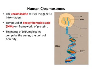
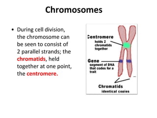
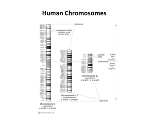
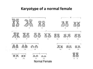
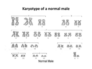
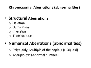
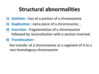
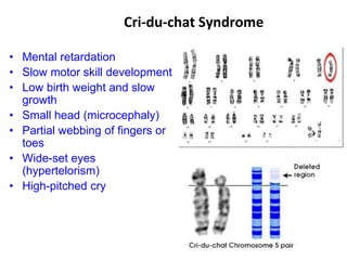
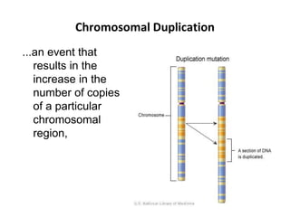
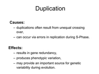
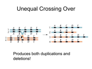
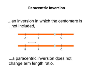
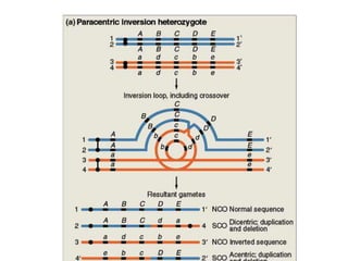
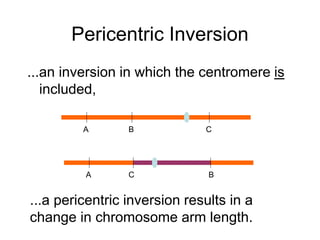
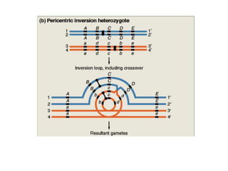
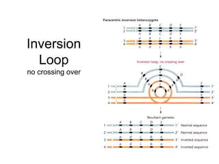
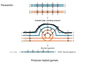
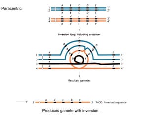
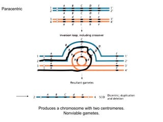
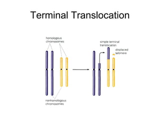
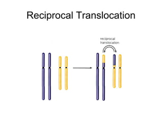
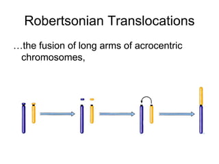
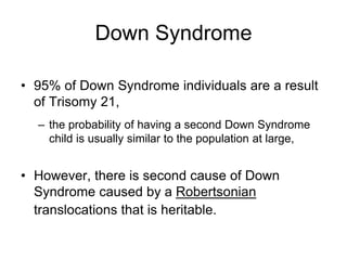
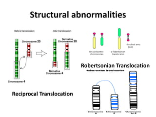
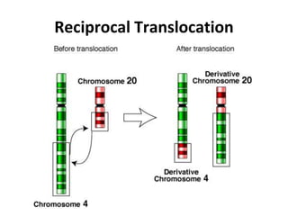
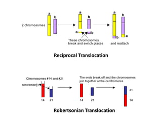
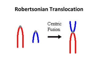
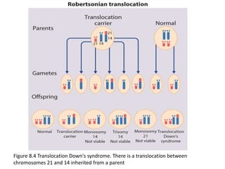
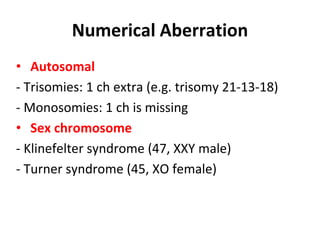
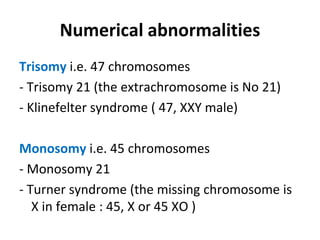
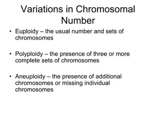
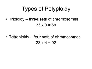
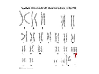
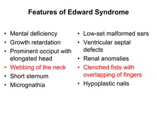
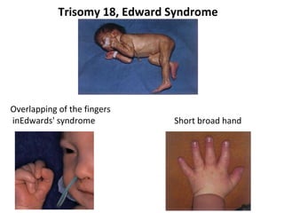
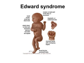
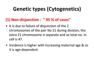
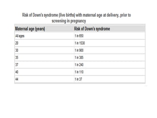
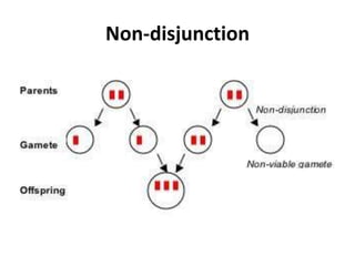
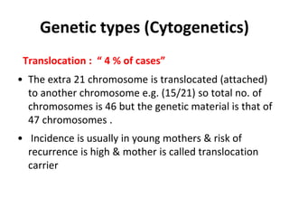
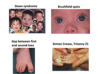
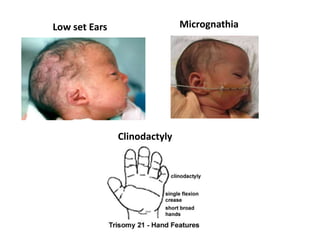
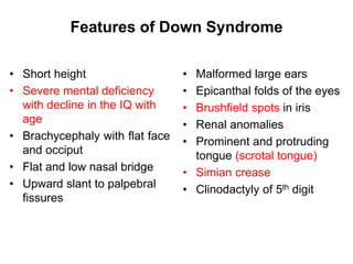
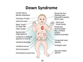
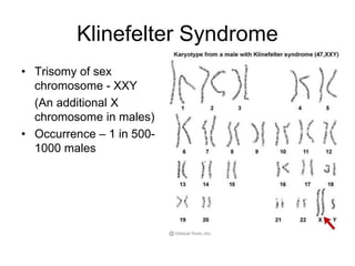
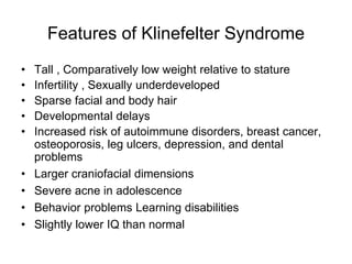
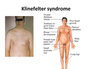
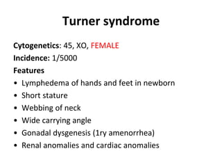
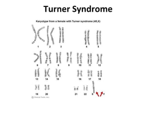
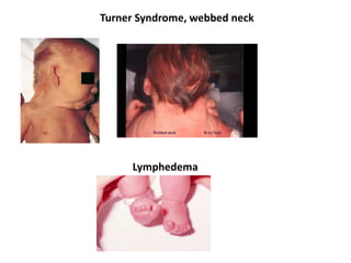
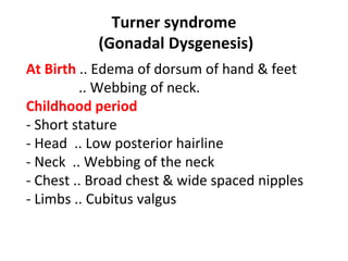
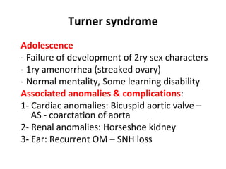
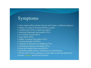
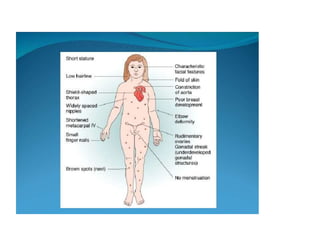
S.Y.B.Sc Semester III
Botany Paper II
Unit II :Cytogenetics
Chromosomal aberrations
By Mrs. Mandakini R Ingle
Department of Botany
Satish Pradhan Dnyanasadhana College, Thane (w)
Basic definitions
• Chromosomes, DNA and genes
• Chromatides, and centromere
• Arms of a chromosome (p and q)
• Karyotype
• Autosomes and sex chromosomes
• Genotype and phenotype
Human Chromosomes
• The chromosome carries the genetic
information.
• composed of deoxyribonucieic acid
(DNA) on framework of protein .
• Segments of DNA molecules
comprise the genes; the units of
heredity.
• During cell division,
the chromosome can
be seen to consist of
2 parallel strands; the
chromatids, held
together at one point,
the centromere.
Chromosomes
Human Chromosomes
Karyotype
• It is the set of chromosomes of an individual.
• It is the systematized arrangement of the
chromosomes of a single cell.
• In the human cell, there are 46 chromosomes or 23
pairs (diploid number); of these 23 pairs, 22 are
similar in both sexes and are called the autosomes.
The remaining pair is called sex chromosomes : XX in
the female cells and XY in the male cells .
• Chromosomes are arranged in groups A to G
according to their shape & size.
Karyotype of a normal female
Karyotype of a normal male
Chromosomal Abnormalities
• Chromosomal abnormalities are either numerical or
structural.
• They are a very common cause of early spontaneous
miscarriage.
• Usually, but not always, cause multiple congenital
anomalies and learning difficulties.
Chromosomal Aberrations (abnormalities)
• Structural Aberrations
o Deletion
o Duplication
o Inversion
o Translocation
• Numerical Aberrations (abnormalities)
o Polyploidy: Multiple of the haploid (> Diploid)
o Aneuploidy: Abnormal number
Structural abnormalities
1) Deletion : loss of a portion of a chromosome
2) Duplication : extra piece of a chromosome. .
3) Inversion : fragmentation of a chromosome
followed by reconstitution with a section inverted.
4) Translocation :
the transfer of a chromosome or a segment of it to a
non-homologous chromosome
Chromosomal Deletions
• a deletion results in a lost portion of a
chromosome,
• Deletion Causative Agents: Heat, radiation,
viruses, chemicals, errors in recombination. result
in partial monosomy,
• the organism is monosomic for the portion of the
chromosome that is deleted,
• as in monosomy, most segmental deletions are
deleterious.
Deletion
Recognizing Deletions
Intercalary
Terminal
Homologous Pairs?
Intercalary
Terminal
Hemizygous
Hemizygous: gene is present in a single dose.
Psuedodominance: hemizygous genes are expressed.
Cri-du-chat Syndrome
• Mental retardation
• Slow motor skill development
• Low birth weight and slow
growth
• Small head (microcephaly)
• Partial webbing of fingers or
toes
• Wide-set eyes
(hypertelorism)
• High-pitched cry
Chromosomal Duplication
...an event that
results in the
increase in the
number of copies
of a particular
chromosomal
region,
Duplication
Causes:
– duplications often result from unequal crossing
over,
– can occur via errors in replication during S-Phase.
Effects:
– results in gene redundancy,
– produces phenotypic variation,
– may provide an important source for genetic
variability during evolution.
Unequal Crossing Over
Produces both duplications and
deletions!
Duplication in Evolution
essential genes do not tolerate mutation,
duplications of essential genes, then subsequent
mutations, confers adaptive potential to the organism,
new gene family members are ‘recruited’ to perform new
functions.
Chromosomal Inversions
inversion: aberration in which a portion of
the chromosome is turned around 180o
.
Paracentric Inversion
...an inversion in which the centomere is
not included,
A B C
...a paracentric inversion does not
change arm length ratio.
B
A C
B A
Pericentric Inversion
...an inversion in which the centromere is
included,
A B C
A B
C
...a pericentric inversion results in a
change in chromosome arm length.
Inversion
Loop
no crossing over
Produces haploid gamete.
Paracentric
Produces gamete with inversion.
Paracentric
Produces a chromosome with two centromeres.
Nonviable gametes.
Paracentric
Paracentric Outcomes
1 Normal Gamete, 1 Inversion Gamete, No Crossover Classes
Recombination is not inhibited, but recombinant gametes are selected against.
Dicentric/Ascentric
…results only when the crossing over
occurs within the region of the paracentric
inversion,
Dicentric
...a chromosome having two centromeres;
Non-Viable (gametes)
Segregate
Acentric
…a chromosome having no centromeres,
…segregates to daughter cells randomly,
or is lost during cell division,
…deletions impart partial monosomy.
Pericentric
Recombination and Inversions
• Paracentric and Pericentric;
– 1 Normal Gamete,
– 1 Inverted Gamete,
– No Crossover Classes = No Recombination,
Inversions select against recombinant
gametes, thus preserves co-segregation of
specific alleles.
Inversions and Evolution
• Inversions ‘lock’ specific alleles
together,
– all offspring get the alleles from either a
wild-type, or inverted chromosome,
• If the ‘set of alleles’ is advantageous,
the set can be maintained in the
population.
Terminal Translocation
Reciprocal Translocation
Robertsonian Translocations
…the fusion of long arms of acrocentric
chromosomes,
Down Syndrome
• 95% of Down Syndrome individuals are a result
of Trisomy 21,
– the probability of having a second Down Syndrome
child is usually similar to the population at large,
• However, there is second cause of Down
Syndrome caused by a Robertsonian
translocations that is heritable.
Structural abnormalities
Reciprocal Translocation
Robertsonian Translocation
Reciprocal translocations
• An exchange of material between two different
chromosomes is called a reciprocal translocation.
When this exchange involves no loss or gain of
chromosomal material, the translocation is 'balanced'
and has no phenotypic effect.
• Balanced reciprocal translocations are relatively
common, occurring in 1 in 500 of the general
population.
Reciprocal translocations
•Finding a balanced translocation in one parent
indicates a recurrence risk for future pregnancies and
antenatal diagnosis by chorionic villus sampling or
amniocentesis should be offered as well as testing of
relatives.
Reciprocal Translocation
Robertsonian Translocation
Figure 8.4 Translocation Down's syndrome. There is a translocation between
chromosomes 21 and 14 inherited from a parent
Numerical Aberration
• Autosomal
- Trisomies: 1 ch extra (e.g. trisomy 21-13-18)
- Monosomies: 1 ch is missing
• Sex chromosome
- Klinefelter syndrome (47, XXY male)
- Turner syndrome (45, XO female)
Overview
I. Chromosomal Variations
A. Polyploidy
B. Aneuploidy
II. Syndromes resulting from Aneuploidy
A. Trisomy 13
B. Trisomy 18
C. Down Syndrome
D. Turner Syndrome
E. Klinefelter Syndrome
F. XYY Syndrome
Numerical abnormalities
Trisomy i.e. 47 chromosomes
- Trisomy 21 (the extrachromosome is No 21)
- Klinefelter syndrome ( 47, XXY male)
Monosomy i.e. 45 chromosomes
- Monosomy 21
- Turner syndrome (the missing chromosome is
X in female : 45, X or 45 XO )
Variations in Chromosomal
Number
• Euploidy – the usual number and sets of
chromosomes
• Polyploidy – the presence of three or more
complete sets of chromosomes
• Aneuploidy – the presence of additional
chromosomes or missing individual
chromosomes
Types of Polyploidy
• Triploidy – three sets of chromosomes
23 x 3 = 69
• Tetraploidy – four sets of chromosomes
23 x 4 = 92
Types of Aneuploidy
• Monosomy – one less chromosome
(23 x 2) – 1 = 45
• Trisomy – one additional chromosome
(23 x 2) + 1 = 47
Aneuploidy
• When aneuploidy occurs in humans,
syndromes can result. Examples include
the following:
1. Trisomy 13
2. Trisomy 18
3. Down Syndrome
3. Turner Syndrome
4. Klinefelter Syndrome
5. XYY Syndrome
Trisomy of Autosomes
• Trisomy 13 or D-trisomy (Patau syndrome)
• Trisomy 18 or E-trisomy (Edward syndrome)
• Trisomy 21 or G-trisomy (Down syndrome)
Trisomy 13 (Patau Syndrome)
• 1st described by Bartholin (1657) & redefined by
Patau (1960).
• Chromosomal complement: 47,XX,+13 (female) or
47,XY,+13 (male)
• Phenotype: Male or female
• Incidence: 1:12,000 (increases with the age of
mother)
Patau syndrome
Features of Patau Syndrome
• Mental deficiency
• Low birth weight
• Abnormal development
of frontal lobe
• Absence of corpus
callosum
• Hypoplasia of
cerebellum
• Sloping forehead
• Scalp defects
• Malformed ears
• Congenital heart
defects
• Renal tract anomalies
• Microphthalmia
• Bilateral cleft lip/palate
• Polydactyly with
rudimentary digits
• Rocker-bottom heel
Patau syndrome
Trisomy 18 (Edward Syndrome)
• Chromosomal complement: 47,XX,+18
(female) or
47,XY,+18
(male)
• Phenotype: Male or female
• Incidence: 1:8000
Features of Edward Syndrome
• Mental deficiency
• Growth retardation
• Prominent occiput with
elongated head
• Webbing of the neck
• Short sternum
• Micrognathia
• Low-set malformed ears
• Ventricular septal
defects
• Renal anomalies
• Clenched fists with
overlapping of fingers
• Hypoplastic nails
Trisomy 18, Edward Syndrome
Overlapping of the fingers
inEdwards' syndrome Short broad hand
Edward syndrome
Pathogenic mechanisms
Disruption
Involves destruction of a fetal part which
initially formed normally; e.g. amniotic
membrane rupture may lead to amniotic bands
which may cause limb reduction defects.
Dysplasia
Refers to abnormal cellular organization or
function of specific tissue types, e.g. skeletal
dysplasias and dysplastic kidney disease.
Down Syndrome
Cardiofaciocutaneous Syndrome
Antimongoloid slant
• Chromosomal Variation –
Trisomy 21 (three copies of
chromosome 21)
• Occurrence – 1 in 800-1000
Down Syndrome (Mongolism) Trisomy 21
Incidence :
• The most common chromosomal aberration
• Incidence 1/700 live birth & 10 % of M.R.
Definition : It is trisomy 21 i.e. the cell contain an extra
chromosome, number 21 i.e. the cell contains three
21 chromosomes instead of two .
Genetic types (Cytogenetics)
(1) Non-disjunction : “ 95 % of cases”
• It is due to failure of disjunction of the 2
chromosomes of the pair No 21 during division, the
extra 21 chromosome is separate and so total no. in
cell is 47.
• Incidence is higher with increasing maternal age & so
it is age-dependent
Non-disjunction
Genetic types (Cytogenetics)
Translocation : “ 4 % of cases”
• The extra 21 chromosome is translocated (attached)
to another chromosome e.g. (15/21) so total no. of
chromosomes is 46 but the genetic material is that of
47 chromosomes .
• Incidence is usually in young mothers & risk of
recurrence is high & mother is called translocation
carrier
Down syndrome
Translocation carrier
• One parent contains a 14/21 translocation
and has only 45 chromosomes, and is a
phenotypically normal carrier.
• 1/4 of the individual's gametes will have
almost 2 copies of chromosome 21.
• The resulting zygote has 46 chromosomes, but
almost 3 copies of chromosome 21, and
exhibits Down syndrome.
Translocation carrier
Recurrence Risk ??
Genetic types (Cytogenetics)
Mosaicism : “ 1 % of cases”
• Some cells are normal (46 chromosomes) & others
are trisomic (47 chromosomes)
Clinical feature are less evident & M.R. is mild .
Characteristic physical features
(in the absence of mental retardation, they should
NEVER be considered diagnostic)
Hands
• Simian crease (transverse palmar crease)
• Clinodactly (incurved little finger)
• Short broad hand
Feet
• Big space between the first and second toes
Clinical Features
Down syndrome Brushfield spots
Simian Crease, Trisomy 21
Gap between first
and second toes
Low set Ears Micrognathia
Clinodactyly
Features of Down Syndrome
• Short height
• Severe mental deficiency
with decline in the IQ with
age
• Brachycephaly with flat face
and occiput
• Flat and low nasal bridge
• Upward slant to palpebral
fissures
• Malformed large ears
• Epicanthal folds of the eyes
• Brushfield spots in iris
• Renal anomalies
• Prominent and protruding
tongue (scrotal tongue)
• Simian crease
• Clinodactyly of 5th digit
Down Syndrome
Klinefelter Syndrome
• Trisomy of sex
chromosome - XXY
(An additional X
chromosome in males)
• Occurrence – 1 in 500-
1000 males
Features of Klinefelter Syndrome
• Tall , Comparatively low weight relative to stature
• Infertility , Sexually underdeveloped
• Sparse facial and body hair
• Developmental delays
• Increased risk of autoimmune disorders, breast cancer,
osteoporosis, leg ulcers, depression, and dental
problems
• Larger craniofacial dimensions
• Severe acne in adolescence
• Behavior problems Learning disabilities
• Slightly lower IQ than normal
Klinefelter syndrome
Turner syndrome
Cytogenetics: 45, XO, FEMALE
Incidence: 1/5000
Features
• Lymphedema of hands and feet in newborn
• Short stature
• Webbing of neck
• Wide carrying angle
• Gonadal dysgenesis (1ry amenorrhea)
• Renal anomalies and cardiac anomalies
Turner Syndrome
Turner Syndrome, webbed neck
Lymphedema
Turner syndrome
(Gonadal Dysgenesis)
At Birth .. Edema of dorsum of hand & feet
.. Webbing of neck.
Childhood period
- Short stature
- Head .. Low posterior hairline
- Neck .. Webbing of the neck
- Chest .. Broad chest & wide spaced nipples
- Limbs .. Cubitus valgus
Turner syndrome
Adolescence
- Failure of development of 2ry sex characters
- 1ry amenorrhea (streaked ovary)
- Normal mentality, Some learning disability
Associated anomalies & complications:
1- Cardiac anomalies: Bicuspid aortic valve –
AS - coarctation of aorta
2- Renal anomalies: Horseshoe kidney
3- Ear: Recurrent OM – SNH loss
Thank you…..

chromosomal aberrations.ppt

  • 1.
    S.Y.B.Sc Semester III BotanyPaper II Unit II :Cytogenetics Chromosomal aberrations By Mrs. Mandakini R Ingle Department of Botany Satish Pradhan Dnyanasadhana College, Thane (w)
  • 2.
    Basic definitions • Chromosomes,DNA and genes • Chromatides, and centromere • Arms of a chromosome (p and q) • Karyotype • Autosomes and sex chromosomes • Genotype and phenotype
  • 3.
    Human Chromosomes • Thechromosome carries the genetic information. • composed of deoxyribonucieic acid (DNA) on framework of protein . • Segments of DNA molecules comprise the genes; the units of heredity.
  • 4.
    • During celldivision, the chromosome can be seen to consist of 2 parallel strands; the chromatids, held together at one point, the centromere. Chromosomes
  • 5.
  • 6.
    Karyotype • It isthe set of chromosomes of an individual. • It is the systematized arrangement of the chromosomes of a single cell. • In the human cell, there are 46 chromosomes or 23 pairs (diploid number); of these 23 pairs, 22 are similar in both sexes and are called the autosomes. The remaining pair is called sex chromosomes : XX in the female cells and XY in the male cells . • Chromosomes are arranged in groups A to G according to their shape & size.
  • 7.
    Karyotype of anormal female
  • 8.
    Karyotype of anormal male
  • 9.
  • 10.
    Chromosomal Abnormalities • Chromosomalabnormalities are either numerical or structural. • They are a very common cause of early spontaneous miscarriage. • Usually, but not always, cause multiple congenital anomalies and learning difficulties.
  • 11.
    Chromosomal Aberrations (abnormalities) •Structural Aberrations o Deletion o Duplication o Inversion o Translocation • Numerical Aberrations (abnormalities) o Polyploidy: Multiple of the haploid (> Diploid) o Aneuploidy: Abnormal number
  • 12.
    Structural abnormalities 1) Deletion: loss of a portion of a chromosome 2) Duplication : extra piece of a chromosome. . 3) Inversion : fragmentation of a chromosome followed by reconstitution with a section inverted. 4) Translocation : the transfer of a chromosome or a segment of it to a non-homologous chromosome
  • 13.
    Chromosomal Deletions • adeletion results in a lost portion of a chromosome, • Deletion Causative Agents: Heat, radiation, viruses, chemicals, errors in recombination. result in partial monosomy, • the organism is monosomic for the portion of the chromosome that is deleted, • as in monosomy, most segmental deletions are deleterious.
  • 14.
  • 15.
  • 16.
    Homologous Pairs? Intercalary Terminal Hemizygous Hemizygous: geneis present in a single dose. Psuedodominance: hemizygous genes are expressed.
  • 17.
    Cri-du-chat Syndrome • Mentalretardation • Slow motor skill development • Low birth weight and slow growth • Small head (microcephaly) • Partial webbing of fingers or toes • Wide-set eyes (hypertelorism) • High-pitched cry
  • 18.
    Chromosomal Duplication ...an eventthat results in the increase in the number of copies of a particular chromosomal region,
  • 19.
    Duplication Causes: – duplications oftenresult from unequal crossing over, – can occur via errors in replication during S-Phase. Effects: – results in gene redundancy, – produces phenotypic variation, – may provide an important source for genetic variability during evolution.
  • 20.
    Unequal Crossing Over Producesboth duplications and deletions!
  • 23.
    Duplication in Evolution essentialgenes do not tolerate mutation, duplications of essential genes, then subsequent mutations, confers adaptive potential to the organism, new gene family members are ‘recruited’ to perform new functions.
  • 24.
    Chromosomal Inversions inversion: aberrationin which a portion of the chromosome is turned around 180o .
  • 25.
    Paracentric Inversion ...an inversionin which the centomere is not included, A B C ...a paracentric inversion does not change arm length ratio. B A C B A
  • 27.
    Pericentric Inversion ...an inversionin which the centromere is included, A B C A B C ...a pericentric inversion results in a change in chromosome arm length.
  • 29.
  • 30.
  • 31.
    Produces gamete withinversion. Paracentric
  • 32.
    Produces a chromosomewith two centromeres. Nonviable gametes. Paracentric
  • 33.
    Paracentric Outcomes 1 NormalGamete, 1 Inversion Gamete, No Crossover Classes Recombination is not inhibited, but recombinant gametes are selected against.
  • 34.
    Dicentric/Ascentric …results only whenthe crossing over occurs within the region of the paracentric inversion,
  • 35.
  • 36.
  • 37.
    Acentric …a chromosome havingno centromeres, …segregates to daughter cells randomly, or is lost during cell division, …deletions impart partial monosomy.
  • 38.
  • 39.
    Recombination and Inversions •Paracentric and Pericentric; – 1 Normal Gamete, – 1 Inverted Gamete, – No Crossover Classes = No Recombination, Inversions select against recombinant gametes, thus preserves co-segregation of specific alleles.
  • 40.
    Inversions and Evolution •Inversions ‘lock’ specific alleles together, – all offspring get the alleles from either a wild-type, or inverted chromosome, • If the ‘set of alleles’ is advantageous, the set can be maintained in the population.
  • 41.
  • 42.
  • 43.
    Robertsonian Translocations …the fusionof long arms of acrocentric chromosomes,
  • 44.
    Down Syndrome • 95%of Down Syndrome individuals are a result of Trisomy 21, – the probability of having a second Down Syndrome child is usually similar to the population at large, • However, there is second cause of Down Syndrome caused by a Robertsonian translocations that is heritable.
  • 45.
  • 46.
    Reciprocal translocations • Anexchange of material between two different chromosomes is called a reciprocal translocation. When this exchange involves no loss or gain of chromosomal material, the translocation is 'balanced' and has no phenotypic effect. • Balanced reciprocal translocations are relatively common, occurring in 1 in 500 of the general population.
  • 47.
    Reciprocal translocations •Finding abalanced translocation in one parent indicates a recurrence risk for future pregnancies and antenatal diagnosis by chorionic villus sampling or amniocentesis should be offered as well as testing of relatives.
  • 48.
  • 49.
  • 50.
  • 51.
    Figure 8.4 TranslocationDown's syndrome. There is a translocation between chromosomes 21 and 14 inherited from a parent
  • 52.
    Numerical Aberration • Autosomal -Trisomies: 1 ch extra (e.g. trisomy 21-13-18) - Monosomies: 1 ch is missing • Sex chromosome - Klinefelter syndrome (47, XXY male) - Turner syndrome (45, XO female)
  • 53.
    Overview I. Chromosomal Variations A.Polyploidy B. Aneuploidy II. Syndromes resulting from Aneuploidy A. Trisomy 13 B. Trisomy 18 C. Down Syndrome D. Turner Syndrome E. Klinefelter Syndrome F. XYY Syndrome
  • 54.
    Numerical abnormalities Trisomy i.e.47 chromosomes - Trisomy 21 (the extrachromosome is No 21) - Klinefelter syndrome ( 47, XXY male) Monosomy i.e. 45 chromosomes - Monosomy 21 - Turner syndrome (the missing chromosome is X in female : 45, X or 45 XO )
  • 55.
    Variations in Chromosomal Number •Euploidy – the usual number and sets of chromosomes • Polyploidy – the presence of three or more complete sets of chromosomes • Aneuploidy – the presence of additional chromosomes or missing individual chromosomes
  • 56.
    Types of Polyploidy •Triploidy – three sets of chromosomes 23 x 3 = 69 • Tetraploidy – four sets of chromosomes 23 x 4 = 92
  • 57.
    Types of Aneuploidy •Monosomy – one less chromosome (23 x 2) – 1 = 45 • Trisomy – one additional chromosome (23 x 2) + 1 = 47
  • 58.
    Aneuploidy • When aneuploidyoccurs in humans, syndromes can result. Examples include the following: 1. Trisomy 13 2. Trisomy 18 3. Down Syndrome 3. Turner Syndrome 4. Klinefelter Syndrome 5. XYY Syndrome
  • 59.
    Trisomy of Autosomes •Trisomy 13 or D-trisomy (Patau syndrome) • Trisomy 18 or E-trisomy (Edward syndrome) • Trisomy 21 or G-trisomy (Down syndrome)
  • 60.
    Trisomy 13 (PatauSyndrome) • 1st described by Bartholin (1657) & redefined by Patau (1960). • Chromosomal complement: 47,XX,+13 (female) or 47,XY,+13 (male) • Phenotype: Male or female • Incidence: 1:12,000 (increases with the age of mother)
  • 61.
  • 62.
    Features of PatauSyndrome • Mental deficiency • Low birth weight • Abnormal development of frontal lobe • Absence of corpus callosum • Hypoplasia of cerebellum • Sloping forehead • Scalp defects • Malformed ears • Congenital heart defects • Renal tract anomalies • Microphthalmia • Bilateral cleft lip/palate • Polydactyly with rudimentary digits • Rocker-bottom heel
  • 63.
  • 64.
    Trisomy 18 (EdwardSyndrome) • Chromosomal complement: 47,XX,+18 (female) or 47,XY,+18 (male) • Phenotype: Male or female • Incidence: 1:8000
  • 66.
    Features of EdwardSyndrome • Mental deficiency • Growth retardation • Prominent occiput with elongated head • Webbing of the neck • Short sternum • Micrognathia • Low-set malformed ears • Ventricular septal defects • Renal anomalies • Clenched fists with overlapping of fingers • Hypoplastic nails
  • 67.
    Trisomy 18, EdwardSyndrome Overlapping of the fingers inEdwards' syndrome Short broad hand
  • 68.
  • 69.
    Pathogenic mechanisms Disruption Involves destructionof a fetal part which initially formed normally; e.g. amniotic membrane rupture may lead to amniotic bands which may cause limb reduction defects. Dysplasia Refers to abnormal cellular organization or function of specific tissue types, e.g. skeletal dysplasias and dysplastic kidney disease.
  • 70.
    Down Syndrome Cardiofaciocutaneous Syndrome Antimongoloidslant • Chromosomal Variation – Trisomy 21 (three copies of chromosome 21) • Occurrence – 1 in 800-1000
  • 71.
    Down Syndrome (Mongolism)Trisomy 21 Incidence : • The most common chromosomal aberration • Incidence 1/700 live birth & 10 % of M.R. Definition : It is trisomy 21 i.e. the cell contain an extra chromosome, number 21 i.e. the cell contains three 21 chromosomes instead of two .
  • 73.
    Genetic types (Cytogenetics) (1)Non-disjunction : “ 95 % of cases” • It is due to failure of disjunction of the 2 chromosomes of the pair No 21 during division, the extra 21 chromosome is separate and so total no. in cell is 47. • Incidence is higher with increasing maternal age & so it is age-dependent
  • 75.
  • 76.
    Genetic types (Cytogenetics) Translocation: “ 4 % of cases” • The extra 21 chromosome is translocated (attached) to another chromosome e.g. (15/21) so total no. of chromosomes is 46 but the genetic material is that of 47 chromosomes . • Incidence is usually in young mothers & risk of recurrence is high & mother is called translocation carrier
  • 77.
    Down syndrome Translocation carrier •One parent contains a 14/21 translocation and has only 45 chromosomes, and is a phenotypically normal carrier. • 1/4 of the individual's gametes will have almost 2 copies of chromosome 21. • The resulting zygote has 46 chromosomes, but almost 3 copies of chromosome 21, and exhibits Down syndrome.
  • 78.
  • 79.
    Genetic types (Cytogenetics) Mosaicism: “ 1 % of cases” • Some cells are normal (46 chromosomes) & others are trisomic (47 chromosomes) Clinical feature are less evident & M.R. is mild . Characteristic physical features (in the absence of mental retardation, they should NEVER be considered diagnostic)
  • 80.
    Hands • Simian crease(transverse palmar crease) • Clinodactly (incurved little finger) • Short broad hand Feet • Big space between the first and second toes Clinical Features
  • 81.
    Down syndrome Brushfieldspots Simian Crease, Trisomy 21 Gap between first and second toes
  • 82.
    Low set EarsMicrognathia Clinodactyly
  • 83.
    Features of DownSyndrome • Short height • Severe mental deficiency with decline in the IQ with age • Brachycephaly with flat face and occiput • Flat and low nasal bridge • Upward slant to palpebral fissures • Malformed large ears • Epicanthal folds of the eyes • Brushfield spots in iris • Renal anomalies • Prominent and protruding tongue (scrotal tongue) • Simian crease • Clinodactyly of 5th digit
  • 84.
  • 85.
    Klinefelter Syndrome • Trisomyof sex chromosome - XXY (An additional X chromosome in males) • Occurrence – 1 in 500- 1000 males
  • 86.
    Features of KlinefelterSyndrome • Tall , Comparatively low weight relative to stature • Infertility , Sexually underdeveloped • Sparse facial and body hair • Developmental delays • Increased risk of autoimmune disorders, breast cancer, osteoporosis, leg ulcers, depression, and dental problems • Larger craniofacial dimensions • Severe acne in adolescence • Behavior problems Learning disabilities • Slightly lower IQ than normal
  • 87.
  • 88.
    Turner syndrome Cytogenetics: 45,XO, FEMALE Incidence: 1/5000 Features • Lymphedema of hands and feet in newborn • Short stature • Webbing of neck • Wide carrying angle • Gonadal dysgenesis (1ry amenorrhea) • Renal anomalies and cardiac anomalies
  • 89.
  • 90.
    Turner Syndrome, webbedneck Lymphedema
  • 91.
    Turner syndrome (Gonadal Dysgenesis) AtBirth .. Edema of dorsum of hand & feet .. Webbing of neck. Childhood period - Short stature - Head .. Low posterior hairline - Neck .. Webbing of the neck - Chest .. Broad chest & wide spaced nipples - Limbs .. Cubitus valgus
  • 92.
    Turner syndrome Adolescence - Failureof development of 2ry sex characters - 1ry amenorrhea (streaked ovary) - Normal mentality, Some learning disability Associated anomalies & complications: 1- Cardiac anomalies: Bicuspid aortic valve – AS - coarctation of aorta 2- Renal anomalies: Horseshoe kidney 3- Ear: Recurrent OM – SNH loss
  • 95.