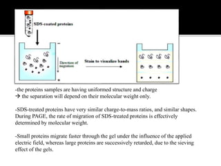
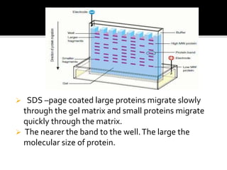
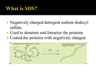
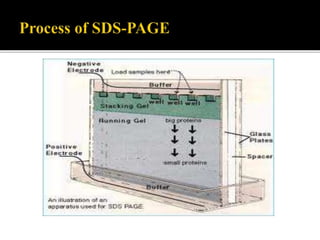
SDS- Polyacrylamide Gel Electrophoresis
 Standard test that used to determine the charged
molecules, mainly proteins and nucleic acids.
 widely used in biochemistry ,forensics, genetics and
molecular biology to separate and identify proteins
according to their molecular weight.
 This method separates proteins based primarily on
their molecular weights.
 Laemmli system of SDS-PAGE was first introduced
in 1970s.
 Separates protein in an electric field.
 Migrate through a liquid or semisolid medium
when subjected to an electric field from anode
to cathode terminal.
 Molecules flow at different rates depend on
the molecular size of proteins.
-the proteins samples are having uniformed structure and charge
 the separation will depend on their molecular weight only.
-SDS-treated proteins have very similar charge-to-mass ratios, and similar shapes.
During PAGE, the rate of migration of SDS-treated proteins is effectively
determined by molecular weight.
-Small proteins migrate faster through the gel under the influence of the applied
electric field, whereas large proteins are successively retarded, due to the sieving
effect of the gels.
 SDS –page coated large proteins migrate slowly
through the gel matrix and small proteins migrate
quickly through the matrix.
 The nearer the band to the well.The large the
molecular size of protein.
 Negatively charged detergent sodium dodecyl
sulfate.
 Used to denature and linearize the proteins
 Coated the proteins with negatively charged.
 SDS-PAGE is differentiated into two systems.
*continuous sds-page
*discontinuous sds-page.
• Polyacrylamide is used to form a gel, a matrix of a
pores which allow the molecules migrate at
different rates.
 The size of pores is determined by the
concentration of acrylamide.
 The higher the concentration, the smaller the
size of pores.
 Discontinuos sds-page consist of two
different gels.
 *stacking gel -4%of acrylamide
 *separating gel-range from 5-15% of
acrylamide.
Preparation of gel
 Clean the plates and combs.
 Set up the plates on the rack.
 Pour the separating gel.
 Pour the stacking gel.
 Gel storage.
 Visualizes the band under UV light.
 Types of stains;
1. Coomassie Blue;
* Coomassie Brilliant Blue staining The Coomassie dyes R-250 and G-250 bind to
proteins stoichiometrically through their sulfonic acid groups.
* . The interactions between dye and protein are Van der Waals and ionic. The sulfonic
acid groups interact with positive amine groups. Therefore coomassie dye binds to
wide range of proteins.
* Limited to ~100ng of protein.
1. Silver stain;
*most sensitive test
*detection limit 0.1-1.0ng of protein
 Determine purity of protein samples
 Determine molecular weight of protein
 Identifying disulfide bonds between protein
 Quantifying proteins
 Blotting applications
 SDS-page is a technique that used to separate
proteins according to their molecular size
through the gel.
 Proteins are unfolded and migrate from cathode
to anode terminal at different rates.
 Molecular weight is determined by compare the
result with a standard curve of relative motility of
standard proteins.
• P.K .Gupta molecular biology second
edition.
• https://www.google.co.in/search?q=sds+page
+principle.
Thank you

SDS- Polyacrylamide Gel Electrophoresis

  • 1.
    SDS- Polyacrylamide GelElectrophoresis
  • 2.
     Standard testthat used to determine the charged molecules, mainly proteins and nucleic acids.  widely used in biochemistry ,forensics, genetics and molecular biology to separate and identify proteins according to their molecular weight.  This method separates proteins based primarily on their molecular weights.  Laemmli system of SDS-PAGE was first introduced in 1970s.
  • 3.
     Separates proteinin an electric field.  Migrate through a liquid or semisolid medium when subjected to an electric field from anode to cathode terminal.  Molecules flow at different rates depend on the molecular size of proteins.
  • 4.
    -the proteins samplesare having uniformed structure and charge  the separation will depend on their molecular weight only. -SDS-treated proteins have very similar charge-to-mass ratios, and similar shapes. During PAGE, the rate of migration of SDS-treated proteins is effectively determined by molecular weight. -Small proteins migrate faster through the gel under the influence of the applied electric field, whereas large proteins are successively retarded, due to the sieving effect of the gels.
  • 5.
     SDS –pagecoated large proteins migrate slowly through the gel matrix and small proteins migrate quickly through the matrix.  The nearer the band to the well.The large the molecular size of protein.
  • 6.
     Negatively chargeddetergent sodium dodecyl sulfate.  Used to denature and linearize the proteins  Coated the proteins with negatively charged.
  • 7.
     SDS-PAGE isdifferentiated into two systems. *continuous sds-page *discontinuous sds-page. • Polyacrylamide is used to form a gel, a matrix of a pores which allow the molecules migrate at different rates.
  • 8.
     The sizeof pores is determined by the concentration of acrylamide.  The higher the concentration, the smaller the size of pores.  Discontinuos sds-page consist of two different gels.  *stacking gel -4%of acrylamide  *separating gel-range from 5-15% of acrylamide.
  • 9.
    Preparation of gel Clean the plates and combs.  Set up the plates on the rack.  Pour the separating gel.  Pour the stacking gel.  Gel storage.
  • 11.
     Visualizes theband under UV light.  Types of stains; 1. Coomassie Blue; * Coomassie Brilliant Blue staining The Coomassie dyes R-250 and G-250 bind to proteins stoichiometrically through their sulfonic acid groups. * . The interactions between dye and protein are Van der Waals and ionic. The sulfonic acid groups interact with positive amine groups. Therefore coomassie dye binds to wide range of proteins. * Limited to ~100ng of protein. 1. Silver stain; *most sensitive test *detection limit 0.1-1.0ng of protein
  • 13.
     Determine purityof protein samples  Determine molecular weight of protein  Identifying disulfide bonds between protein  Quantifying proteins  Blotting applications
  • 14.
     SDS-page isa technique that used to separate proteins according to their molecular size through the gel.  Proteins are unfolded and migrate from cathode to anode terminal at different rates.  Molecular weight is determined by compare the result with a standard curve of relative motility of standard proteins.
  • 15.
    • P.K .Guptamolecular biology second edition. • https://www.google.co.in/search?q=sds+page +principle.
  • 16.