This document provides information about a case study presentation on chronic obstructive pulmonary disease (COPD). It includes sections on the symptoms of emphysema and chronic bronchitis, an overview of the patient's history and exam findings, and assessments from a nutrition consult. COPD is characterized by inflammation and narrowing of the airways and destruction of lung tissue, making it difficult to breathe over time. Smoking is the leading cause of COPD.
COPD exacerbation case presentation and disease overview farah al souheil
management of a simulated case scenario: patient presenting with COPD exacerbation: what's the best next step? summary of the guideline is then described
oxygen is a medication. oxygen therapy must be known to all health professionals for optimum management of patient and optimum use of resourses. even more oxygen can cause oxygen toxicity and can harm the patient in many ways. There are various methods for giving oxygen,varieties of face masks, cylinders. also there is criteria when to give oxygen ,how to give oxygen,what are the benefits and mechanism of oxygen therapy.
Critically ill patients requiring noninvasive or invasive ventilation often present to emergency departments, and due to hospital crowding and constrained critical care services, may remain in the emergency department for a prolonged duration. Compared with their intensive care unit counterparts, emergency department clinicians may have variable exposure to management of this patient population and may lack knowledge and expertise, particularly in their
longitudinal management beyond initial stabilization. This
review has discussed several key aspects of management
of noninvasive and invasive ventilation, with a particular emphasis on initiation and ongoing monitoring priorities,
and focused on maintaining patient safety and improving
patient outcomes.
Case presentation on bronchial asthma, respiratory disorder, Introduction-Definition-History collection-Physical examination-lab diagnosis- nursing diagnosis of asthma, treatment of asthma
Case presentation on bronchial asthma, respiratory disorder, Introduction-Definition-History collection-Physical examination-lab diagnosis- nursing diagnosis of asthma, treatment of asthma
Thank you for selecting our 𝐂𝐎𝐏𝐃 PPT
This medical PowerPoint template about 𝐂𝐎𝐏𝐃
You can download our template by visiting our website:
https://www.rxslides.com/product/copd-powerpoint-template
copy and paste this URL into the browser and download the full editable template.
This 𝐂𝐎𝐏𝐃 animated template is designed by RxSlides, a medical professional team covering the following topics about 𝐂𝐎𝐏𝐃
𝐓𝐡𝐞 𝐭𝐞𝐦𝐩𝐥𝐚𝐭𝐞 𝐜𝐨𝐯𝐞𝐫𝐬 𝐭𝐡𝐞 𝐟𝐨𝐥𝐥𝐨𝐰𝐢𝐧𝐠 𝐭𝐨𝐩𝐢𝐜𝐬
𝐃𝐞𝐟𝐢𝐧𝐢𝐭𝐢𝐨𝐧
Chronic obstructive pulmonary disease (𝐂𝐎𝐏𝐃) is a group of lung diseases that cause airflow blockage and breathing-related problems.
𝐩𝐫𝐞𝐯𝐚𝐥𝐞𝐧𝐜𝐞
COPD is more prevalent in developing countries, but it is also a growing problem in developed countries. In the United States, COPD is the third leading cause of death.
Forms of 𝐂𝐎𝐏𝐃
• Chronic bronchitis
• Emphysema
𝐏𝐚𝐭𝐡𝐨𝐩𝐡𝐲𝐬𝐢𝐨𝐥𝐨𝐠𝐲
In normal lungs, air flows freely in and out of the bronchi and alveoli. However, in people with COPD, the airflow is blocked. This can be caused by inflammation of the airways, mucus production, or damage to the air sacs.
𝐑𝐢𝐬𝐤 𝐅𝐚𝐜𝐭𝐨𝐫𝐬
There are several risk factors for COPD, including smoking, air pollution, and genetics. Smoking is the most common risk factor for COPD. Smoking is responsible for about 80% of COPD cases. Air pollution, particularly indoor air pollution from cooking and burning fuels, can also increase the risk of COPD. Genetics can also play a role in COPD. People with a family history of COPD are more likely to develop the disease.
𝐜𝐚𝐮𝐬𝐞𝐬
The main causes of COPD are smoking and air pollution. Smoking damages the lungs and makes it difficult to breathe
𝐒𝐭𝐚𝐠𝐞𝐬
• Stage 1
• Stage 2
• Stage 3
.
𝐒𝐲𝐦𝐩𝐭𝐨𝐦𝐬
The most common symptoms of COPD are:
• Shortness of breath
• Cough
• Wheezing
• Chest tightness
• Fatigue
𝐂𝐨𝐦𝐩𝐥𝐢𝐜𝐚𝐭𝐢𝐨𝐧𝐬
COPD can lead to several complications, including:
• Anemia
• Right-sided heart failure
• Muscle weakness
• Lung infections
• Bone thinning
• Collapsed lungs
𝐃𝐢𝐚𝐠𝐧𝐨𝐬𝐭𝐢𝐜 𝐦𝐞𝐭𝐡𝐨𝐝𝐬
COPD is diagnosed with a spirometry test, which measures how much air a person can exhale.
𝐓𝐫𝐞𝐚𝐭𝐦𝐞𝐧𝐭 𝐎𝐩𝐭𝐢𝐨𝐧𝐬
There is no cure for COPD, but there are treatments that can help manage the symptoms and slow the progression of the disease. Treatment options include:
• Bronchodilators
• Antibiotics
• Supplemental oxygen
• Vaccination
𝐏𝐫𝐞𝐯𝐞𝐧𝐭𝐢𝐯𝐞 𝐌𝐞𝐭𝐡𝐨𝐝𝐬
The best way to prevent COPD is to avoid smoking and air pollution. Several lifestyle changes can help reduce the risk of COPD, such as eating a healthy diet and exercising regularly.
Visit our site for more animated templates
𝗵𝘁𝘁𝗽𝘀://𝘄𝘄𝘄.𝗿𝘅𝘀𝗹𝗶𝗱𝗲𝘀.𝗰𝗼𝗺
RxSlides PowerPoint icons and illustrations related to 𝐂𝐎𝐏𝐃 will help you customize the content of this editable presentation according to your content and audience interest.
COPD (chronic obstructive pulmonary disease) is a group of lung diseases that make it hard to breathe and get worse over time.
Normally, the airways and air sacs in the lungs are elastic or stretchy.
When we breathe in, the airways bring air to the air sacs.
The air sacs fill up with air, like a small balloons.
When we breathe out, the air sacs deflate, and the air goes out.
In COPD, less air flows in and out of airways because of one or more problems:
-The airways and air sacs in the lungs become less elastic
-The walls between many of the air sacs are destroyed
-The walls of the airways become inflamed
-The airways make more mucus than usual and can become clogged
Biology For Engineers Module 3 / HUMAN ORGAN SYSTEMS AND BIO-DESIGNS - 2 Dr. Pavan Kundur
An organ system is a group of organs that work together in the body to perform a complex function, such as pumping blood or processing and utilizing nutrients.
Respiratory system
This presentation covers three section: Anatomy, physiology and the pathology. Anatomy covers the structure and the specialized tissues. Physiology section covers the process of breathing(inspiration and expiration) and the process of gas exchange(alveoli and diffusion).The last section pathology covers Asthma, Emphysema, Bronchitis and Cystic fibrosis and their symptoms and treatment in detail.
How to Create Map Views in the Odoo 17 ERPCeline George
The map views are useful for providing a geographical representation of data. They allow users to visualize and analyze the data in a more intuitive manner.
Synthetic Fiber Construction in lab .pptxPavel ( NSTU)
Synthetic fiber production is a fascinating and complex field that blends chemistry, engineering, and environmental science. By understanding these aspects, students can gain a comprehensive view of synthetic fiber production, its impact on society and the environment, and the potential for future innovations. Synthetic fibers play a crucial role in modern society, impacting various aspects of daily life, industry, and the environment. ynthetic fibers are integral to modern life, offering a range of benefits from cost-effectiveness and versatility to innovative applications and performance characteristics. While they pose environmental challenges, ongoing research and development aim to create more sustainable and eco-friendly alternatives. Understanding the importance of synthetic fibers helps in appreciating their role in the economy, industry, and daily life, while also emphasizing the need for sustainable practices and innovation.
2024.06.01 Introducing a competency framework for languag learning materials ...Sandy Millin
http://sandymillin.wordpress.com/iateflwebinar2024
Published classroom materials form the basis of syllabuses, drive teacher professional development, and have a potentially huge influence on learners, teachers and education systems. All teachers also create their own materials, whether a few sentences on a blackboard, a highly-structured fully-realised online course, or anything in between. Despite this, the knowledge and skills needed to create effective language learning materials are rarely part of teacher training, and are mostly learnt by trial and error.
Knowledge and skills frameworks, generally called competency frameworks, for ELT teachers, trainers and managers have existed for a few years now. However, until I created one for my MA dissertation, there wasn’t one drawing together what we need to know and do to be able to effectively produce language learning materials.
This webinar will introduce you to my framework, highlighting the key competencies I identified from my research. It will also show how anybody involved in language teaching (any language, not just English!), teacher training, managing schools or developing language learning materials can benefit from using the framework.
How to Make a Field invisible in Odoo 17Celine George
It is possible to hide or invisible some fields in odoo. Commonly using “invisible” attribute in the field definition to invisible the fields. This slide will show how to make a field invisible in odoo 17.
We all have good and bad thoughts from time to time and situation to situation. We are bombarded daily with spiraling thoughts(both negative and positive) creating all-consuming feel , making us difficult to manage with associated suffering. Good thoughts are like our Mob Signal (Positive thought) amidst noise(negative thought) in the atmosphere. Negative thoughts like noise outweigh positive thoughts. These thoughts often create unwanted confusion, trouble, stress and frustration in our mind as well as chaos in our physical world. Negative thoughts are also known as “distorted thinking”.
The French Revolution, which began in 1789, was a period of radical social and political upheaval in France. It marked the decline of absolute monarchies, the rise of secular and democratic republics, and the eventual rise of Napoleon Bonaparte. This revolutionary period is crucial in understanding the transition from feudalism to modernity in Europe.
For more information, visit-www.vavaclasses.com
The Roman Empire A Historical Colossus.pdfkaushalkr1407
The Roman Empire, a vast and enduring power, stands as one of history's most remarkable civilizations, leaving an indelible imprint on the world. It emerged from the Roman Republic, transitioning into an imperial powerhouse under the leadership of Augustus Caesar in 27 BCE. This transformation marked the beginning of an era defined by unprecedented territorial expansion, architectural marvels, and profound cultural influence.
The empire's roots lie in the city of Rome, founded, according to legend, by Romulus in 753 BCE. Over centuries, Rome evolved from a small settlement to a formidable republic, characterized by a complex political system with elected officials and checks on power. However, internal strife, class conflicts, and military ambitions paved the way for the end of the Republic. Julius Caesar’s dictatorship and subsequent assassination in 44 BCE created a power vacuum, leading to a civil war. Octavian, later Augustus, emerged victorious, heralding the Roman Empire’s birth.
Under Augustus, the empire experienced the Pax Romana, a 200-year period of relative peace and stability. Augustus reformed the military, established efficient administrative systems, and initiated grand construction projects. The empire's borders expanded, encompassing territories from Britain to Egypt and from Spain to the Euphrates. Roman legions, renowned for their discipline and engineering prowess, secured and maintained these vast territories, building roads, fortifications, and cities that facilitated control and integration.
The Roman Empire’s society was hierarchical, with a rigid class system. At the top were the patricians, wealthy elites who held significant political power. Below them were the plebeians, free citizens with limited political influence, and the vast numbers of slaves who formed the backbone of the economy. The family unit was central, governed by the paterfamilias, the male head who held absolute authority.
Culturally, the Romans were eclectic, absorbing and adapting elements from the civilizations they encountered, particularly the Greeks. Roman art, literature, and philosophy reflected this synthesis, creating a rich cultural tapestry. Latin, the Roman language, became the lingua franca of the Western world, influencing numerous modern languages.
Roman architecture and engineering achievements were monumental. They perfected the arch, vault, and dome, constructing enduring structures like the Colosseum, Pantheon, and aqueducts. These engineering marvels not only showcased Roman ingenuity but also served practical purposes, from public entertainment to water supply.
This is a presentation by Dada Robert in a Your Skill Boost masterclass organised by the Excellence Foundation for South Sudan (EFSS) on Saturday, the 25th and Sunday, the 26th of May 2024.
He discussed the concept of quality improvement, emphasizing its applicability to various aspects of life, including personal, project, and program improvements. He defined quality as doing the right thing at the right time in the right way to achieve the best possible results and discussed the concept of the "gap" between what we know and what we do, and how this gap represents the areas we need to improve. He explained the scientific approach to quality improvement, which involves systematic performance analysis, testing and learning, and implementing change ideas. He also highlighted the importance of client focus and a team approach to quality improvement.
1. Homework Help
https://www.homeworkping.com/
Research Paper help
https://www.homeworkping.com/
Online Tutoring
https://www.homeworkping.com/
click here for freelancing tutoring sites
CASE STUDY PRESENTATION
CHRONIC OBSTRUCTIVE PULMONARY DISEASE
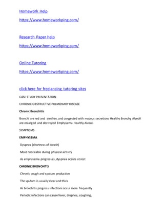
Chronic Bronchitis
Bronchi are red and swollen, and congested with mucous secretions Healthy Bronchy Alveoli
are enlarged and destroyed Emphysema Healthy Alveoli
SYMPTOMS
EMPHYSEMA
Dyspnea (shortness of breath)
Most noticeable during physical activity
As emphysema progresses, dyspnea occurs at rest
CHRONIC BRONCHITIS
Chronic cough and sputum production
The sputum is usually clear and thick
As bronchitis progress infections occur more frequently
Periodic infections can cause fever, dyspnea, coughing,
2. production of purulent sputum and wheezing
OVERVIEW OF THE CASE
Patient history:
• Medical Dx: stage 1 COPD (emphysema) 5 years ago
• Meds: Combivent (ipratropium bromide and albuterol sulfate)
• PMH: Bronchitis and upper respiratory infections during winter
• Smoker: 1 ppd for 46 years – has quit 1 year ago
• Family hx: CA – mother, 2 aunts died from lung cancer
OVERVIEW OF THE CASE
Physical exam:
• General appearance: 62-years-old female in no acute distress
• Vitals: Temp. 98.8ºF HR 92 bpm RR 22bpm BP 130/88
• Heart: Regular rate and rhythm; mild jugular distension noted
• Extremities: 1+ bilateral pitting edema. No cyanosis or clubbing
OVERVIEW OF THE CASE
• Neurologic: Alert, oriented; cranial nerves intact
• Skin: Warm, dry
• Chest/lungs: Decreased breath sounds, percussion hyperresonant; prolonged expiration
with wheezing; ronchi throughout; using accessory muscles at rest
• Abdomen: Liver, spleen palpable; nondistended, nontender, normal bowel sounds Hospital
course:
• Admitting dx: Acute exacerbation of COPD, increasing dyspnea, hypercapnia, r/o pneumonia
• Tx plan: O2 1L/min via nasal cannula, O2 saturation 90-91% IVF D5 ½ NS with 20mEq KCL @
75 cc/hr
Corticosteroid -Methylprednisone: Solumedrol
Antibiotic -Chephalosporin: Ancef
Bronchodilator: Ipratropium bromide, Albuterol sulfate
ABGs q 6 hours, CXR, sputum cultures and Gram stain
3. OVERVIEW OF THE CASE
The Physician ordered a nutrition consult
Nutrition history:
• Appetite is poor, fast satiety
• Meal preparations are difficult
• In the previous 2 days, she has eaten very little
• Coughing has made eating difficult
• Food doesn’t taste good, it has a bitter taste
• 5 years ago she weighted 145-150lb, now she is 119lb, 5’3”
• She didn’t weight herself for a while, but clothes are bigger, dentures fit
loosely her family tells her how thin she has gotten
• Avoids milk: “People say it will increase mucus production”
• No previous MNT
• No vit/min supplementation
ASSESSMENT
Physical Examination Data
Head and neck
Teeth: Poorly fitting dentures
Skin
Skin: dry
Edema, peripheral: 1+ bilateral
Vital Signs
↑ Temperature: 98.8ºF
↑ Respiratory Rate: 22 bpm
Shortness of breath
ASSESSMENT
Client History Data
4. Social history
Physical activity, easy fatigue with increased activity; unable to
achieve desired levels
Medical/ Health history
Chronic Obstructive Pulmonary Disease
Upper respiratory infections or pneumonia
Signs and symptoms
Shortness of breath or dyspnea on exertion or at rest
Meds and supplements
Medications that cause anorexia: Albuterol sulfate
What Is COPD?
COPD, or chronic obstructive pulmonary (PULL-mun-ary) disease, is a progressive disease that
makes it hard to breathe. "Progressive" means the disease gets worse over time.
COPD can cause coughing that produces large amounts of mucus (a slimy substance), wheezing,
shortness of breath, chest tightness, and other symptoms.
Cigarette smoking is the leading cause of COPD. Most people who have COPD smoke or used to
smoke. Long-term exposure to other lung irritants—such as air pollution, chemical fumes, or
dust—also may contribute to COPD.
Overview
To understand COPD, it helps to understand how the lungs work. The air that you breathe goes
down your windpipe into tubes in your lungs called bronchial (BRONG-ke-al) tubes or airways.
Within the lungs, your bronchial tubes branch into thousands of smaller, thinner tubes called
bronchioles (BRONG-ke-ols). These tubes end in bunches of tiny round air sacs called alveoli (al-
VEE-uhl-eye).
Small blood vessels called capillaries (KAP-ih-lare-ees) run through the walls of the air sacs.
When air reaches the air sacs, oxygen passes through the air sac walls into the blood in the
capillaries. At the same time, carbon dioxide (a waste gas) moves from the capillaries into the
air sacs. This process is called gas exchange.
The airways and air sacs are elastic (stretchy). When you breathe in, each air sac fills up with air
like a small balloon. When you breathe out, the air sacs deflate and the air goes out.
In COPD, less air flows in and out of the airways because of one or more of the following:
5. The airways and air sacs lose their elastic quality.
The walls between many of the air sacs are destroyed.
The walls of the airways become thick and inflamed.
The airways make more mucus than usual, which can clog them.
Normal Lungs and Lungs With COPD
Figure A shows the location of the lungs and airways in the body. The inset image shows a
detailed cross-section of the bronchioles and alveoli. Figure B shows lungs damaged by COPD.
The inset image shows a detailed cross-section of the damaged bronchioles and alveolar
walls.
In the United States, the term "COPD" includes two main conditions—emphysema (em-fih-
SE-ma) and chronic bronchitis (bron-KI-tis). (Note: The Health Topics article about bronchitis
discusses both acute and chronic bronchitis.)
In emphysema, the walls between many of the air sacs are damaged. As a result, the air sacs
lose their shape and become floppy. This damage also can destroy the walls of the air sacs,
6. leading to fewer and larger air sacs instead of many tiny ones. If this happens, the amount of
gas exchange in the lungs is reduced.
In chronic bronchitis, the lining of the airways is constantly irritated and inflamed. This causes
the lining to thicken. Lots of thick mucus forms in the airways, making it hard to breathe.
Most people who have COPD have both emphysema and chronic bronchitis. Thus, the general
term "COPD" is more accurate.
Outlook
COPD is a major cause of disability, and it's the third leading cause of death in the United
States. Currently, millions of people are diagnosed with COPD. Many more people may have the
disease and not even know it.
COPD develops slowly. Symptoms often worsen over time and can limit your ability to do
routine activities. Severe COPD may prevent you from doing even basic activities like walking,
cooking, or taking care of yourself.
Most of the time, COPD is diagnosed in middle-aged or older adults. The disease isn't passed
from person to person—you can't catch it from someone else.
COPD has no cure yet, and doctors don't know how to reverse the damage to the airways and
lungs. However, treatments and lifestyle changes can help you feel better, stay more active,
and slow the progress of the disease.
Other Names for COPD
Chronic bronchitis - Chronic productive cough for 3 months in 2 successive years
• Emphysema – Defined as a pathological term referring to permanent changes in terminal
bronchioles & alveoli
• Asthma – Inflammatory disease of airways, with reversible changes
Chronic bronchitis
Chronic obstructive airway disease
Chronic obstructive lung disease
Emphysema
What Causes COPD?
• Exposure to pipe, cigar, tobacco smoke
• Exposure to second hand smoke
• Exposure to heavy air pollution
• Exposure to heavy dust
7. • Exposure to chemical/toxic fumes
• Genetic conditions
Risk Factors
• Major
• • Age
• • Male
• • Occupation
• • Smoking
• • Alpha – one – antitrypsin
• Deficiency
Risk Factors
• Minor
• • Air Pollution
• • Alcohol
• • Race
• • Nutritional Status
• • FH
• • Bronchial Reactivity
What Are the Signs and Symptoms of COPD?
• Wheezing
• Coughing
• Sputum production
• Shortness of breath
• Chest tightness
• ANATOMY AND PHYSIOLOGY:
• The respiratory system consists of all the organs involved in breathing. These include the
nose, pharynx, larynx, trachea, bronchi and lungs. The respiratory system does two very
important things: it brings oxygen into our bodies, which we need for our cells to live and
8. function properly; and it helps us get rid of carbon dioxide, which is a waste product of
cellular function. The nose, pharynx, larynx, trachea and bronchi all work like a system
of pipes through which the air is funneled down into our lungs. There, in very small air
sacs called alveoli, oxygen is brought into the bloodstream and carbon dioxide is pushed
from the blood out into the air. When something goes wrong with part of the respiratory
system, such as an infection like pneumonia, chronic obstructive pulmonary diseases, it
makes it harder for us to get the oxygen we need and to get rid of the waste product
carbon dioxide. Common respiratory symptoms include breathlessness, cough, and
chest pain
• The Upper Airway and Trachea
• When you breathe in, air enters your body through your nose or mouth. From there, it
travels down your throat through the larynx (or voice box) and into the trachea (or
windpipe) before entering your lungs. All these structures act to funnel fresh air down
from the outside world into your body. The upper airway is important because it must
always stay open for you to be able to breathe. It also helps to moisten and warm the air
before it reaches your lungs.
• The Lungs
• Structure
• The lungs are paired, cone-shaped organs which take up most of the space in our chests,
along with the heart. Their role is to take oxygen into the body, which we need for our
cells to live and function properly, and to help us get rid of carbon dioxide, which is a
waste product. We each have two lungs, a left lung and a right lung. These are divided up
into µlobes¶, or big sections of tissue separated by µfissures¶ or dividers. The right lung
has three lobes but the left lung has only two, because the heart takes up some of the
space in the left side of our chest. The lungs can also be divided up into even smaller
portions, called broncho pulmonary segments .These are pyramidal-shaped areas which
are also separated from each other by membranes.There are about 10 of them in each
lung. Each segment receives its own blood supply and air supply.
• Blood supply The lungs are very vascular organs, meaning they receive a very large
blood supply. This is because the pulmonary arteries, which supply the lungs, come
directly from the right side of your heart. They carry blood which is low in oxygen and
high in carbon dioxide into your lungs so that the carbon dioxide can be blown off, and
more oxygen can be absorbed into the bloodstream. The newly oxygen-rich blood then
travels back through the paired pulmonary veins into the left side of your heart. From
there, it is pumped all around your body to supply oxygen to cells and organs.
• The Work ofBreathing The Pleurae
• The lungs are covered by smooth membranes that we call pleurae. The pleurae have two
layers, a visceral layer which sticks closely to the outside surface of your lungs, and a
µparietal layer which lines the inside of your chest wall (ribcage). The pleurae
are important because they help you breathe in and out smoothly, without any friction.
They also make sure that when your ribcage expands on breathing in, your lungs expand
as well to fill the extra space.
• The Diaphragm and Intercostal Muscles
• When you breathe in (inspiration), your muscles need to work to fill your lungs with air.
Thediaphragm, a large, sheet-like muscle which stretches across your chest under the
ribcage, doesmuch of this work. At rest, it is shaped like a dome curving up into your
chest. When you breathein, the diaphragm contracts and flattens out, expanding the
9. space in your chest and drawing air into your lungs. Other muscles, including the muscles
between your ribs (the intercostal muscles)also help by moving your ribcage in and out.
Breathing out (expiration) does not normallyrequire your muscles to work. This is
because your lungs are very elastic, and when your
• muscles relax at the end of inspiration your lungs simply recoil back into their resting
position, pushing the air out as they go.
• The Respiratory Systemand Ageing
• The normal process of ageing is associated with a number of changes in both the
structure andfunction of the respiratory system. These include:
• Enlargement of the alveoli. The air spaces get bigger and lose their elasticity,
meaningthat there is less area for gases to be exchanged across. This change is sometimes
referredto as ¶senile emphysema¶.
• The compliance (or springiness) of the chest wall decreases, so that it takes more effort
to breathe in and out.
• The strength of the respiratory muscles (the diaphragm and intercostal muscles)decreases.
This change is closely connected to the general health of the person.All of these changes
mean that an older person might have more difficulty coping with increasedstress on their
respiratory system, such as with an infection like pneumonia, than a younger person
would.
• DIAGNOSTIC EVALUATION
• PFTs demonstrative airflow obstruction ± reduced forced vital capacity
(FVC), FEV1,FEV1 to FVC ration; increased residual volume to total lung capacity
(TLC) ratio, possibly increased TLC.2.
• ABG levels- decreased PaO2, pH, and increased CO2.3.
• Chest X-ray ± in late stages, hyperinflation, flattened diaphragm, increased
rettrosternalspace, decreased vascular markings, possible bullae.4.
• Alpa1-antitrypsin assay useful in identifying genetically determined deficiency
inemphysema
• ABG’s
•Mild decrease pO2 early, gradual
decrease
•Gradual increase pCO2
•May change during sleepErythrocytosis with Hct > 55 as pO2 levels fall; non
specific
CT Scan
•High resolution 1-2 mm slices
•More sensitive than CXR
•Does not alter basic therapyCXR Not useful for diagnosing early COPD
Findings
•Over distended lungs
•Flattened diaphragms
•Long narrow cardiac silhouette
•Increased retro sternal airspace
•Bullae
10. PATHOPHYSIOLOGY In COPD
, the airflow limitation isboth progressive and associatedwith anabnormal inflammatory response of
the lungs to noxious particles or gases.The inflammatory response occurs throughout the airways,
parenchyma, and pulmonary vasculature. Because of the chronic inflammation and the body
attempts to repairit, narrowing occurs inthe small peripheral airways. Over time, this injury-and-repair
processcauses scartissue formation and narrowing ofthe airway lumen. Airflow obstruction mayalso
becaused by parenchymal destruction, asisseenwith emphysema, a diseaseof the alveoli orgas
exchange units. In additionto inflammation, processes related to imbalances of proteinases and anti
proteinases inthe lung mayberesponsible for airflow limitation. When activated by chronic
inflammation, proteiness and other substances may bereleased, damagingthe parenchymal of the
lung. The parenchymal changes may occur asaconsequence of inflammation or environmental or
genetic factors (eg. Alpha1-antitrypsin deficiency)
Early in the course of COPD , the inflammatory response causes pulmonary vasculature changes
that are characterized by thickening of the vessel wall. These changes may result from exposure to
cigarette smoke, use of tobaccoproducts, and the release ofinflammatory medicators.
CHRONIC BRONCHITIS
Lung damageand inflammation inthe large airways results inchronic bronchitis. Chronic bronchitis is
defined inclinical terms asa cough withsputum production onmost days for 3months of a year, for
2 consecutive years. In the airways ofthe lung, the hallmark of chronic bronchitis isanincreased
number (hyperplasia)and increased size (hypertrophy) of the goblet cells and mucous glands of the
airway. Asa result, there ismore mucus than usual inthe airways, contributing to narrowing of the
airways and causing a cough with sputum. Microscopically there isinfiltration of the airwaywalls with
inflammatory cells. Inflammation isfollowed by scarring and remodeling that thickens the walls and
also results innarrowing of the airways. Aschronic bronchitis progresses, there issquamous
metaplasia (anabnormal change inthe tissue lining the insideof the airway) and fibrosis(further
thickening and scarring of the airwaywall). The consequence of these changes isalimitation of
airflow. Patients with advanced COPD that have primarily chronic bronchitis rather than emphysema
were commonly referred to as³blue bloaters´ because of the bluish color of the skin and lips
(cyanosis) seen in them. The hypoxia and fluid retention leads to them beingcalled ³Blue
Bloaters.´
ACUTE BRONCHITIS
PHYSICAL MANIFESTATIONS
One of the most common symptoms of COPD
isshortness of breath(dyspnea). Peoplewith COPD
commonly describe this as: ³My breathing requires effort´, ³I feel out of breath´, or ³I can
not getenough airin´. Peoplewith COPD typically first notice dyspnea during vigorous exercise when
11. the demands onthe lungs aregreatest. Over the years, dyspnea tends to getgradually worse sothat
itcan occur during milder, everyday activities such as housework. In the advanced stages of COPD ,
dyspnea can becomesobad that itoccurs during rest and isconstantly present. Other symptoms of
COPD are a persistent cough, sputum or mucus production, wheezing, chest tightness, and
tiredness. Peoplewith advanced (very severe) COPD sometimes develop respiratory failure. When
this happens, cyanosis, abluish discoloration of the lips caused byalack of oxygen inthe blood, can
occur. Anexcess of carbon dioxideinthe blood cancause headaches, drowsiness or twitching
(asterixis). Acomplicationof advanced COPD is corpulmonal, astrain onthe heart due tothe extra
work required by the heart to pump bloodthrough the affected lungs.
Symptoms of cor pulmonale areperipheral edema, seenasswelling of the ankles, and dyspnea.
There are afew signsof COPD that ahealthcare worker may detect although they can beseenin
other diseases.Somepeople have COPD and have none of these signs. Commonsigns are:
tachypnea, arapidbreathing rate wheezing sounds orcrackles in the lungs heard through a
stethoscope breathing out taking a longer timethan breathing in enlargement of the chest, particularly
the front-to-back distance (hyperinflation) active use of muscles inthe neck to help with breathing
breathing through pursed lips increased antero posterior tolateral ratio of the chest(i.e. barrel chest).
Emphysema
isachronic obstructive pulmonary isease (COPD,asit isotherwise known, formerly termed achronic
obstructive lung disease).It isoften caused by exposure to toxic chemicals ,including long-term
exposure to tobacco smoke. Emphysema ischaracterized byloss of elasticity(increased pulmonary
compliance) of the lung tissue caused by destruction of structures feeding the alveoli, owing to the
actionof alpha 1antitrypsin deficiency. This causes the small airways to collapse during forced
exhalation, asalveolar collapsibility has decreased.As aresult, airflow is impededand airbecomes
trapped inthe lungs, inthe same way asother obstructive lung diseases. Symptoms include
shortness of breathon exertion, and anexpanded chest. However, the constriction of airpassages
isnt always immediately deadly, and treatment isavailable.
• NURSINGINTERVENTIONS
• Monitoring
1.Monitor for adverse effects of bronchodilators ± tremulousness, tachycardia, cardiac
arrhythmias, central nervous system stimulation, hypertension.2.
• Monitor condition after administration of aerosol bronchodilators to assess for improved
aeration, reduced adventitious sounds, reduced dyspnea.3.
• Monitor serum theophylline level, as ordered, to ensure therapeutic level and
preventtoxicity.4.
• Monitor oxygen saturation at rest and with activity.
•
• Supportive Care
• Eliminate all pulmonary irritants, particularly cigarette smoke. Smoking cessation usually
reduces pulmonary irritation, sputum production, and cough. Keep the patients room
asdust-free as possible.
• 2.
12. • Use postural drainage positions to help clear secretions responsible
for airwayobstructions.3.
• Teach controlled coughing.4.
• Encourage high level of fluid intake ( 8 to 10 glasses; 2 to 2.5 liters daily) within level
of cardiac reserve.5.
• Give inhalations of nebulized saline to humidify bronchial tree and liquefy sputum. Add
moisture (humidifier, vaporizer) to indoor air.6.
• Avoid dairy products if these increases sputum production.7.
• Encourage the patient to assume comfortable position to decrease dyspnea.8.
• Instruct and supervise patient breathing retraining exercises.9.
• Use pursed lip breathing at intervals and during periods of dyspnea to control rate and
depth of respiration and improve respiratory muscle coordination.10.
• Discuss and demonstrate relaxation exercises to reduce stress, tension, and anxiety.11.
• Maintain the patient nutritional status.12.
• Reemphasize the importance of graded exercise and physical conditioning programs.13.
• Encourage use of portable oxygen system for ambulation for patients with hypoxemia
and marked disability.14.
• Train the patient in energy conservation technique.15.
• Assess the patient for reactive-behaviors such as anger, depression and acceptance.
Education and heaLthmaintenance
• Review with the patient the objectives of treatment and nursing management.2.
• Advise the patient to avoid respiratory irritants. Suggest that high efficiency particular the
air filter may have some benefit.3.
• Warn patient to stay out of extremely hot or cold weather and to avoid
aggravating bronchial obstruction and sputum obstruction.4.
• Warn patient to avoid persons with respiratory infections, and to avoid crowds and areas
with poor ventilation.5.
• Teach the patient how to recognize and report evidence of respiratory infection promptly
such as chest pain, changes in character of sputum (amount, color and
consistency),increasing difficulty in raising sputum, increasing coughing and wheezing,
increasing of shortness of breath.
Homework Help
https://www.homeworkping.com/
Math homework help
https://www.homeworkping.com/
Research Paper help