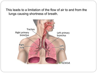
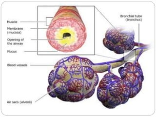
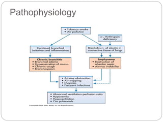
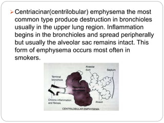
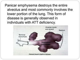
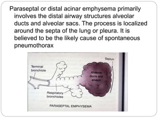
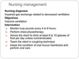
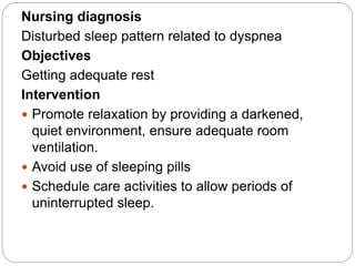
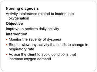
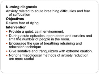
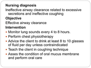
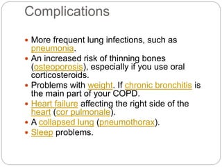
Chronic obstructive pulmonary disease (COPD) refers to two lung diseases, chronic bronchitis and emphysema, that are typically caused by smoking. In COPD, airflow to the lungs is limited by inflammation and damage to airways and lung tissue. Symptoms include cough, sputum production, wheezing, shortness of breath, and weight loss. Treatment focuses on smoking cessation, bronchodilators, oxygen therapy, and managing exacerbations. Nursing care aims to improve ventilation and gas exchange, manage anxiety, and ensure effective airway clearance and rest.
Despite modern anti-tuberculous chemotherapy, approximately 2% of all cases of pulmonary mycobacterial infection require surgical treatment.Therefore, surgical treatment of pulmonary mycobacterial disease is rarely necessary.Types of surgical procedures for PTB include: Collapse therapy, pulmonary resection, lung decortication, drainage procedures such as closed tube thoracostomy, rib resection and open window thoracotomy beside pulmonary resection+ collapse therapy (thoracoplasty). The decreasing morbidity and mortality of pulmonary resection for PTB is due to careful patient selection ( failure of chemotherapy, massive haemoptysis, BPF), improved anaesthetic techniques, stapling devices and better chemotherapy.The prognosis after successful resection is excellent ( 90% survive and remain disease free).
It is the comprehensive note of abdominal tuberculosis..Students can take help from it to study and preparing notes. It is totally a adequate notes to learn and teach both for the bsc nursing and msc nursing students.
Atelectasis is a complete or partial collapse of the entire lung or area (lobe) of the lung. It occurs when the tiny air sacs (alveoli) within the lung become deflated or possibly filled with alveolar fluid.
Bronchitis is an inflammation of the bronchial tubes, the airways that carry air to your lungs. It causes a cough that often brings up mucus. It can also cause shortness of breath, wheezing, a low fever, and chest tightness. There are two main types of bronchitis: acute and chronic.
Pneumonia is an infection that inflames the air sacs in one or both lungs. The air sacs may fill with fluid or pus (purulent material), causing cough with phlegm or pus, fever, chills, and difficulty breathing. A variety of organisms, including bacteria, viruses and fungi, can cause pneumonia.
Pneumonia can range in seriousness from mild to life-threatening. It is most serious for infants and young children, people older than age 65, and people with health problems or weakened immune systems.
What is emphysema?
Emphysema is a condition that forms part of chronic obstructive pulmonary disease (COPD) and involves the enlargement of the air sacs in the lung.
The alveoli at the end of the bronchioles of the lung become enlarged because of the breakdown of their walls. The fewer and larger damaged sacs that result mean there is a reduced surface area for the exchange of oxygen into the blood and carbon dioxide out of it.
Definition
Emphysema is a condition in which the alveoli become stiff expands and continuously filled the air even after expiration. Emphysema is a chronic obstructive disease due to lack of elasticity in the lungs and alveoli surface area.
Classification
Panlobular (panacinar)
It is damage to the respiratory bronchi, alveolar ducts and alveoli. All air space in the little lobes much enlarged, with little inflammatory disease. The characteristics that have chest hyperinflation, and is characterized by dyspnea on exertion, and weight loss.
CENTRILOBULAR (CENTROACINAR)
The pathological changes mainly occur in the centre of the secondary lobes, and peripheral of acini remain good. Often there is chaos-ventilation perfusion ratio, which lead to hypoxia, hypercapnia (increased CO2 in the arterial blood), polycythaemia and heart failure episodes right. The condition leads to cyanosis, peripheral oedema, and respiratory failure.
CAUSES OF EMPHYSEMA
The biggest known cause or risk factor for emphysema - and for COPD - is smoking. Cigarette smoking is responsible for around 90% of cases of COPD. However, COPD will develop only in smokers who are genetically susceptible - smoking does not always lead to the disease.
CHRONIC OBUSTRUCTIVE PULMONARY DISEASE POWER POINT.pptxAgbaMakuochi
This describes a whole lot more of what Chronic Obstructive Pulmonary Disease is with their pathophysiology and management both medical and nursing management
Thank you for selecting our 𝐂𝐎𝐏𝐃 PPT
This medical PowerPoint template about 𝐂𝐎𝐏𝐃
You can download our template by visiting our website:
https://www.rxslides.com/product/copd-powerpoint-template
copy and paste this URL into the browser and download the full editable template.
This 𝐂𝐎𝐏𝐃 animated template is designed by RxSlides, a medical professional team covering the following topics about 𝐂𝐎𝐏𝐃
𝐓𝐡𝐞 𝐭𝐞𝐦𝐩𝐥𝐚𝐭𝐞 𝐜𝐨𝐯𝐞𝐫𝐬 𝐭𝐡𝐞 𝐟𝐨𝐥𝐥𝐨𝐰𝐢𝐧𝐠 𝐭𝐨𝐩𝐢𝐜𝐬
𝐃𝐞𝐟𝐢𝐧𝐢𝐭𝐢𝐨𝐧
Chronic obstructive pulmonary disease (𝐂𝐎𝐏𝐃) is a group of lung diseases that cause airflow blockage and breathing-related problems.
𝐩𝐫𝐞𝐯𝐚𝐥𝐞𝐧𝐜𝐞
COPD is more prevalent in developing countries, but it is also a growing problem in developed countries. In the United States, COPD is the third leading cause of death.
Forms of 𝐂𝐎𝐏𝐃
• Chronic bronchitis
• Emphysema
𝐏𝐚𝐭𝐡𝐨𝐩𝐡𝐲𝐬𝐢𝐨𝐥𝐨𝐠𝐲
In normal lungs, air flows freely in and out of the bronchi and alveoli. However, in people with COPD, the airflow is blocked. This can be caused by inflammation of the airways, mucus production, or damage to the air sacs.
𝐑𝐢𝐬𝐤 𝐅𝐚𝐜𝐭𝐨𝐫𝐬
There are several risk factors for COPD, including smoking, air pollution, and genetics. Smoking is the most common risk factor for COPD. Smoking is responsible for about 80% of COPD cases. Air pollution, particularly indoor air pollution from cooking and burning fuels, can also increase the risk of COPD. Genetics can also play a role in COPD. People with a family history of COPD are more likely to develop the disease.
𝐜𝐚𝐮𝐬𝐞𝐬
The main causes of COPD are smoking and air pollution. Smoking damages the lungs and makes it difficult to breathe
𝐒𝐭𝐚𝐠𝐞𝐬
• Stage 1
• Stage 2
• Stage 3
.
𝐒𝐲𝐦𝐩𝐭𝐨𝐦𝐬
The most common symptoms of COPD are:
• Shortness of breath
• Cough
• Wheezing
• Chest tightness
• Fatigue
𝐂𝐨𝐦𝐩𝐥𝐢𝐜𝐚𝐭𝐢𝐨𝐧𝐬
COPD can lead to several complications, including:
• Anemia
• Right-sided heart failure
• Muscle weakness
• Lung infections
• Bone thinning
• Collapsed lungs
𝐃𝐢𝐚𝐠𝐧𝐨𝐬𝐭𝐢𝐜 𝐦𝐞𝐭𝐡𝐨𝐝𝐬
COPD is diagnosed with a spirometry test, which measures how much air a person can exhale.
𝐓𝐫𝐞𝐚𝐭𝐦𝐞𝐧𝐭 𝐎𝐩𝐭𝐢𝐨𝐧𝐬
There is no cure for COPD, but there are treatments that can help manage the symptoms and slow the progression of the disease. Treatment options include:
• Bronchodilators
• Antibiotics
• Supplemental oxygen
• Vaccination
𝐏𝐫𝐞𝐯𝐞𝐧𝐭𝐢𝐯𝐞 𝐌𝐞𝐭𝐡𝐨𝐝𝐬
The best way to prevent COPD is to avoid smoking and air pollution. Several lifestyle changes can help reduce the risk of COPD, such as eating a healthy diet and exercising regularly.
Visit our site for more animated templates
𝗵𝘁𝘁𝗽𝘀://𝘄𝘄𝘄.𝗿𝘅𝘀𝗹𝗶𝗱𝗲𝘀.𝗰𝗼𝗺
RxSlides PowerPoint icons and illustrations related to 𝐂𝐎𝐏𝐃 will help you customize the content of this editable presentation according to your content and audience interest.
This is about the sickness or diseases in the health of your lungs the most. On how it affects the airways, alveoli, interstitium, your blood vessels, the pleurae and the chest wall. Also about the respiratory system procedures, that we should mainly know for us to prevent those respiratory system
This presentation is about Emphysema and Chronic Bronchitis made by CHAKRAPANI BHUVANESH.
Bronchitis is an inflammation of the lining of your bronchial tubes, which carry air to and from your lungs. People who have bronchitis often cough up thickened mucus, which can be discolored. Bronchitis may be either acute or chronic.
NIDM (National Institute Of Digital Marketing) Bangalore Is One Of The Leading & best Digital Marketing Institute In Bangalore, India And We Have Brand Value For The Quality Of Education Which We Provide.
www.nidmindia.com
New Explore Careers and College Majors 2024.pdfDr. Mary Askew
Explore Careers and College Majors is a new online, interactive, self-guided career, major and college planning system.
The career system works on all devices!
For more Information, go to https://bit.ly/3SW5w8W
1. Mr. Yahye Sheikh Abdulle
Msc Nursing 1st year
Kle college of Nursing
Chronic obstructive
pulmonary diseases
2. Chronic obstructive pulmonary
diseases
COPD is also known as chronic obstructive
lung disease (COLD), chronic obstructive
airway disease (COAD), chronic airflow
limitation (CAL) and chronic obstructive
respiratory disease (CORD)
Chronic obstructive pulmonary disease
(COPD) refers to chronic bronchitis and
emphysema, a pair of two commonly co-
existing diseases of the lungs in which the
airways become narrowed.
3. This leads to a limitation of the flow of air to and from the
lungs causing shortness of breath.
4. In COPD, less air flows in and out of the
airways because of one or more of the
following:
The airways and air sacs lose their elastic
quality.
The walls between many of the air sacs are
destroyed.
The walls of the airways become thick and
inflamed.
The airways make more mucus than usual,
which tends to clog them.
5.
6. Incidence
It is the 4th leading cause of mortality
and 12th leading cause of disability in the
united states.
In 2020 COPD is the 3rd leading cause
of death.
7. CAUSES
Smoking. Smoking is primary risk factors for
COPD. The numerous irritants found in cigarette
smoke stimulate excess mucus production and
coughing, destroy ciliary function and lead to
inflammation and damage of bronchiolar and
alveolar walls.
8.
9. Air pollution high levels of urban air
pollution are harmful to persons with
existing lung disease. However, the
effect of outdoor air pollution as a risk
factor for COPD. Another risk factor
for COPD development is fossil fuels
that used for indoor heating and
cooking.
10. Occupational exposures- exposure
to workplace dusts found in coal
mining, gold mining, and the cotton
textile industry and chemicals such as
cadmium, and fumes from welding
have been implicated in the
development of airflow obstruction.
Exposure of these irritants causes the
airway to be hyper responsive.
11. Infection :infections is risk factor for
developing COPD. Severe recurring
respiratory tract infection in childhood
have been associated with reduced
lung function and increased
respiratory symptoms in adulthood.
Recurring infections impair normal
defense mechanisms, making
bronchioles and alveoli more
susceptible to injury.
12. Genetics-Alpha 1-antitrypsin deficiency
is a genetic condition that is responsible for
about 2% of cases of COPD. In this
condition, the body does not make enough
of a protein, alpha 1-antitrypsin. Alpha 1-
antitrypsin protects the lungs from damage
caused by protease enzymes, such as
elastase and trypsin, that can be released
as a result of an inflammatory response to
tobacco smoke
15. pathophysiology
COPD is characterized by airflow limitation that
is poorly reversible. chronic exposure to
cigarette smoking is the number one cause of
the disease, but repeated exposure to
secondhand smoke, air pollution and
occupational exposure (to coal, cotton, grain)
are also important risk factors.
16. Smoking and other airway irritants cause
neutrophils, T-lymphocytes, and other
inflammatory cells to accumulate in the
airways. Once activated, they trigger an
inflammatory response in which an influx of
molecules, known as inflammatory
mediators, navigate to the site in an
attempt to destroy and remove inhaled
foreign debris.
17. Under normal circumstances, the
inflammatory response is useful and leads
to healing. In fact, without it, the body
would never recover from injury. In COPD,
repeated exposure to airway irritants
perpetuates an ongoing inflammatory
response that never seems to shut itself
off. Over time, this process causes
structural and physiological lung changes
that get progressively worse.
18. As inflammation continues, the airways
constrict, becoming excessively narrow
and swollen. This leads to excess mucus
production and poorly functioning cilia, a
combination that makes airway clearance
especially difficult. When people with
COPD cannot clear their secretions, they
develop the hallmark symptoms of COPD,
including a chronic, productive cough,
wheezing and dyspnea.
19. Finally, the build-up of mucus attracts a host of
bacteria that thrive and multiply in the warm, moist
environment of the airway and lungs.
The end result is further inflammation, the formation of
diverticula in the bronchial tree, and bacterial lung
infection, a common cause of COPD exacerbation.
20. CLINICAL FEATURES
Chronic cough
Sputum production
Wheezing
Chest tightness
Dyspnoea on exertion
Wt.loss
Respiratory insufficiency
Respiratory infections
Barrel chest- chronic hyperinflation leads to
loss of lung elasticity.
21. Bronchitis
Bronchitis results from inflammation of bronchi
leading to increased musus production, cough
and eventual scaring of the bronchial lining.
acute (short term) Infections or lung irritants
cause acute bronchitis.
chronic is an ongoing, serious condition. It
occurs if the lining of the bronchial tubes is
constantly irritated and inflamed, causing a
long-term cough with mucus
It is defined as the presence of cough and
sputum production for atleast 3 months.
22. Chronic bronchitis is characterized by
the following :
A increased in size and number of
submucus glands in the large bronchi,
which increase mucus production.
An increased number of goblet cells which
also secrete mucus.
Impaired cillary function which reduce
mucus clearance.
23.
24. PATHOPHYSOPLOGY
Irritants irrritate the airway
Excess mucus production
Inflammation
Cause the mucus secreting glands and goblet cells to
increase in number.
Ciliary function is reduced
25. More mucus production
Bronchial walls become thickened and lumen
narrows and mucus plug the airway
Alveoli adjacent tto the bronchioles may become
damaged and fibrosed.
Alter function of alveolar macrophages.
Infection
26. Signs and symptoms-Acute
sore throat,
fatigue (tiredness),
fever, body aches,
stuffy or runny nose,
vomiting, and
Diarrhea
persistent cough
cough may produce clear mucus
shortness of breath
27. Chronic symptoms
coughing,
wheezing, and
chest discomfort.
The coughing may produce large amounts of
mucus. This type of cough often is called a
smoker's cough.
28. EMPHYSEMA
Definition:-
Emphysema is defined as enlargement of
the air spaces distal to the terminal
bronchioles, with destruction of their walls
of the alveoli.
As the alveoli are destroyed the alveolar
surface area in contact with the capillaries
decreases.
Causing dead spaces (no gas exchange
takes place) Leads to hypoxia.
29. In later stages:
CO2 elimination is disturbed and increase in CO2
tension in arterial blood causing respiratory
acidosis
30. There are three types of emphysema
1. Centriacinar
2. Panacinar
3. Paraseptal
31. Centriacinar(centrilobular) emphysema the most
common type produce destruction in bronchioles
usually in the upper lung region. Inflammation
begins in the bronchioles and spread peripherally
but usually the alveolar sac remains intact. This
form of emphysema occurs most often in
smokers.
32. Panicar emphysema destroys the entire
alveolus and most commonly involves the
lower portion of the lung. This form of
disease is generally observed in
individuals with ATT deficiency.
33. Paraseptal or distal acinar emphysema primarily
involves the distal airway structures alveolar
ducts and alveolar sacs. The process is localized
around the septa of the lung or pleura. It is
believed to be the likely cause of spontaneous
pneumothorax
34. DIAGNOSIS
Blood Test
Blood tests can help determine if symptoms are
being caused by an infection.
An arterial blood gas test will measure the
amount of oxygen in blood. This is one
indication of how well lungs are working. This
can help doctor determine how severe COPD
is and whether need oxygen therapy.
35. . Chest X-ray or CT scan
A CT scan is a type of X-ray that creates a more
detailed image than a standard X-ray. Whichever
type doctor chooses, an X-ray will give a picture
of the structures inside chest. These include your
heart, lungs, and blood vessels. Your doctor will
be able to see if client has evidence of COPD. If
symptoms are being caused by another condition
such as heart failure, the doctor will be able
identify that as well
36. Sputum Examination
Doctor may order a sputum examination,
especially if the client has a productive cough.
Sputum is the mucus the client cough up.
Analyzing sputum can help identify the cause
of breathing difficulties and may rule out some
lung cancers. If there is a bacterial infection,
it can be identified and treated.
ECG
The doctor might request an ECG to determine
if your shortness of breath is being caused by
a heart condition.
37. Pulmonary Function Test
pulmonary function tests (PFTs) measure
how well the lungs are moving air in and
out. They also measure how well the lungs
are moving oxygen to the blood.
Spirometry (meaning the measuring of
breath) is the most common of the
pulmonary function tests (PFTs). It
measures lung function, specifically the
amount (volume) and/or speed (flow) of air
that can be inhaled and exhaled.
40. MEDICAL MANAGEMENT
The treatment goal for the client with COPD
are:
To improve ventilation
To facilitate the removal of bronchial
secretions
To promote health maintenance
To reduce complications, and
To slow progression of the disease
41. Smoking cessation
Cessation of cigarette smoking is single
most effective and cost effective
intervention to reduce the risk of
developing COPD and stop the
progression of the disease. After
discontinuation of smoking, the
accelerated decline in pulmonary function
slows and pulmonary function usually
improves.
42. Drug therapy
Medications for COPD:
Reduce symptoms
Increase the capacity of exercise,
Improve overall health and
Reduce the severity of exacerbations.
43. Bronchiodilator drug therapy are agents
that widen the air passages by relaxing the
bronchial smooth muscle and improve the
ventilation of lungs. They are usually
administered via inhalation route but in
rare occasions may be given orally or
administered intravenously. Regular
treatment with long acting bronchiodilators
is more effective and convenient than
treatment with short acting forms.
44. The principal bronchiodilator treatment are Beta2
agonists, anticholinergics, and
methylxanthines used singly or combination.
The choice of bronchioldilators depends on
availability and patient’s response.
Beta2 agonists are sympathomimetic drugs that
acts on the beta-adrenoceptors in the smooth
muscles of the airway and cause
bronchioldilation. These drugs may also enhance
mucus clearance and improve the endurance of
respiratory muscles.
45. Short acting Beta2 agonists(e.g., albuterol)have
minimal adverse effects with rapid onset of
action, a peak effect in 60 to 90 minutes and
duration of 4 to 6 hour.
Side effects that may develop with the use of
these drugs are tachycardia, tremor,
nervousness, and nausea.
Anticholinergic agents offer greater
bronchiodilator effect and fewer side effect than
short acting inhaled beta2 agonists.
These drugs work by blocking the cholinergic
receptors located in the larger airways, resulting
in bronchiodilation. Ipratropium(atrovent) is the
most commonly used drug in this category.
46. Oxygen therapy
O2 therapy is frequently used in the treatment of
COPD another problem associated with
hypoxemia. Long term o2 therapy improves
survival, exercise capacity, cognitive
performance and sleep in hypoxemic patients.O2
is colorless, odorless testless gas that
constitutes 20.95% of the atmosphere. O2 raises
the partial pressure of oxygen(PO2) in inspired
air.
Indications for use: goals for 02 therapy are
To reduce the work of breathing,
To maintain the PaO2
To reduce the workload of heart
To keep the SaO2 more than 90% during rest
47. Oxygen is usually administered to treat
hypoxemia caused by
Respiratory disorders such as COPD
pulmonary hypertension pneumonia and
pulmonary emboli
Cardiovascular disorders such as
myocardial infraction, angina pectoris and
cardiogenic shock
Central nervous system disorders such as
overdose of opiods
48. SURGICAL MANAGEMENT
bullectomy
Bullae are enlarged airspaces that do not
contribute to ventillation but occupy space in
the thorax,these areas may be surgically
excised
lung volume reduction surgery
It involves the removal of a portion of the
diseased lung parenchyma.this allows the
functional tissue to expand.
lung transplantation
49. Nursing management
Nursing diagnosis
Impaired gas exchange related to decreased ventilation
Objectives
Improve ventilation
Intervention
a. Monitor lung sounds every 4 to 8 hours.
b. Perform chest physiotherapy
c. Advice the client to drink at least 8 to 10 glasses of
fluid per day unless contraindicated
d. Teach the client in coughing technique
e. Asses the condition of oral mucus membrane and
perform oral care
50. Nursing diagnosis
Disturbed sleep pattern related to dyspnea
Objectives
Getting adequate rest
Intervention
Promote relaxation by providing a darkened,
quiet environment, ensure adequate room
ventilation.
Avoid use of sleeping pills
Schedule care activities to allow periods of
uninterrupted sleep.
51. Nursing diagnosis
Activity intolerance related to inadequate
oxygenation
Objective
Improve to perform daily activity
Intervention
Monitor the severity of dyspnea
Stop or slow any activity that leads to change in
respiratory rate
Advice the client to avoid conditions that
increase oxygen demand
52. Nursing diagnosis
Anxiety related to acute breathing difficulties and fear
of suffocation
Objectives
Relieve fear of dying
Intervention
Provide a quiet, calm environment.
During acute episodes, open doors and curtains and
limit the number of people in the room.
Encourage the use of breathing retraining and
relaxation technique
Give sedative and tranquilizers with extreme caution.
Nonpharmacological methods of anxiety reduction
are more useful
53. Nursing diagnosis
Ineffective airway clearance related to excessive
secretions and ineffective coughing
Objective
Effective airway clearance
Intervention
Monitor lung sounds every 4 to 8 hours.
Perform chest physiotherapy
Advice the client to drink at least 8 to 10 glasses
of fluid per day unless contraindicated
Teach the client in coughing technique
Asses the condition of oral mucus membrane
and perform oral care
54. Complications
More frequent lung infections, such as
pneumonia.
An increased risk of thinning bones
(osteoporosis), especially if you use oral
corticosteroids.
Problems with weight. If chronic bronchitis is
the main part of your COPD.
Heart failure affecting the right side of the
heart (cor pulmonale).
A collapsed lung (pneumothorax).
Sleep problems.